2099
Automatic segmentation of spinal cord nerve rootlets1NeuroPoly Lab, Polytechnique Montreal, Montreal, QC, Canada, 2Mila - Quebec AI Institute, Montreal, QC, Canada, 3Department of Neurosurgery, Faculty of Medicine and Dentistry, Palacký University Olomouc, Olomouc, Czech Republic, 4Department of Neurology, Faculty of Medicine and Dentistry, Palacký University Olomouc, Olomouc, Czech Republic, 5Laboratoire de Neurosciences Cognitives (UMR 7291), CNRS – Aix Marseille Université, Marseille, France, 6Department of Neuroimaging, Institute of Psychiatry, Psychology & Neuroscience, King’s College London, London, United Kingdom, 7Wellcome Centre for Human Neuroimaging, University College London, London, United Kingdom, 8Functional Neuroimaging Unit, CRIUGM, Université de Montréal, Montreal, QC, Canada, 9Centre de Recherche du CHU Sainte-Justine, Université de Montréal, Montreal, QC, Canada
Synopsis
Keywords: Analysis/Processing, Spinal Cord, Deep Learning; Nerve Rootlets; Segmentation
Motivation: Precise identification of spinal nerve rootlets is relevant for studying functional activity in the spinal cord.
Goal(s): Our goal was to develop a deep learning-based tool for the automatic segmentation of spinal nerve rootlets from multi-site T2-w images coupled with a method for the automatic identification of spinal levels.
Approach: Active learning was employed to iteratively train a nnUNet model to perform multi-class spinal nerve rootlets segmentation.
Results: The code/model is available on GitHub and is currently being validated by several laboratories worldwide.
Impact: Currently, most spinal cord fMRI studies use vertebral levels for groupwise registration, which is inaccurate. This new tool enables researchers to identify spinal levels via the automatic segmentation of nerve rootlets, improving fMRI analysis pipeline accuracy.
Introduction
Currently, most fMRI studies in the spinal cord utilize vertebral levels for the registration to the template to perform group-level analysis. However, the vertebral levels are not necessarily aligned with spinal levels studied by fMRI1,2. Due to the absence of tools for automatic spinal level identification, researchers either have to perform manual time-consuming and error-prone landmark identification or rely on the vertebral levels defined based on vertebral bodies, introducing potential bias.In this study, we developed a deep learning-based tool for the automatic segmentation of cervical spinal cord nerve rootlets together with a subsequent method for the identification of corresponding spinal levels.
Methods
3T T2-weighted isotropic 0.6 to 0.8 mm MRI scans from two open-access datasets (Spinal Cord Head Position MRI dataset (OpenNeuro ds004507) and spine-generic multi-subject dataset3) were used.Active learning4 was employed to iteratively train a self-configuring nnUNet5 v2 3D model. During each iteration, automatically segmented spinal cord nerve rootlets were manually corrected using the FSLeyes image viewer, and the model was re-trained. The final dataset consisted of 33 images and was used to train the multi-class nnUNet 3D model, with each class representing a specific nerve rootlet (e.g., 2 for C2 rootlets, 3 for C3 rootlets, and so forth). This model was trained over five folds and 2000 epochs.
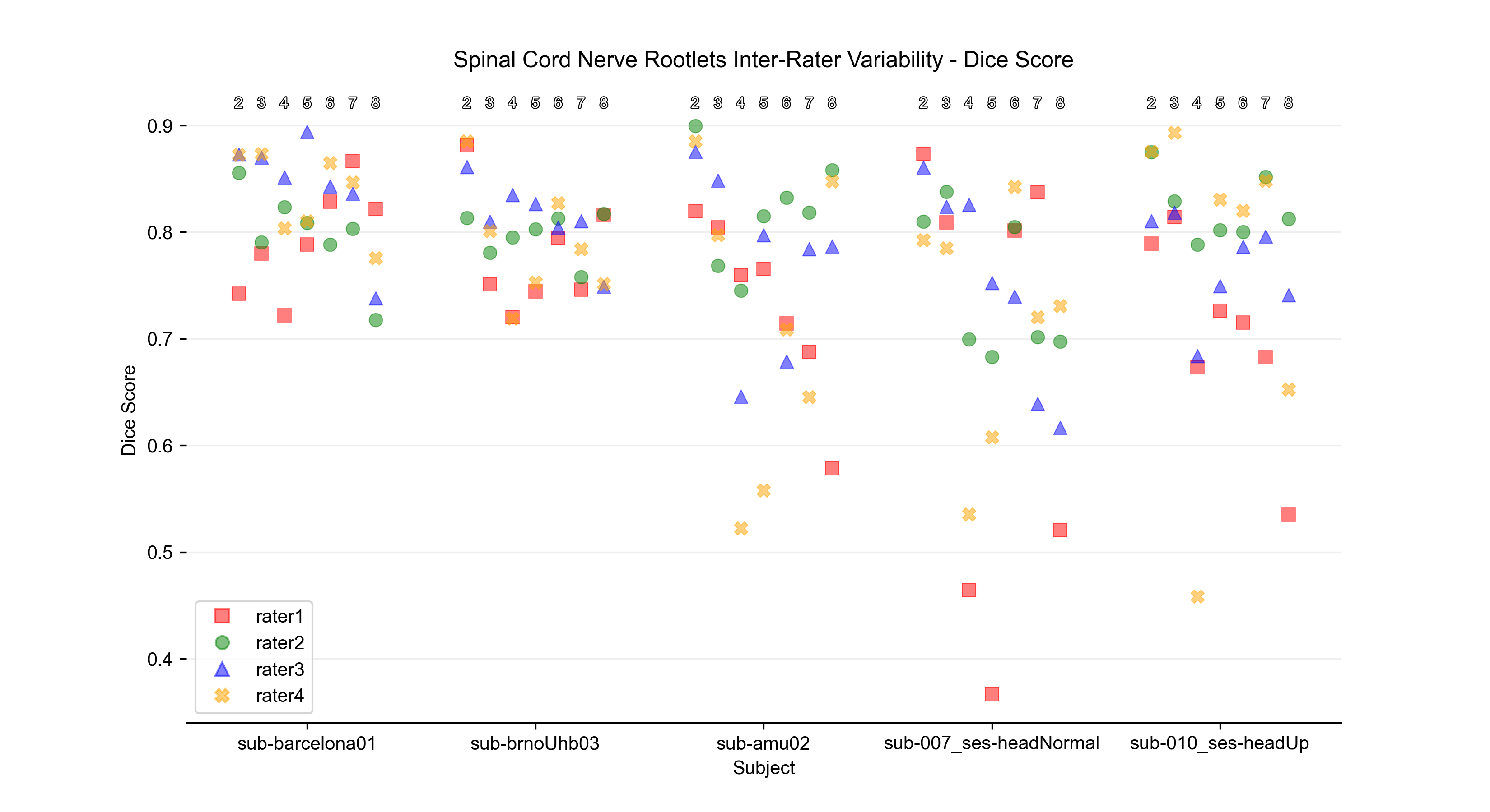
To assess inter-rater variability, 4 raters from 3 different sites manually segmented the nerve rootlets masks in 5 randomly chosen images. A consensus reference segmentation mask for each image was produced using the STAPLE algorithm6. The Dice coefficient was then calculated between the reference segmentation and manual segmentation from each rater.
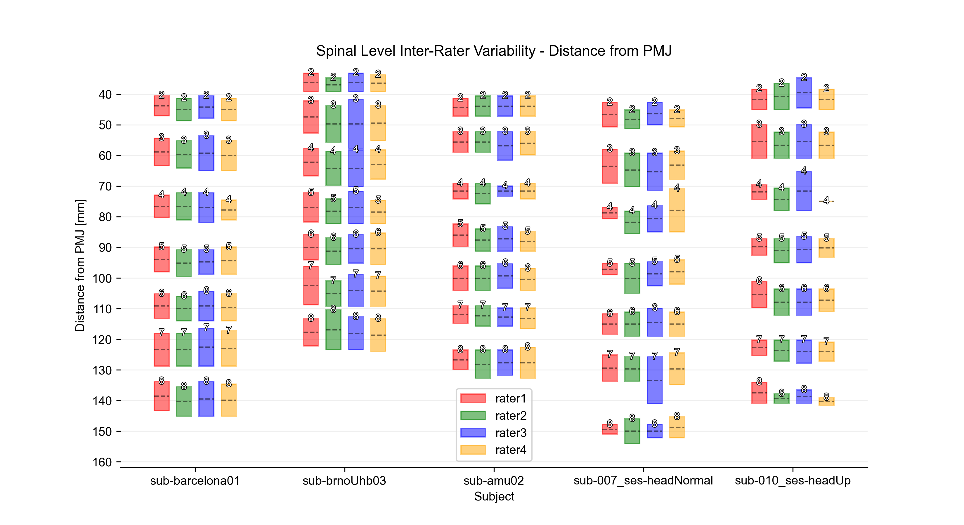
Spinal levels were automatically identified based on an intersection of the spinal nerve rootlet segmentation and automatically segmented7 spinal cord mask dilated by 3 voxels. The distance between the automatically identified pontomedullary junction8 and the middle of each spinal level was computed along the spinal cord centerline9,10 to assess the inter-rater coefficient of variation (COV). Then, the mean COV was computed.
Results
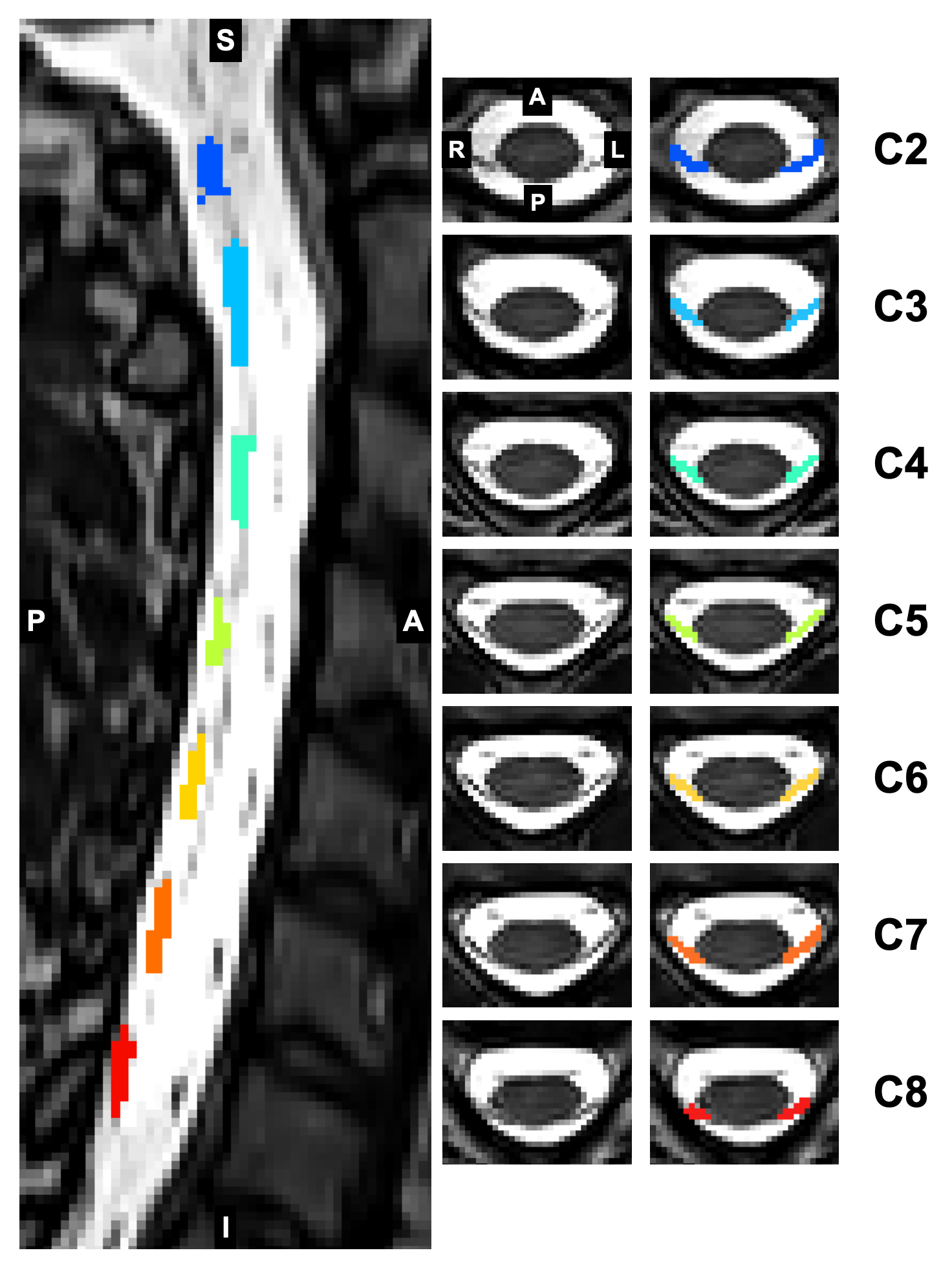
The active learning was successfully used to interactively improve the model performance reaching nnUNet validation pseudo Dice up to 0.6 (based on the fold).Figure 1 illustrates an example of the spinal cord nerve rootlet segmentation obtained using the proposed 3D nnUNet model. Note that the segmentation is multi-class, with each class representing a specific nerve rootlet.
Figure 2 shows the Dice coefficient between manual segmentation from each rater and the reference STAPLE segmentation. Overall, the Dice was higher for upper cervical nerve rootlets vs. lower cervical nerve rootlets (C2 mean Dice: 0.84 vs. C8 mean Dice: 0.72).
Figure 3 shows spinal level inter-rater variability assessed as the distance between the pontomedullary junction and the middle of each spinal level. The mean COV across subjects and raters is between 0.55% and 1.49%, depending on the spinal level.
Discussion
We observed low inter-rater variability and good agreement between raters represented by high Dice and low COV across subjects. Higher Dice for upper rootlets can be attributed to the fact that rostally located nerve rootlets are, due to their anatomy, better visible in MRI scans and, thus, easier to identify. Based on our preliminary application of the model to other datasets from collaborators (not shown here), the proposed model can generalize well on new unseen datasets.There are remaining limitations in the identification of the spinal levels based on the intersection of the rootlets and spinal cord segmentation. For example, if the segmentation does not extend enough towards the medial plane, it is not picked up by the dilation and the level is not marked. We are currently working on a more robust method based on the PAM50 spinal cord template.
Conclusion
This study introduces an automatic method for the segmentation of spinal nerve rootlets coupled with spinal level estimation. Based on preliminary experiments, the proposed model generalizes well to unseen T2w datasets, providing the spatial resolution is sufficient to see the nerve rootlets (0.8mm isotropic or better). The code/model is open source and available at: github.com/ivadomed/model-spinal-rootlets. Subsequent research will focus on the improvement of spinal level estimation, model validation on test-retest datasets, and application to pathologies.Acknowledgements
Jan Valošek and Theo Mathieu contributed equally and share co-first authorship.
Funded by the Canada Research Chair in Quantitative Magnetic Resonance Imaging [CRC-2020-00179], the Canadian Institute of Health Research [PJT-190258], the Canada Foundation for Innovation [32454, 34824], the Fonds de Recherche du Québec - Santé [322736, 324636], the Natural Sciences and Engineering Research Council of Canada [RGPIN-2019-07244], the Canada First Research Excellence Fund (IVADO and TransMedTech), the Courtois NeuroMod project, the Quebec BioImaging Network [5886, 35450], INSPIRED (Spinal Research, UK; Wings for Life, Austria; Craig H. Neilsen Foundation, USA), Mila - Tech Transfer Funding Program. Supported by the Ministry of Health of the Czech Republic, grant nr. NU22-04-00024. All rights reserved. JV has received funding from the European Union's Horizon Europe research and innovation programme under the Marie Sktodowska-Curie grant agreement No 101107932.
References
1. Kinany, N., Pirondini, E., Micera, S. & Van De Ville, D. Spinal Cord fMRI: A New Window into the Central Nervous System. Neuroscientist 10738584221101827 (2022).
2. Cadotte, D. W. et al. Characterizing the location of spinal and vertebral levels in the human cervical spinal cord. AJNR Am. J. Neuroradiol. 36, 803–810 (2015).
3. Cohen-Adad, J. et al. Open-access quantitative MRI data of the spinal cord and reproducibility across participants, sites and manufacturers. Scientific Data 8, 219 (2021).
4. Budd, S., Robinson, E. C. & Kainz, B. A survey on active learning and human-in-the-loop deep learning for medical image analysis. Med. Image Anal. 71, 102062 (2021).
5. Isensee, F., Jaeger, P. F., Kohl, S. A. A., Petersen, J. & Maier-Hein, K. H. nnU-Net: a self-configuring method for deep learning-based biomedical image segmentation. Nat. Methods 18, 203–211 (2021).
6. Warfield, S. K., Zou, K. H. & Wells, W. M. Simultaneous truth and performance level estimation (STAPLE): an algorithm for the validation of image segmentation. IEEE Trans. Med. Imaging 23, 903–921 (2004).
7. Gros, C. et al. Automatic segmentation of the spinal cord and intramedullary multiple sclerosis lesions with convolutional neural networks. Neuroimage 184, 901–915 (2019).
8. Gros, C. et al. Automatic spinal cord localization, robust to MRI contrasts using global curve optimization. Med. Image Anal. 44, - (2018).
9. Bédard, S. & Cohen-Adad, J. Automatic measure and normalization of spinal cord cross-sectional area using the pontomedullary junction. Frontiers in Neuroimaging 1, 43 (2022).
10. Bédard, S., Bouthillier, M. & Cohen-Adad, J. Pontomedullary junction as a reference for spinal cord cross-sectional area: validation across neck positions. Sci. Rep. 13, 13527 (2023).
Figures