2098
Robust Rat Liver Lobe Segmentation in Low-SNR T2-weighted Datasets Using a 2.5D Approach1Neuroradiology Division, Institute of Diagnostic and Interventional Radiology, Jena University Hospital, Jena, Germany, 2Medical Physics Group, Institute of Diagnostic and Interventional Radiology, Jena University Hospital, Jena, Germany, 3Visualization and Explorative Data Analysis Group, Faculty of Mathematics and Computer Science, Friedrich Schiller University Jena, Jena, Germany, 4Experimental Transplantation Surgery, Department of General, Visceral and Vascular Surgery, Jena University Hospital, Jena, Germany
Synopsis
Keywords: Analysis/Processing, Segmentation, portal vein ligation (PVL), signal-to-noise ratio (SNR)
Motivation: Our project was motivated by the lack of an efficient way of segmenting ligated and non-ligated liver lobes in portal vein ligation (PVL) experiments.
Goal(s): Our goal was to demonstrate that a 2.5D segmentation approach can achieve precise and robust lobe segmentation in experimental PVL volumetry to reduce manual annotation work.
Approach: We stacked adjacent slices as input and trained a U-Net to segment the rat liver lobes using 15 rat T2-weighted datasets.
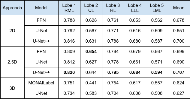
Results: An average Dice score of 0.707 was reached by 5-fold cross validation on 15 datasets, showing the robustness in low-SNR MR images with high intensity variation.
Impact: We demonstrate the 2.5D approach is robust in segmenting liver lobes with varied intensity in low-SNR MR images. The framework can greatly reduce manual annotation work even with limited datasets.
Introduction
Portal vein ligation (PVL)1 is an initial step to induce hypertrophy prior to liver transplantation. In rats, PVL generates inter-lobal contrast as the hypertrophy develops. It also allows a direct assessment of liver volumetry that facilitates monitoring the volume changes. For segmentation purposes, we enhance the inter-lobal water contrast by extending the nominal TE, which however, concurrently reduces image signal to noise ratio (SNR). The resulting low SNR complicates the use of conventional segmentation algorithms such as region-growing or thresholding, and necessitates manual segmentation, a process that is both labor-intensitve and time-consuming.In this study, we introduced a 2.5-dimension (2.5D) approach using convolutional networks to segment the rat liver lobes. Our method has demonstrated superior performance and robustness compared to 2D and 3D approaches under the constraint of limited datasets.
Material and Methods
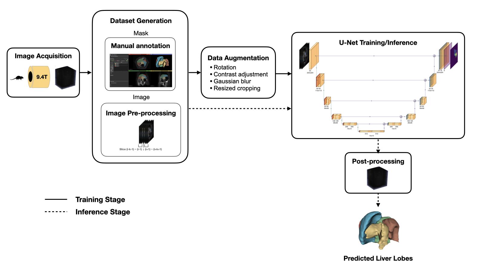
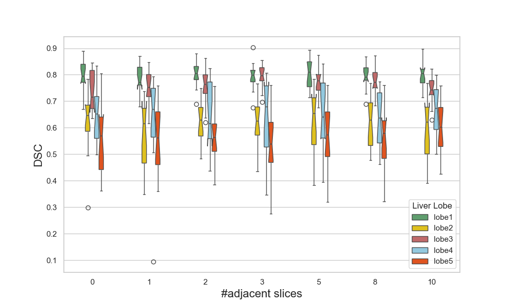
MRI data were collected on a Bruker BioSpec 94/20USR AVIII scanner using an 86 mm quadrature transceiver coil. We studied 23 rats: 3 controls and the remaining 20 underwent PVL on post-operative days (POD) 1, 2, 3, or 5, respectively2. T2-weighted 3D VFA-RARE sequence was acquired with TR/TE = 1000/1.97 ms, MTX = 200x200x160, RARE factor = 100, Resl = 350μm3, BW = 250 kHz, TA ~ 7m30s, and the nominal TE = 90.98 ms. Manual segmentation of the five lobes (right middle lobe, caudate lobe, right lobe, left lateral lobe, left middle lobe) was performed in 3DSlicer and served as ground truth.We employed a 2D U-Net3 as the base network for segmentation. We adopted the 2.5D approach, which processes the data slice-wise like 2D segmentation, but includes the context from the neighboring slices. The inputs are 3-channel image stacks of each coronal slice and the mean of $$$k$$$ adjacent slices above and below each slice, where k is a positive integer. If $$$k=0$$$, then it reduces to the 2D approach. To overcome the class imbalance, we used the weighted cross entropy loss. The training process was specified by 20 epochs, a batch size of 16, learning rate of 0.001, and AdamW optimization. Data augmentation was applied to make the model more robust to data heterogeneity. The pipeline is demonstrated in Figure 1. We selected 15 out of the 23 rats, such that there were exactly three rats measured on POD 0 (control), 1, 2, 3, and 5. With limited datasets, we implemented 5-fold cross validation in which the rats in each test set belong to the same POD category (similar tissue contrast), and evaluated per lobe by Dice similarity coefficient (DSC).
Results
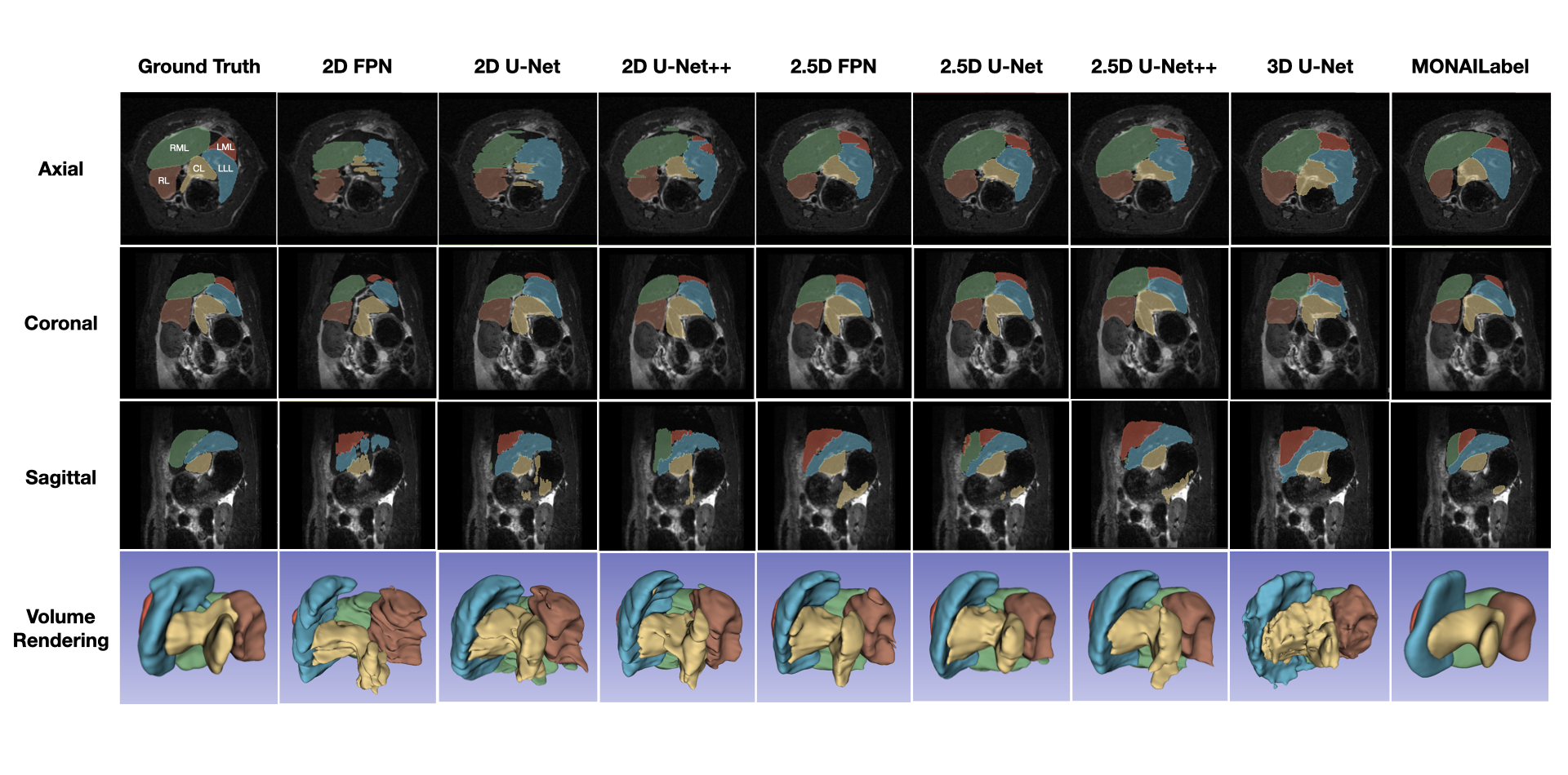
Figure 2 qualitatively evaluate the segmentation result of the five liver lobes using 2D, 2.5D and 3D approaches. Table 1 shows the quantitative result in DSC. 2.5D approaches, U-Net++ in particular, performed the best both quantitatively and qualitatively. The models include: feature pyramid network (FPN)4, U-Net3, U-Net++5, and SegResNet6 in MONAILabel7.Discussion and Conclusion
Accurate liver lobe segmentation is essential for evaluating regenerated liver size and weight, facilitating treatment planning and improving patient outcomes. This study served as a proof of concept for liver lobe segmentation in low-SNR MR images to reduce manual work. We demonstrated that the 2.5D approach provided a robust way to identify different liver lobes with similar and varied intensity, given only 15 datasets available.Intuitively, the 3D approach should perform better at segmenting volumetric structures because of more spatial context. In the case of a limited number of datasets with small objects for segmentation, the 2.5D approach could converge faster and perform better. The 2D approach is restricted by the small receptive field and limited spatial context. U-Net++ was superior to U-Net for its nested skip and dense skipped pathways that can capture multi-scale context. One limitation of our work is that manual work is still required to refine the labels for accurate analysis of ligated/non-ligated liver lobes, especially at the tissue boundary. Also, MONAILabel and our approach might be incomparable, as it is aimed for interactive segmentation and is equipped with generic post-processing steps that over-smoothed the predicted masks. Strategies such as multi-stage segmentation (liver detection and lobe segmentation) or boundary-based loss function may be considered in future work to improve the segmentation result.
Acknowledgements
This work was supported by the German Research Foundation (DFG) within the Research Unit Programme FOR5151 "QuaLiPerF (Quantifying Liver Perfusion–Function Relationship in Complex Resection - A Systems Medicine Approach)" grant number 436883643.References
1. Aragiusto, G., Ceriello, A., Giuliani, A., Migliaccio, C., & Calise, F. (2013). Portal Vein Ligation 23. Minimally Invasive Surgery of the Liver, 177-180.
2. Zhao, W.-T., Wei, W., Herrmann, K.-H., Krämer, M., Dahmen, U., & Reichenbach, J. R. (2023) Adaptive Response of Liver Perfusion in Preclinical Portal Vein Ligation. Poster#LB280 of 39th European Society of Magnetic Resonance in Medicine and Biology. Basel, Switzerland.
3. Ronneberger, O., Fischer, P., & Brox, T. (2015). U-net: Convolutional networks for biomedical image segmentation. In Medical Image Computing and Computer-Assisted Intervention–MICCAI 2015: 18th International Conference, Munich, Germany, October 5-9, 2015, Proceedings, Part III 18 (pp. 234-241). Springer International Publishing.
4. Lin, T. Y., Dollár, P., Girshick, R., He, K., Hariharan, B., & Belongie, S. (2017). Feature pyramid networks for object detection. In Proceedings of the IEEE conference on computer vision and pattern recognition (pp. 2117-2125).
5. Zhou, Z., Rahman Siddiquee, M. M., Tajbakhsh, N., & Liang, J. (2018). Unet++: A nested u-net architecture for medical image segmentation. In Deep Learning in Medical Image Analysis and Multimodal Learning for Clinical Decision Support: 4th International Workshop, DLMIA 2018, and 8th International Workshop, ML-CDS 2018, Held in Conjunction with MICCAI 2018, Granada, Spain, September 20, 2018, Proceedings 4 (pp. 3-11). Springer International Publishing.
6. Myronenko, A. (2019). 3D MRI brain tumor segmentation using autoencoder regularization. In Brainlesion: Glioma, Multiple Sclerosis, Stroke and Traumatic Brain Injuries: 4th International Workshop, BrainLes 2018, Held in Conjunction with MICCAI 2018, Granada, Spain, September 16, 2018, Revised Selected Papers, Part II 4 (pp. 311-320).
7. Diaz-Pinto, A., Alle, S., Nath, V., Tang, Y., Ihsani, A., Asad, M., ... & Cardoso, M. J. (2022). Monai label: A framework for ai-assisted interactive labeling of 3d medical images. arXiv preprint arXiv:2203.12362.
Figures