2087
Neuroinflammatory Imaging Markers: Insights into the Cerebral Consequences of Post-Acute Sequelae of COVID-19 (PASC)1Radiology and Imaging Sciences, Emory University, Atlanta, GA, United States, 2Biomedical Engineering, Emory and Georgia Institute of Technology, Atlanta, GA, United States, 3Biomedical Engineering, Stony Brook University, Stony Brook, NY, United States, 4Medicine, Stony Brook University, Stony Brook, NY, United States, 5Family, Population, and Preventive Medicine, Stony Brook University, Stony Brook, NY, United States, 6Radiology, Stony Brook University, Stony Brook, NY, United States, 7World Trade Center Health and Wellness Program, Stony Brook University, Stony Brook, NY, United States
Synopsis
Keywords: Infectious Disease, Diffusion/other diffusion imaging techniques
Motivation: The motivation behind this study stems from the urgent need to decipher the neuroinflammatory processes contributing to Post-Acute Sequelae of COVID-19 (PASC) and to identify potential biomarkers for this condition through neuroimaging.
Goal(s): The primary goal is to investigate the presence of cerebral imaging markers indicative of neuroinflammation in individuals experiencing neurological PASC.
Approach: The study employed multi-shell diffusion MRI, to investigate changes in individuals with PASC. Diffusion parameters indicative of neuroinflammation were analyzed, by comparing among PASC patients, never-infected individuals, and acute-COVID controls.
Results: PASC patients demonstrated increased whole-brain alterations consistent with axonal injury with inflammation.
Impact: The findings of altered diffusion parameters in PASC patients shed light on the presence of axonal injuries with inflammation. These insights can potentially impact the diagnosing and treating long-term neurological symptoms of COVID-19, ultimately improving patient care and recovery strategies.
Introduction
COVID-19 can cause symptoms that persist for months or years after infection called the Post-Acute Sequelae of COVID-19 (PASC) [1]. While PASC is a heterogeneous disorder, it can cause neuropsychological symptoms including brain fog, fatigue, and anxiety that can also induce cognitive and physical functional impairment [2,3]. Prior studies [4] support the view that neuro-PASC might signify the presence of cerebral changes, the prognosis of the disease is unclear. Symptoms of fatigue, brain fog, executive dysfunction, and slowed response speed in PASC might reflect the presence of axonal or cortical injury that may be lasting. The objective of the present study was to study white matter diffusion markers associated with neuroinflammation among individuals with mild to moderate COVID-19 who developed neurological PASC by comparing them to individuals who were never infected and who did not develop PASC.Methods
Participants were recruited from a monitoring program for essential workers who work in construction and law enforcement agencies [5] that has an active neuroimaging program [6].Diffusion parameters from whole-brain were examined using multi-shell diffusion MRI data. All images were acquired on a 3T Siemens Biograph mMR. Diffusion MRI images were acquired using a state-of-the-art multi-band diffusion-weighted imaging sequence [7] with TE/TR=121.4/6300 ms, voxel size = 2x2x2 mm3, multi-band factor = 3, four b-values = 1000 - 4000 s/mm². T1-weighted magnetization-prepared rapid gradient echo (MPRAGE) images were also acquired (TE/TR/TI = 2.49/1900/900 ms, flip angle =9°, isotropic voxel size = 0.9x0.9x0.9 mm3, grappa factor = 2).
Brain parcellation was performed with FreeSurfer (7.3.0) using T1-MPRAGE images. We calculated fractional anisotropy (FA), mean diffusivity (MD), axonal diffusivity (AD), and radial diffusivity (RD) using diffusion images acquired with b-value=0; 1,000 s/mm². To generate the spin distribution function, q-space diffeomorphic reconstruction was performed. Correlational tractography was utilized to dissect the nature and extent of white matter changes.
Results
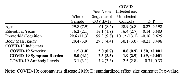
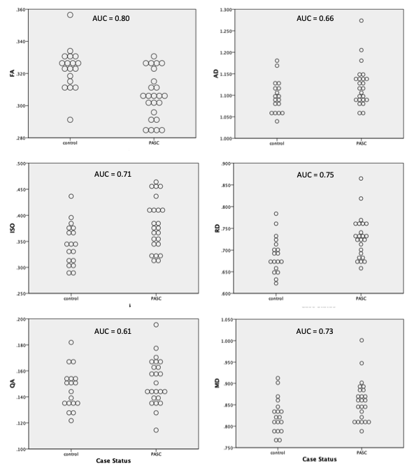
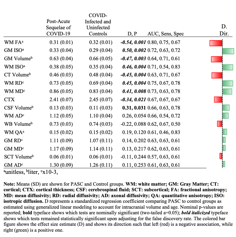
Participant Characteristics: We examined data from 42 male participants with characteristics shown in Figure 1.Whole-brain analysis: Figure 2 shows the dot plots of diffusion parameters stratified by PASC status. Overall, these results show that diffusion parameters were often quite different and were relatively good at identifying PASC cases. As shown in Figure 3, whole-brain white matter averages of MD, RD, and ISO were found to be significantly increased in PASC, while the average FA is significantly lower in PASC, with many surviving multiple comparison corrections.
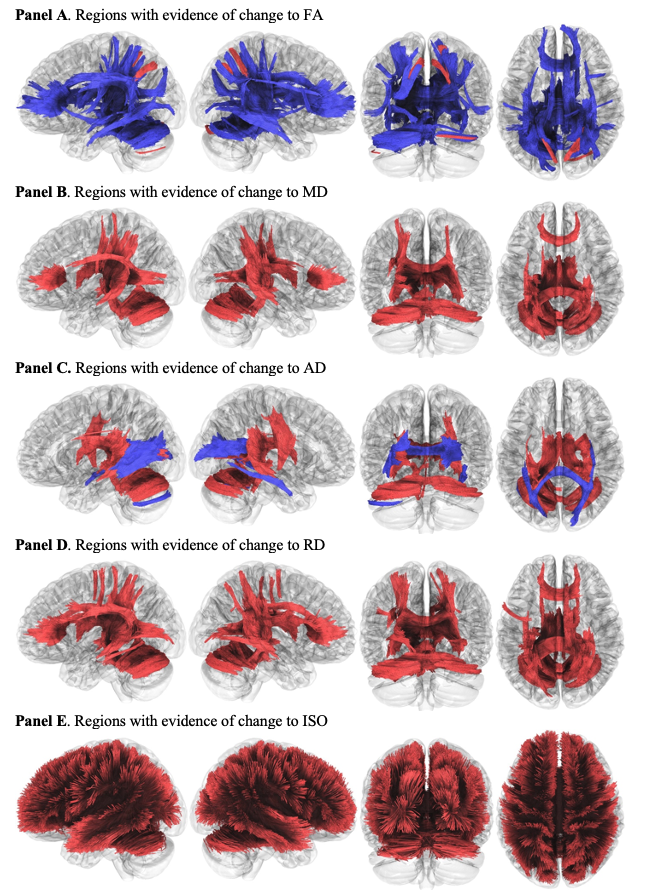
Correlational Tractography: To understand the scope of white matter changes, correlational tractography was performed to identify white matter tracts with a diffusion parameter correlated with the group variable (i.e., PASC versus controls). Figure 4A-E shows white matter tracts throughout the brain with differences in tractography, with decreases in red, and increases in blue, only tracts with false discovery rate<0.05 are shown. All measures had evidence of some levels of change.
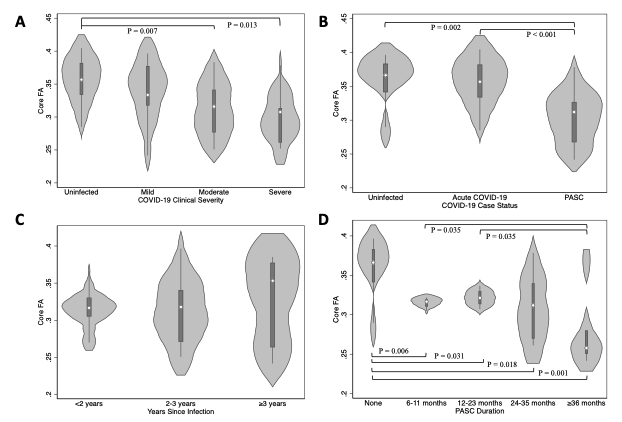
CoreFA: We further estimated a parameter we called “CoreFA”, which was parametrized as the average FA values of the key ROI derived from the most affected tracts. Our CoreFA measure was found to have an even higher effect size with Cohen’s D = -0.59 (P<0.001), AUC=0.81.
Figure 5 shows the association between clinical indicators of COVID-19 severity and CoreFA.
Sensitivity Analyses: None of the whole-brain diffusion parameters was found to have a noticeable association with vaccination/COVID severity/APOE4 status (data not shown).
Discussions
The presence of neurological PASC is a new problem emerging after acute COVID-19 and may persist for years after initial infections. Replicating and expanding on prior neuroimaging work among COVID-19 patients, our comprehensive analysis revealed pronounced changes in white matter diffusion parameters in PASC patients compared to controls. Through correlational tractography, we elucidated the presence of widespread neuroinflammation, with findings indicative of symmetric axonal injury. Elevated values of white matter MD, RD, and ISO coupled with diminished FA values and insignificant AD and QA changes observed in PASC patients are strongly suggestive of axonal injuries with neuroinflammation. Our results bridge the current knowledge gap by highlighting the pervasive inflammatory landscape for individuals with neurological PASC.Controls with a history of acute COVID-19 and individuals lacking COVID-19 exposure did not show marked differences in the diffusion parameters evaluated here. This observation strengthens the premise that the neurocognitive sequelae in PASC, often recognized by the presence of brain fog, are intricately tied to neuroinflammation.
The results of this study imply that we might be able to positively diagnose neurological PASC using a neuroimaging-based signature in the white matter with a high degree of accuracy.
Given the changes evident here, therapeutic strategies targeting neuroinflammation might prove beneficial for PASC patients.
Acknowledgements
This work was in part supported by CDC/NIOSHU01 OH012275 and NIH/NIA R01 AG049953.References
1. Wu Y, Xu X, Chen Z, Duan J, Hashimoto K, Yang L, Liu C, Yang C. Nervous system involvement after infection with COVID-19 and other coronaviruses. Brain, Behavior, and Immunity. 2020. doi: https://doi.org/10.1016/j.bbi.2020.03.031.
2. Vanichkachorn G, Newcomb R, Cowl CT, Murad MH, Breeher L, Miller S, Trenary M, Neveau D, Higgins S, editors. Post–COVID-19 syndrome (long haul syndrome): description of a multidisciplinary clinic at Mayo Clinic and characteristics of the initial patient cohort. Mayo clinic proceedings; 2021: Elsevier.
3. Nalbandian A, Sehgal K, Gupta A, Madhavan MV, McGroder C, Stevens JS, Cook JR, Nordvig AS, Shalev D, Sehrawat TS. Post-acute COVID-19 syndrome. Nature medicine. 2021;27(4):601-15.
4. Petersen M, Nägele FL, Mayer C, Schell M, Petersen E, Kühn S, Gallinat J, Fiehler J, Pasternak O, Matschke J. Brain imaging and neuropsychological assessment of individuals recovered from a mild to moderate SARS-CoV-2 infection. Proceedings of the National Academy of Sciences. 2023;120(22):e2217232120.
5. Luft BJ, Schechter C, Kotov R, Broihier J, Reissman D, Guerrera K, Udasin I, Moline J, Harrison D, Friedman-Jimenez G, Pietrzak RH, Southwick SM, Bromet EJ. Exposure, probable PTSD and lower respiratory illness among World Trade Center rescue, recovery and clean-up workers. Psychol Med. 2012;42(5):1069-79
6. Clouston SAP, Deri Y, Horton M, Tang C, Diminich ED, Pellecchia A, Carr M, Gandy S, Sano M, Bromet E. Reduced cortical thickness in World Trade Center responders with cognitive impairment: Neuroimaging/differential diagnosis. Alzheimer's & Dementia. 2020;16:e039996.
7. Setsompop K, Cohen-Adad J, Gagoski BA, Raij T, Yendiki A, Keil B, Wedeen VJ, Wald LL. Improving diffusion MRI using simultaneous multi-slice echo planar imaging. Neuroimage. 2012;63(1):569-80.
Figures