2086
The long-term cerebral change of patient transformed from mildly into severely emotional problem after multiple COVID-19 infections: 3-6 months1Radiology, Second Affiliated Hospital of Naval Medical University, Shanghai, China, 2Second Affiliated Hospital of Naval Medical University, Shanghai, China
Synopsis
Keywords: Infectious Disease, COVID-19
Motivation: It was unclear that if the brain function would be changed after multiple COVID-19 infections when the psychological burden incremented.
Goal(s): To evaluate the long-term brain brain change of the patients who transformed from mildly emotional problem after the first COVID-19 infection into severely one after multiple infections.
Approach: The patients with mildly emotional abnormality after once infection (control group) and others transformed from mild into severe abnormality after multiple COVID infections (patients group) were included. The parameters derived from BOLD and DTI were compared after 3-6 months.
Results: The patients group had a decreased FA and increased ALFF in some brain areas.
Impact: The abnormalities in these brain areas could help clinicians to understand the potential machine of the transformation of mild emotion problem into severe emotion problem after another COVID infection.
INTRODUCTION
The relationship between the emotion problem and cerebral change after COVID-19 infection had been reported. It was observed that the psychological burden was incremented for some patients when they suffered multiple COVIS-19 infections. It was unclear that if the brain function and structure would also be changed or damaged. The aim of the study was to evaluate the long-term brain change of the patients who transformed from mildly emotional problem after the first COVID-19 infection into severely one after multiple infections.METHODS
The patients with mildly emotional abnormality after once infection (control group) and others transformed from mild into severe abnormality after multiple COVID infections (patient group) were included. The parameters derived from BOLD and DTI were compared after 3-6 months. The PSS, PHQ, and GAD were evaluated for emotion analysis. The ALFF, ReHo and DC were calculated. TBSS were used to analysis the FA, MD, AD and RD. Unpaired two-sample t tests and Kruskal‒Wallis tests (nonnormally distributed samples) were carried out for the cross-sectional comparison of BOLD-fMRI and DTI metrics. A chi-square test was performed for sex. Unpaired two-sample t tests or Kruskal‒Wallis tests (nonnormally distributed samples) were carried out for age.RESULTS
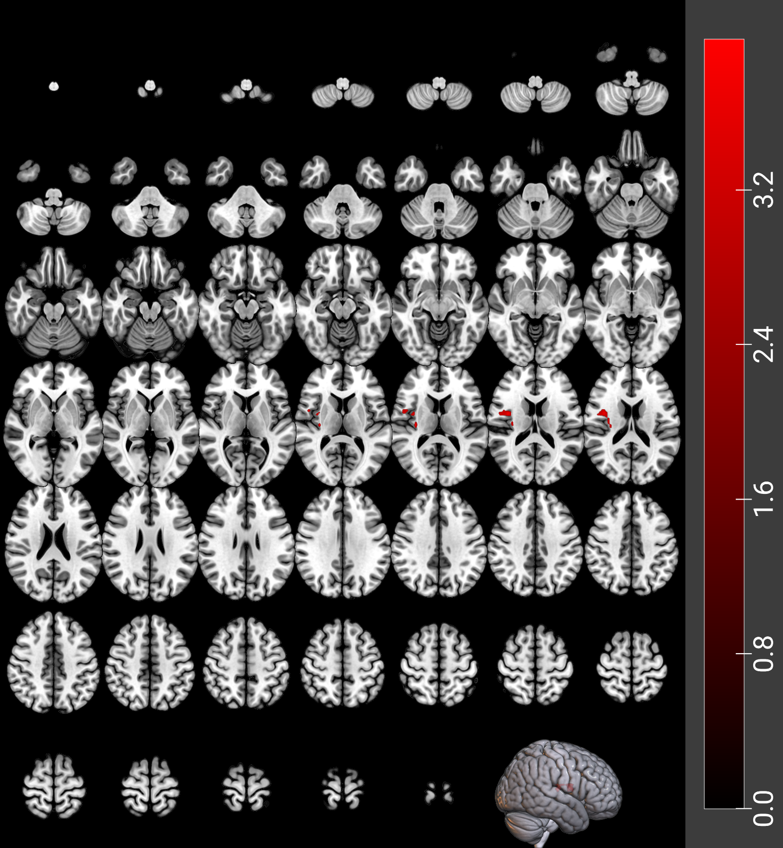
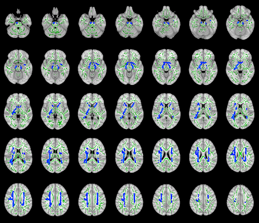
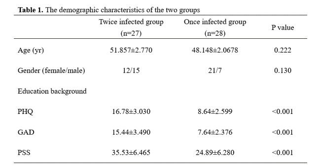
There were 27 patients were included in the patient group. Another 28 patients were included in control group. There was no difference between two groups in age (51.857±2.770 vs. 48.148±2.0678,p=0.222), gender (female: 75.0% vs. 55.6%, P=0.130). The PSS, PHQ, and GAD of control group were higher than those of once infected group (all p < 0.001). (Table 1). Compared to once infected group, the twice infected group showed significantly increased ALFF in the right insula (INS.R) (number of voxels = 18) and rolandic operculum (ROL.L) (number of voxels = 21) (p GRF < 0.05) (Figure 1). Compared to control group, the patient group showed significantly decreased FA in several white matter tracts (p FWE < 0.05). These white matter tracts included the bilateral corticospinal tract, anterior thalamic radiation, and right superior longitudinal fasciculus (Fig. 2) (p FWE < 0.05).CONCLUSION:
The present study revealed that when the patients with mild emotional problem after the first COVID infection transformed into a severely emotional problem after another COVID infection their white matter would suffer an obvious brain damage in cerebral function and the integrity of whiter matter after 3 to 6 months.Acknowledgements
Funding: This work was supported by the National Key R&D Program of China (Grant NO. 2022YFC2010000), the Key Program of National Natural Science Foundation of China (Grant NO. 81930049), the National Natural Science Foundation of China (Grant NO. 92259203), Shanghai Science and Technology Innovation Action Plan Program (Grant NO. 19411951300), the National Natural Science Foundation of China (Grant NO. 82271994), and National Health Commission Radiological Imaging Database Construction Project (Grant NO. YXFSC2022JJSJ010) .References
[1] Chen N, Zhou M, Dong X, et al. Epidemiological and clinical characteristics of 99 cases of 2019 novel coronavirus pneumonia in Wuhan, China: a descriptive study[J]. Lancet, 2020; 395(10223): 507-13. DOI: 10.1016/S0140-6736(20)30211-7.
[2] Nalbandian A, Sehgal K, Gupta A, et al. Post-acute COVID-19 syndrome[J]. Nat Med, 2021; 27(4): 601-15. DOI: 10.1038/s41591-021-01283-z.
[3] Muccioli L, Pensato U, Cani I, et al. COVID-19-Associated Encephalopathy and Cytokine-Mediated Neuroinflammation[J]. Annals of nuerology, 2020; 88(4): 860-61.
[4] Benameur K, Agarwal A, Auld S C, et al. Encephalopathy and Encephalitis Associated with Cerebrospinal Fluid Cytokine Alterations and Coronavirus Disease, Atlanta, Georgia, USA, 2020[J]. Emerg Infect Dis, 2020; 26(9): 2016-21. DOI: 10.3201/eid2609.202122.
[5] Du Y, Zhao W, Du L, et al. Neuropsychiatric symptoms associated with the COVID-19 and its potential nervous system infection mechanism the role of imaging in the study[J]. Psychoradiology, 2021; 1(2): 199-211. DOI: 10.1093/psyrad/kkab019.
[6] Qin Y, Wu J, Chen T, et al. Long-term microstructure and cerebral blood flow changes in patients recovered from COVID-19 without neurological manifestations[J]. J Clin Invest, 2021; 131(8): e147329. DOI: 10.1172/JCI147329.
[7] Lu Y, Li X, Geng D, et al. Cerebral Micro-Structural Changes in COVID-19 Patients - An MRI-based 3-month Follow-up Study[J]. EClinicalMedicine, 2020; 25: 100484-84. DOI: 10.1016/j.eclinm.2020.100484.
[8] Douaud G, Lee S, Alfaro-Almagro F, et al. SARS-CoV-2 is associated with changes in brain structure in UK Biobank[J]. Nature, 2022; 604(7907): 697-707. DOI: 10.1038/s41586-022-04569-5.
[9] Reiken S, Sittenfeld L, Dridi H, et al. Alzheimer's-like signaling in brains of COVID-19 patients[J]. Alzheimers Dement, 2022: 10.1002/alz.12558. DOI: 10.1002/alz.12558.
Figures