2085
Insights into Neurological Long COVID Syndrome with Quantitative Susceptibility Mapping in Basal Ganglia1Radiology and Imaging Sciences, Emory University School of Medicine, Atlanta, GA, United States, 2Biomedical Engineering, Stony Brook University, Stony Brook, NY, United States, 3Biomedical Engineering, Georgia Institute of Technology, Atlanta, GA, United States, 4Medicine, Stony Brook University, Stony Brook, NY, United States, 5Family, Population, and Preventive Medicine, Stony Brook University, Stony Brook, NY, United States, 6World Trade Center Health and Wellness Program, Stony Brook University, Stony Brook, NY, United States
Synopsis
Keywords: Infectious Disease, Quantitative Susceptibility mapping, PASC, COVID-19
Motivation: The etiology of persistent and enigmatic neurological symptoms in post-acute sequelae of COVID-19 (PASC) remains unclear.
Goal(s): To utilize quantitative susceptibility mapping (QSM) to compare iron levels in the basal ganglia of individuals with neurological symptoms of PASC, recovered COVID-19 patients, and uninfected controls, thereby identifying potential biomarkers for the condition.
Approach: QSM imaging was performed on three distinct cohorts: PASC, post-COVID non-PASC, and uninfected subjects
Results: Elevated QSM values, indicative of iron accumulation, in the putamen and caudate nucleus were observed in PASC patients.
Impact: The study aims to offer a novel imaging perspective on PASC, potentially leading to a better understanding of its neurological aspects and aiding in the development of targeted therapies for affected patients.
Introduction
Post-acute sequelae of COVID-19 (PASC), also referred to as Long COVID, is characterized by a multitude of persistent symptoms that can last for months or years following the initial infection. The neurological symptoms of PASC include fatigue, limited cognitive function (commonly known as "brain fog"), and heightened anxiety. While the etiology of PASC remains unclear, prevailing hypotheses predominantly center on virus-induced autoimmune responses or cellular dysfunction as potential underlying mechanisms 1.Previous studies have indicated that alterations in iron homeostasis can impact the immune system's ability to mount a response against viral infections and subsequently contribute to autoimmune reactions 2, 3. Iron distribution within the brain exhibits marked heterogeneity, with the highest concentrations discerned in the basal ganglia region 4. The basal ganglia regions have garnered considerable attention in various brain disorders 5, 6. Furthermore, early findings in patients with PASC indicate a constellation of symptoms closely resembling another chronic illness—myalgic encephalomyelitis /chronic fatigue syndrome (ME/CFS) 7, 8, where central fatigue may result from decreased basal ganglia function 9.
Visualizing and quantifying brain iron content can be achieved through magnetic resonance imaging (MRI) using quantitative susceptibility mapping (QSM), which measures the spatial distribution of magnetic susceptibility within biological tissues 10.
In this study, we focus on basal ganglia and utilize QSM to investigate iron accumulation in individuals who have experienced neurological symptoms of PASC, comparing them to both uninfected controls and those recovered from COVID-19 without symptoms of PASC.
Methods
Study PopulationParticipants were recruited from a monitoring program for essential workers who work in construction and law enforcement agencies 11 that have an active neuroimaging program 12. We included neuroimaging data from three distinct participant groups: i) post COVID-19 with neurological PASC; ii) post COVID-19 non-PASC (recovered); and iii) uninfected control.
MRI Acquisition
All participants were scanned on a 3T Siemens Biograph mMR scanner. QSM images were generated using a multi-echo gradient echo sequence (8 echoes, TEs = 6.2, 12.4, 18.6, 24.8, 31, 37.2, 43.6, and 49.8 ms, TR = 71 ms, in-plane resolution = 1x1 mm2, slice thickness = 2 mm, flip angle = 15°). T1-weighted magnetization-prepared rapid gradient echo (T1-MPRAGE) images were also acquired under the following parameters: TE/TR/TI = 2.49/1900/900 ms, isotropic voxel size = 0.9x0.9x0.9 mm3, flip angle = 9°.
Image Processing
QSM was generated using the MEDI toolbox 13. In brief, the phase image was derived from the multi-echo phase data, followed by phase unwrapping via the Laplacian operation 14, and the removal of the background field by projecting onto dipole fields 15. QSM was then reconstructed using morphology-enabled dipole inversion zero-referenced to the cerebrospinal fluid across the entire brain (MEDI+0) 16. Affine registration of T1-weighted images to QSM images was achieved using the FMRIB Software Library FLIRT tool 16.
For region of interest (ROI) analysis, brain parcellation was performed with FreeSurfer (v7.3.2) using T1-MPRAGE images. The resultant masks were registered to QSM data by applying the deformation obtained in the earlier registration step.
Statistical Analysis
A two-sample t-test was used to compare the mean magnetic susceptibility of groups, with a significance level set at 0.05.
Results
The study included 39 male participants with ages ranging from 47–80 years. The demographic characteristics can be found in Figure 1.Figure 2 shows representative reconstructed QSM maps from participants in the PASC, recovered, and uninfected groups.
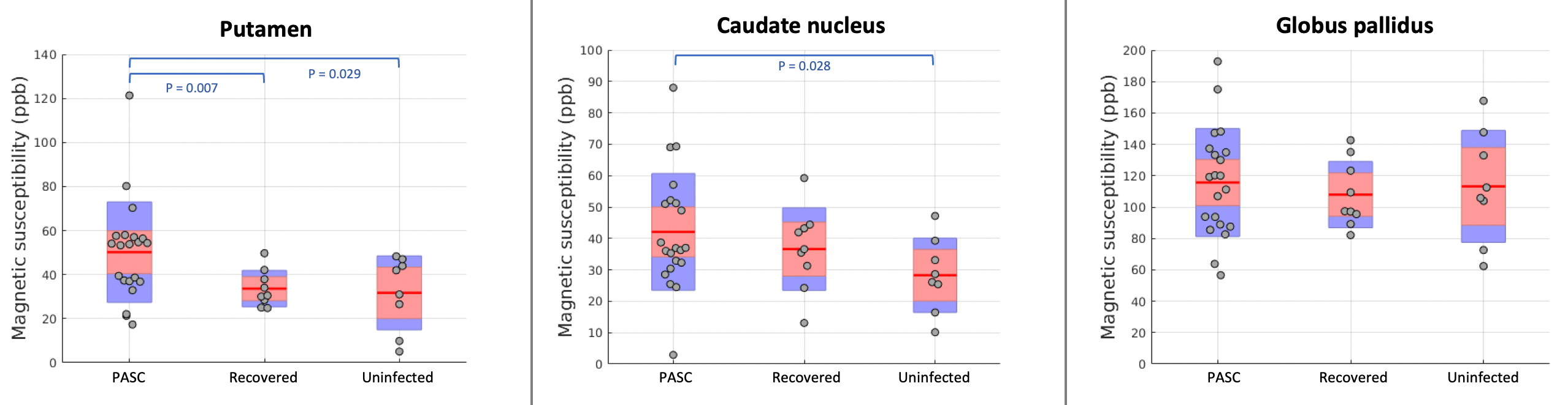
Among the 3 ROIs examined, elevated QSM values were observed in the putamen and caudate nucleus in the PASC group when compared to the uninfected group (Figure 3), with P = 0.029 and 0.028, respectively. When comparing the values between the PASC and the recovered groups, only the putamen exhibited a statistically significant difference (P = 0.007). No statistically significant difference was noted in the pallidum for either comparison.
Discussion
Our findings demonstrated increased magnetic susceptibility, which is indicative of iron accumulation, in the putamen and caudate nucleus among individuals who are experiencing PASC. This observation prompts inquiries regarding the clinical implications of basal ganglia iron accumulation in PASC patients. As mentioned above, altered basal ganglia function has been known to be a primary mechanism underlying central fatigue 9; this study provides further evidence that long COVID shares some mechanistic and phenotypic characteristics with ME/CFS. As such, QSM could be a useful imaging marker in elucidating the neurobiological underpinnings of PASC.Acknowledgements
This work was in part supported by CDC/NIOSHU01 OH012275 and NIH/NIA R01 AG049953.
References
1. Sundaresan B, Shirafkan F, Ripperger K, Rattay K. The role of viral infections in the onset of autoimmune diseases. Viruses. 2023;15(3):782.
2. Ward RJ, Crichton RR, Taylor DL, Corte LD, Srai SK, Dexter DT. Iron and the immune system. Journal of neural transmission. 2011;118:315-28.
3. Urrutia PJ, Bórquez DA, Núñez MT. Inflaming the brain with iron. Antioxidants. 2021;10(1):61.
4. Ward RJ, Zucca FA, Duyn JH, Crichton RR, Zecca L. The role of iron in brain ageing and neurodegenerative disorders. The Lancet Neurology. 2014;13(10):1045-60.
5. Li KR, Avecillas‐Chasin J, Nguyen TD, Gillen KM, Dimov A, Chang E, et al. Quantitative evaluation of brain iron accumulation in different stages of Parkinson's disease. Journal of Neuroimaging. 2022;32(2):363-71.
6. Tellez N, Alonso J, Rio J, Tintore M, Nos C, Montalban X, et al. The basal ganglia: a substrate for fatigue in multiple sclerosis. Neuroradiology. 2008;50:17-23.
7. Proal AD, VanElzakker MB. Long COVID or post-acute sequelae of COVID-19 (PASC): an overview of biological factors that may contribute to persistent symptoms. Frontiers in microbiology. 2021;12:1494.
8. Davis HE, McCorkell L, Vogel JM, Topol EJ. Long COVID: major findings, mechanisms and recommendations. Nature Reviews Microbiology. 2023;21(3):133-46.
9. Chaudhuri A, Behan PO. Fatigue and basal ganglia. Journal of the neurological sciences. 2000;179(1-2):34-42.
10. Liu C, Wei H, Gong N-J, Cronin M, Dibb R, Decker K. Quantitative susceptibility mapping: contrast mechanisms and clinical applications. Tomography. 2015;1(1):3-17.
11. Luft B, Schechter C, Kotov R, Broihier J, Reissman D, Guerrera K, et al. Exposure, probable PTSD and lower respiratory illness among World Trade Center rescue, recovery and clean-up workers. Psychological medicine. 2012;42(5):1069-79.
12. Clouston SA, Deri Y, Horton M, Tang C, Diminich E, DeLorenzo C, et al. Reduced cortical thickness in World Trade Center responders with cognitive impairment. Alzheimer's & Dementia: Diagnosis, Assessment & Disease Monitoring. 2020;12(1):e12059.
13. Liu J, Liu T, de Rochefort L, Ledoux J, Khalidov I, Chen W, et al. Morphology enabled dipole inversion for quantitative susceptibility mapping using structural consistency between the magnitude image and the susceptibility map. Neuroimage. 2012;59(3):2560-8.
14. Schofield MA, Zhu Y. Fast phase unwrapping algorithm for interferometric applications. Optics letters. 2003;28(14):1194-6.
15. Liu T, Khalidov I, de Rochefort L, Spincemaille P, Liu J, Tsiouris AJ, et al. A novel background field removal method for MRI using projection onto dipole fields. NMR in Biomedicine. 2011;24(9):1129-36.
16. Liu Z, Spincemaille P, Yao Y, Zhang Y, Wang Y. MEDI+ 0: Morphology enabled dipole inversion with automatic uniform cerebrospinal fluid zero reference for quantitative susceptibility mapping. Magnetic resonance in medicine. 2018;79(5):2795-803.
Figures
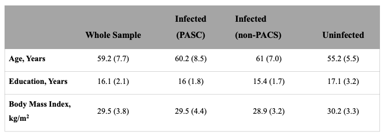
Figure 1. The characteristics of the participants.
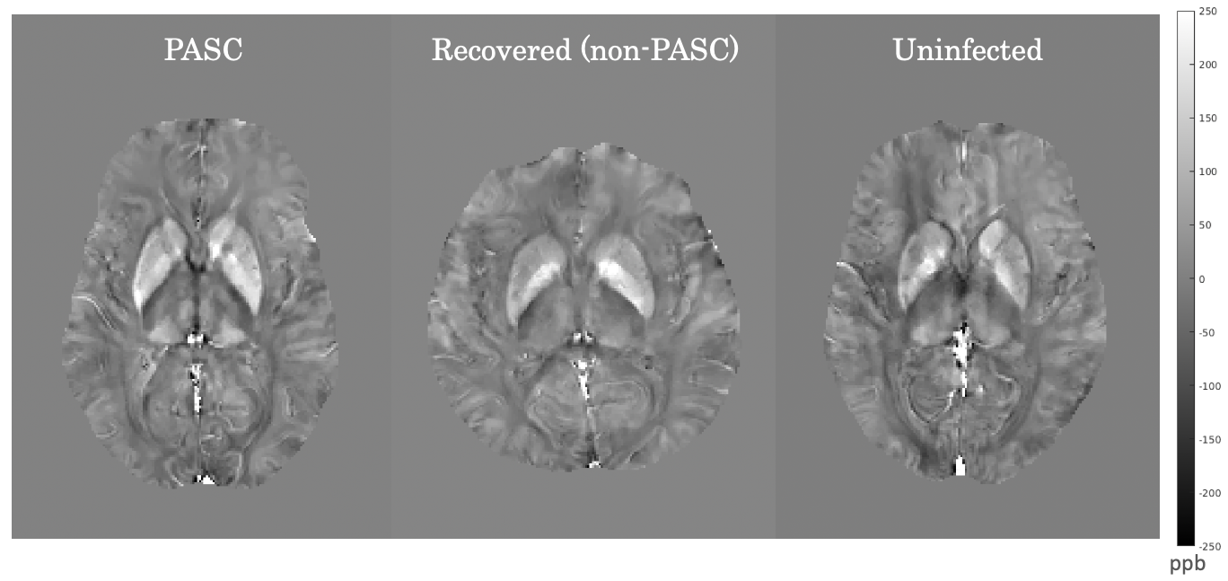
Figure 2. Representative axial QSM images from (left) PASC, (middle) recovered, and (right) uninfected groups. An overall higher signal intensity is apparent within the basal ganglia region in the PASC patient, indicative of increased iron accumulation. QSM: quantitative susceptibility mapping; PASC: Post-Acute Sequelae of COVID-19.