2081
Independent Component Analysis in Long Covid during cognitive exertion reveals widespread deficits in BOLD activity1NCNED, Griffith University, Southport, Australia, 2Medicine, Georgetown University, Washington, DC, United States
Synopsis
Keywords: Infectious Disease, COVID-19, Independent Component Analysis
Motivation: To understand the cognitive deficits of Long Covid (LCov).
Goal(s): What are the brain locations with different BOLD activity in LCov?
Approach: Two consecutive fMRI were acquired in 19 LCov and 16 healthy controls (HC) with a 7 Tesla scanner during the cognitive color-word Stroop task. Run2 was affected by fatigue induced by Run1. BOLD time series were processed with the CONN toolbox and submitted to independent component analysis (group ICA).
Results: ICA detected widespread deficits in LCov activity and sensorimotor excesses. The extent of LCov activity differences supports the hypothesis that global Covid19 infection affects brain-wide BOLD activity and regulatory function.
Impact: Our discovery of brain-wide changes in Long Covid BOLD activity supports the mechanism of brain-wide Covid-19 infection inducing cognitive deficits. Research should be directed to therapies that eliminate cerebral infection and facilitate and monitor recovery from virus inflicted damage.
In both runs five ICA components of BOLD differences between LCov and HC groups were detected. Run2 ICA components were spatially larger than Run 1 with higher statistical inference (Tables 1 and 2). No DMN or Dorsal Attention network differences were found in either run. A component in each run, which involved sensorimotor and visual networks, showed increased activity in LCov (Fig 2). The other components showed decreased LCov activity. Run 1 ICA revealed deficits in LCov connectivity in the Salience (Fig 1 left above) and Language (Fig 1 left below). Anterior cingulate cortex (ACC) deficits were found in the Fronto-Parietal network (not shown). Run 2 ICA detected a component (Fig 1 right) with more than 20 focal LCov deficits which were widely distributed and involved Salience, Language and Frontoparietal networks. Most deficits were on the right in frontal and parietal (angular gyrus) areas. On the left, deficits in Language (Broca’s and Wernicke’s) areas are seen. Another component, identified by CONN as FrontoParietal network, also showed LCov deficits. was again detected in The Sensorimotor network component again showed increased LCov activity as did a Visual network component (Fig 2). More LCov deficits were seen in the fatigue-affected Run 2. The Stroop task demanded increased LCov Sensorimotor activity in both runs. These connectivity results are consistent with widespread brain function deficits in LCov which have greater significance when affected by cognitive fatigue.
CONN ICA initially analyses single subjects. Group ICA then computes components (15 here) for the pooled cohort and the components for group differences. The pooled components are used to associate components with 8 intrinsic networks according to their spatial overlap. Some networks are associated with more than one component, and a component may involve more than one network. Noise and artifact components are also detected and isolated. The surviving network components are then a powerful tool for characterising cerebral BOLD activity and connectivity. CONN ICA here was enhanced by
a) High quality fMRI from a 7T scanner.
b) Acquisition during a cognitive task that exercised all intrinsic networks.
c) Acquisition of sequential fMRI such that Run 2 was affected by cognitive fatigue induced by Run 1.
An important feature of the results was their widespread nature, especially in the fatigue-affected Run 2. This supports a hypothesis that Long Covid derives from brain-wide COVID-19 infection that compromises brain activity, such as from viral fusogens (1).
Acknowledgements
No acknowledgement found.References
1. Martínez-Mármol R, Giordano-Santini R, Kaulich E, Cho AN, Przybyla M, Riyadh MA, et al. SARS-CoV-2 infection and viral fusogens cause neuronal and glial fusion that compromises neuronal activity. Science Advances [Internet]. 2023 Jun 7 [cited 2023 Jun 13];9(23):eadg2248. Available from: https://www.science.org/doi/full/10.1126/sciadv.adg2248
2. Barnden L, Thapaliya K, Eaton-Fitch N, Barth M, Marshall-Gradisnik S. Altered brain connectivity in Long Covid during cognitive exertion: a pilot study. Front Neurosci. 2023;17:1182607.
3. Nieto-Castanon A. Handbook of functional connectivity Magnetic Resonance Imaging methods in CONN [Internet]. Hilbert Press; 2020. Available from: https://www.researchgate.net/publication/339460691_Handbook_of_functional_connectivity_Magnetic_Resonance_Imaging_methods_in_CONN
4. Whitfield-Gabrieli S, Nieto-Castanon A. Conn: a functional connectivity toolbox for correlated and anticorrelated brain networks. Brain connectivity. 2012;2(3):125–41.
Figures
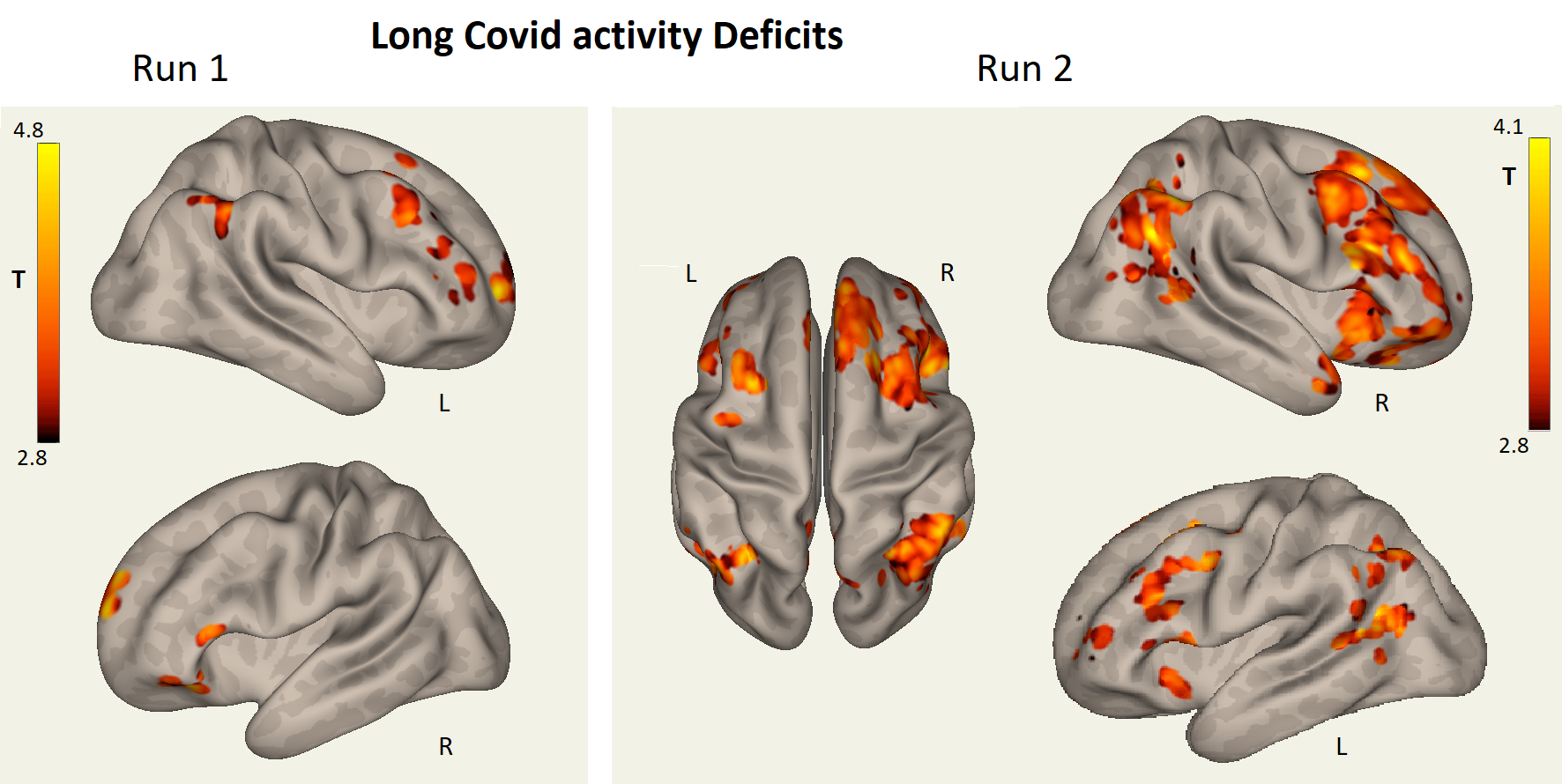
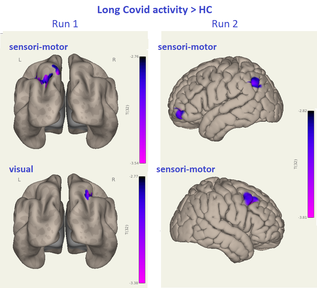
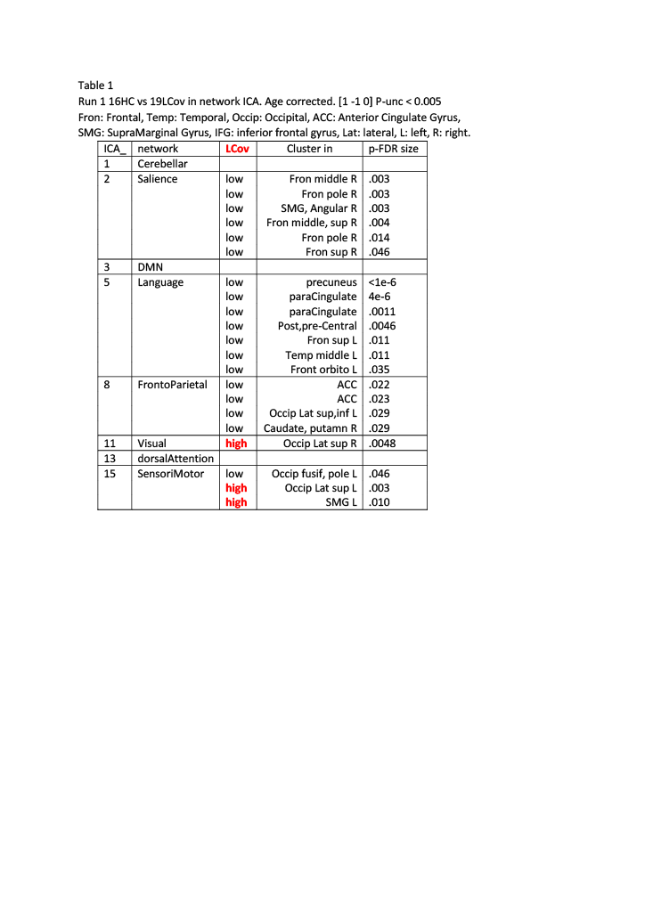
Table 1
Run 1 16HC vs 19LCov ICA components in intrinsic networks. Age corrected. [1 -1 0]. Voxel p-unc < 0.005.
Fron: Frontal, Temp: Temporal, Occip: Occipital, ACC: Anterior Cingulate Gyrus,SMG: SupraMarginal Gyrus, IFG: inferior frontal gyrus, Lat: lateral, L: left, R: right.
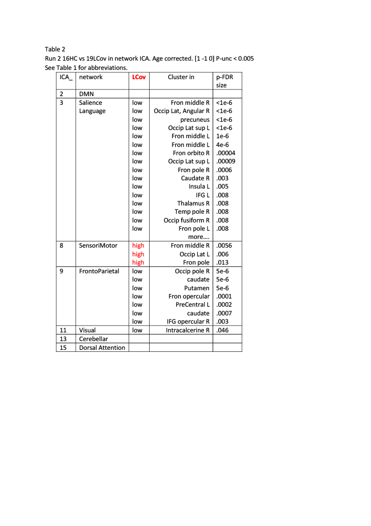
Table 2 Run 2 16HC vs 19LCov ICA components in intrinsic networks. Age corrected. [1 -1 0]. Voxel p-unc < 0.005. See Table 1 for abbreviations.