2080
Subtle Brain Structure Alternations in Young Adult Patients infected with SARS-CoV-2 Omicron and Mild Symptom: A Case Control Study Using MRI1Center for Biomedical Imaging Research, Department of Biomedical Engineering, School of Medicine, Tsinghua University, Beijing, China, 2Department of Radiology, the Affiliated BenQ Hospital of Nanjing Medical University, Nanjing, China
Synopsis
Keywords: Infectious Disease, COVID-19, brain changes, Omicron, young adults, mild symptom, SARS-CoV-2
Motivation: The existence of brain structural abnormalities in young adults with mild Omicron infections remains unclear.
Goal(s): This study aimed to investigate short-term brain structure changes in this specific patient group.
Approach: Young adult patients and age- and sex-matched controls were enrolled, and brain structures were assessed on T1-MPRAGE images. One-way ANOVA, Kruskal-Wallis tests, and logistic regression were employed to determine differences among groups.
Results: While whole brain volume didn't significantly differ among groups, subtle alterations in various regions within cerebral cortex, white matter, and cerebrospinal fluid were observed during the acute phase, with a tendency of recovery over time.
Impact: Short-term structural changes in brain were observed in this study. The findings suggest cerebral impairments at the early stage of infection with SARS-CoV-2 Omicron even in individuals with mild symptoms.
Introduction
SARS-CoV-2 has been linked to changes in brain structure 1. With the rise of the Omicron variant, most infected patients experienced only mild symptoms 2,3. It's unclear if these individuals, particularly young adults who received little attention in previous studies, are at risk of brain structural abnormalities. Therefore, this study aimed to investigate the short-term brain structural changes in young adults infected with SARS-CoV-2 Omicron and experienced mild symptoms using brain magnetic resonance imaging (MRI).Methods
Study population: Young adults with two consecutive positive SARS-CoV-2 antigen tests between December 2022 and February 2023 were enrolled as patients (PT). Age- and sex-matched asymptomatic controls (AC) without SARS-CoV-2 positives or flu-like symptoms during that period were also recruited. Individuals with incidental findings or MR examination contraindications were excluded. The study was approved by the local Institutional Review Board, and written consents were obtained from participants. MR imaging protocol: Participants underwent structural imaging by acquiring T1-MPRAGE sequence on a 3.0T MR scanner (Ingenia TX, Philips Healthcare, Best, The Netherlands) with a 32-channel head coil. The imaging parameters were: turbo field echo sequence, repetition time 6.6 ms, echo time 3 ms, field of view 250×250×180 mm3 and 1mm isotropic resolution. MR imaging processing: Brain region volumes were measured using in-house MATLAB scripts and MRICloud (Johns Hopkins University, Baltimore, MD, USA) with the Adult_286labels_10atlases_V5L atlas.Whole brain volume was measured and the regional volumes were normalized for following analysis. Statistical analysis: The infected patients were categorized as ≤30 days (≤30d PT) and >30 days (>30d PT) groups according to the time from infection to MR examination. Brain volume differences among groups were assessed using one-way ANOVA or Kruskal-Wallis tests with Bonferroni adjustment. Univariate and multivariate logistic regressions were employed to determine the association between brain volumes and SARS-CoV-2 infection, adjusting for confounding factors.Results
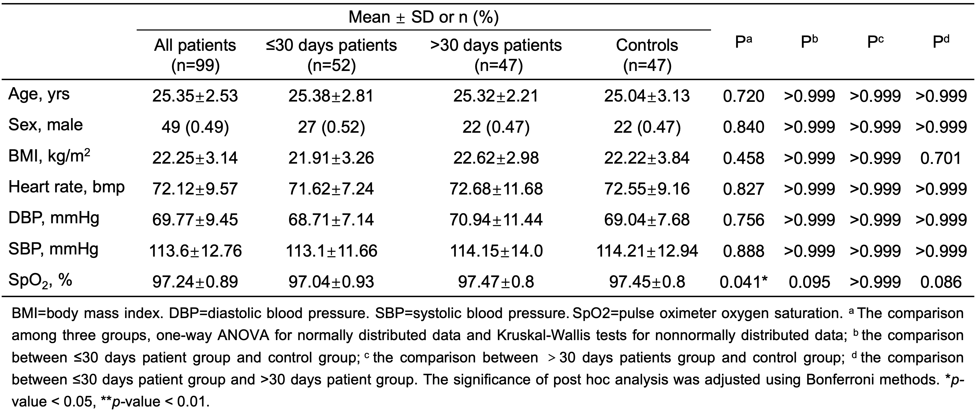
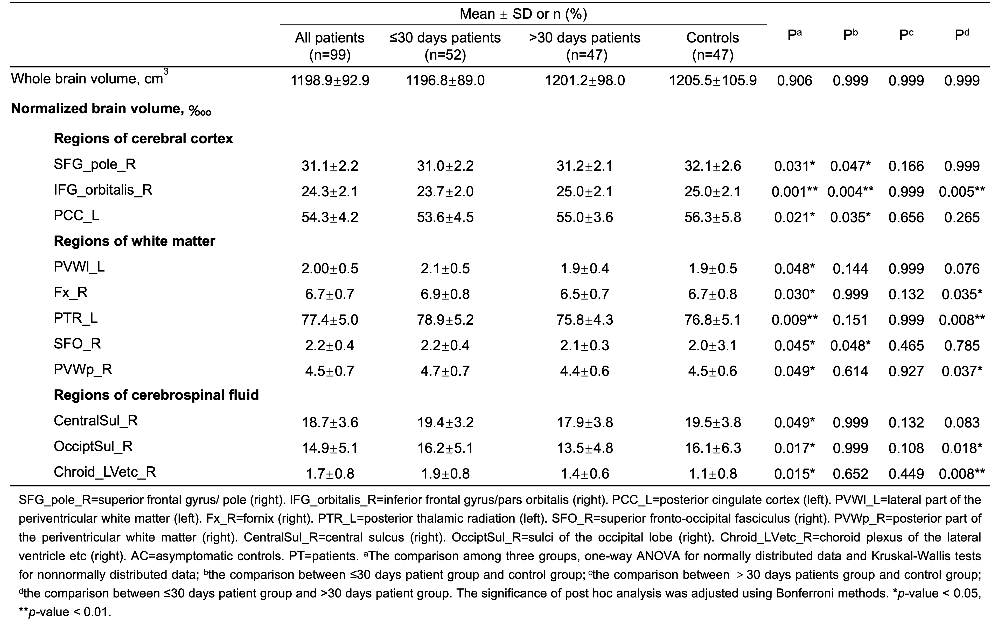
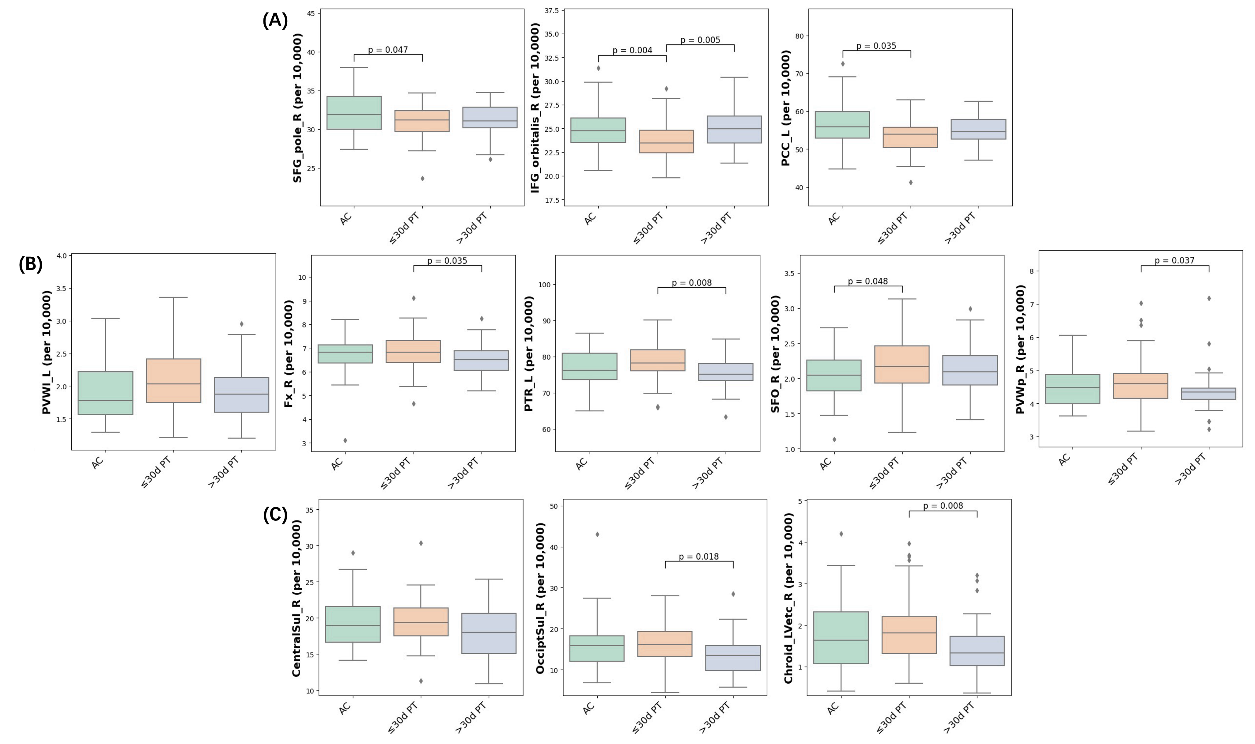
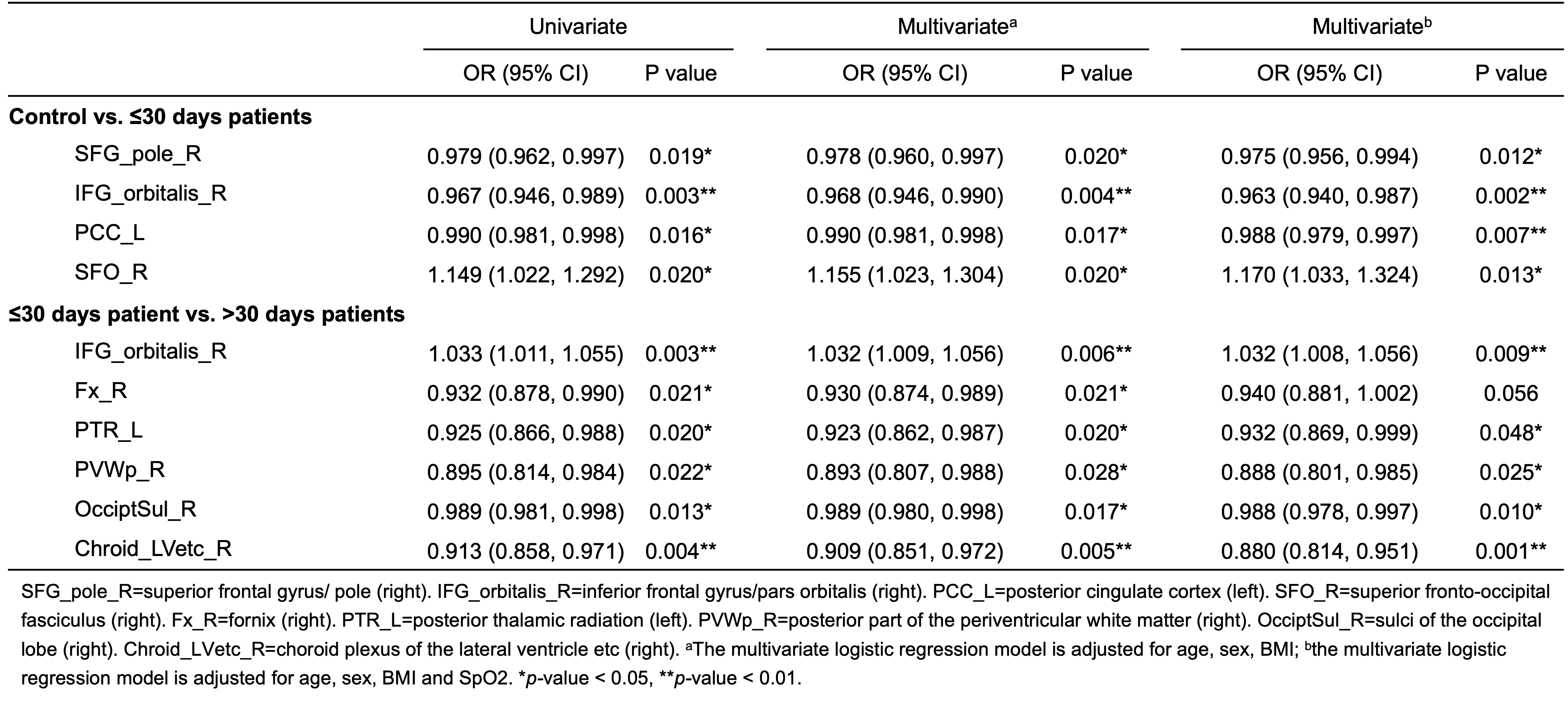
A total of 99 patients (mean age: 25.35 ± 2.53 years; 49 males) and 47 age- and sex-matched controls (mean age: 25.04 ± 3.13; 22 males) were included in the study. The clinical characteristics were summarized in Table 1. A significant difference in SpO2 was observed among three groups, while no significance was reserved in the post hoc analysis. The comparison of whole brain volume and normalized brain volumes, which appeared statisticaly significant among three groups, were summarized in Table 2 and Figure 1. Whole brain volume showed no significant difference among three groups (1196.8±89.0 cm3 vs. 1201.2±98.0 cm3 vs. 1205.5±105.9 cm3, p=0.906). In the ≤30 days patient group, significant declines in SFG_pole_R (32.1±2.6 ‱ vs. 31.0±2.2 ‱, p=0.047), IFG_orbitalis (25.0±2.1 ‱ vs. 23.7±2.0 ‱, p=0.004) and PCC_L (56.3±5.8 ‱ vs. 53.6±4.5 ‱, p=0.035), as well as a significant increase of SFO_R (2.0±3.1 ‱ vs. 2.2±0.4 ‱, p=0.048), were found compared to the control group. Meanwhile, the >30 days patient group showed significant decline of Fx_R (6.9±0.8 ‱ vs. 6.5±0.7 ‱, p=0.035), PTR_L (78.9±5.2 ‱ vs. 75.8±4.3 ‱, p=0.008), PVWp_R (4.7±0.7 ‱ vs. 4.4±0.6 ‱, p=0.037), OcciptSul_R (16.2±5.1 ‱ vs. 13.5±4.8 ‱, p=0.018) and Chroid_LVetc_R (1.9±0.8 ‱ vs. 1.4±0.6 ‱, p=0.008), along with a significant increase of IFG_orbitalis (23.7±2.0 ‱ vs. 25.0±2.1 ‱, p=0.005), were observed compared to the ≤30 days patient group. Notably, most differences remained statistically significant in multivariate logistic regression analyses (except for Fx_R) after adjusting for: model 1: age, sex and BMI; model 2: age, sex, BMI and SpO2 (Table 3).Discussion
While young adult patients did not exhibit significant differences in whole brain volume when compared to controls, subtle alterations were observed in various brain regions, each following a distinct pattern. Regions within cerebral cortex showed slight atrophy in the acute phase, while white matter and CSF volumes exhibited short-term increases. Previous studies have widely reported white matter abnormalities, including demyelination and encephalitis 4,5. In this study, although no apparent radiological findings are observed, there may still exist inflammatory responses in white matter and results in a subtle increase in local volumes. Regional brain damage, particularly in the orbitofrontal cortex and posterior cingulate cortex, was consistent with prior studies 6,7. However, this damage displayed a tendency towards recovery and did not show a significant difference when compared to controls in post-acute phase young adult patients.Conclusion
Young adult patients infected with SARS-CoV-2 Omicron show no significant difference in whole brain volume compared to asymptomatic controls. However, subtle alterations in the cerebral cortex, white matter, and cerebrospinal fluid can be observed, displaying distinct patterns during the acute phase, accompanied with a tendency towards recovery over time.Acknowledgements
No acknowledgement found.References
1.Douaud G, Lee S, Alfaro-Almagro F, et al. SARS-CoV-2 is associated with changes in brain structure in UK Biobank. Nature 2022; 604: 697-707.
2. Sigal A, Milo R, Jassat W. Estimating disease severity of Omicron and Delta SARS-CoV-2 infections. Nat Rev Immunol 2022; 22: 267-269.
3. Abdullah F, Myers J, Basu D, et al. Decreased severity of disease during the first global omicron variant covid-19 outbreak in a large hospital in tshwane, south africa. Int J Infect Dis 2022; 116: 38-42.
4. Andalib S, Biller J, Di Napoli M, et al. Peripheral Nervous System Manifestations Associated with COVID-19. Curr Neurol Neurosci Rep 2021; 21: 9.
5. Kremer S, Lersy F, Anheim M, et al. Neurologic and neuroimaging findings in patients with COVID-19: A retrospective multicenter study. Neurology 2020; 95: e1868-e1882.
6.Qin Y, Wu J, Chen T, et al. Long-term microstructure and cerebral blood flow changes in patients recovered from COVID-19 without neurological manifestations. J Clin Invest 2021; 131: e147329.
7. Najt P, Richards HL, Fortune DG. Brain imaging in patients with COVID-19: A systematic review. Brain Behav Immun Health. 2021 Oct;16:100290.
Figures