2079
Altered Volumes of the Limbic Structures in COVID-19 Patients with neuroPASC revealed by Ultrahigh Field 7T MRI1Diagnostic, Molecular and Interventional Radiology, Icahn School of Medicine at Mount Sinai, New York, NY, United States, 2BioMedical Engineering and Imaging Institute (BMEII), Icahn School of Medicine at Mount Sinai, New York, NY, United States, 3Neurology, Icahn School of Medicine at Mount Sinai, New York, NY, United States, 4General Internal Medicine, Icahn School of Medicine at Mount Sinai, New York, NY, United States
Synopsis
Keywords: Infectious Disease, COVID-19
Motivation: Post-Acute Sequelae of COVID-19 of neurological origin (neuroPASC), such as brain fog and anxiety, are not well understood.
Goal(s): To study volumetric changes in the brain’s limbic structures in COVID-19 patients with neuroPASC such as brain fog.
Approach: 29 COVID-19 patients with neuroPASC and 29 matched controls underwent ultrahigh field brain MRI at 7T. Hippocampal subfields and amygdala subnuclei were segmented with FreeSurfer and studied across neuroPASC subgroups.
Results: Several subunit volumes were significantly decreased in COVID patients compared to controls across neuroPASC subgroups. Sex-based differences were also present.
Impact: There may be subtle changes in the volumes of the brain’s limbic structures in COVID patients with neuroPASC compared to controls. These changes may be related to neuroPASC symptoms and may depend on sex differences.
Introduction
While the Coronavirus Disease 2019 (COVID-19) is primarily a respiratory illness1, a significant proportion of COVID patients continue to suffer persistent, debilitating symptoms2 which are termed Post-Acute Sequelae of COVID-19 (PASC)3 and also termed “long COVID”. Several of these sequelae are neurological in origin (neuroPASC), such as brain fog (impaired cognition, executive function and memory), fatigue, altered smell or taste, anxiety and others. These manifestations are not well understood. Brain fog has been hypothesized to arise from neuronal injury to the hippocampus4,5. Here, we report our preliminary findings of altered volumes in the limbic structures, including the hippocampus and amygdala, of neuroPASC patients using high-resolution, ultrahigh field MRI at 7T.Methods
29 COVID-19 patients (age: 43±14 years , 14 female) who experienced neuroPASC were included in this study, as well as 29 age- and sex-matched healthy control subjects (age: 42±13 years , 14 female). Healthy control subjects were identified as individuals who either A) never contracted COVID, or B) had asymptomatic or mildly symptomatic COVID more than 12 months prior to MRI with no neuroPASC and were never hospitalized for COVID. All subjects underwent whole brain MRI on a Siemens Magnetom 7T scanner using a 1Tx/32Rx head coil. The COVID-19-specific MRI protocol included axial T1-weighted MP2RAGE, FLAIR, T2-weighted TSE, susceptibility weighted imaging (SWI). Self-reported neuroPASC data were collected from questionnaires administered at time of MRI.FreeSurfer6 (version 6) was used to carry out cortical image reconstruction and automated segmentation of the hippocampal subfields, amygdala subnuclei and hypothalamic subunits. All volumes were normalized to the subject’s estimated intracranial volume. Metrics were compared between COVID and control cohorts for all subjects as well as for subgroups based on sex and neuroPASC criteria of brain fog and anxiety. Statistical analysis of segmentation volumes between COVID patients and controls was carried out in MATLAB using a two-tailed t-test for normally distributed volumes or using a Wilcoxon rank sum test for non-normally distributed volumes. Normality was tested using the Shapiro-Wilk test. Corrections for multiple comparisons were not done.
Results
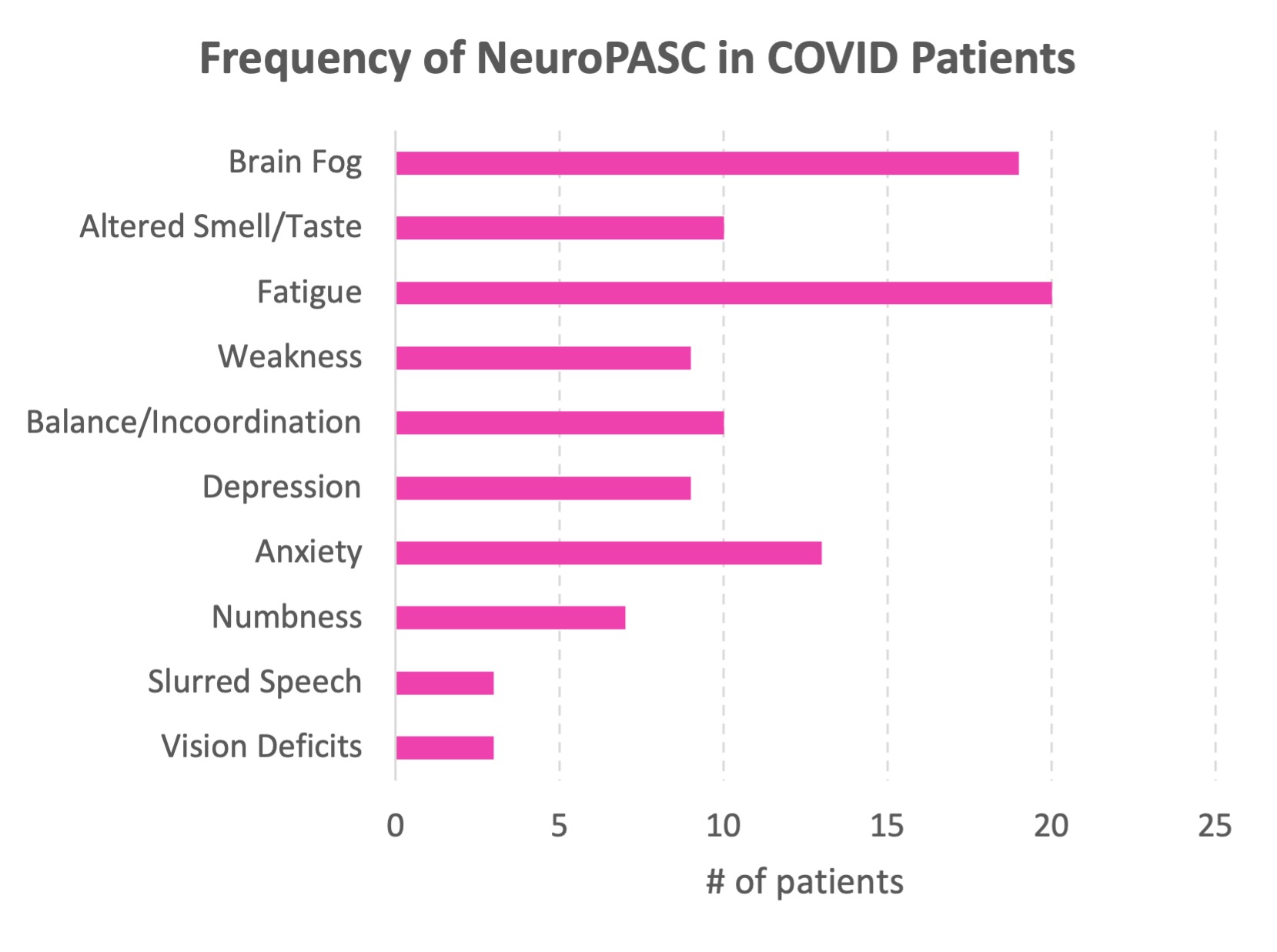
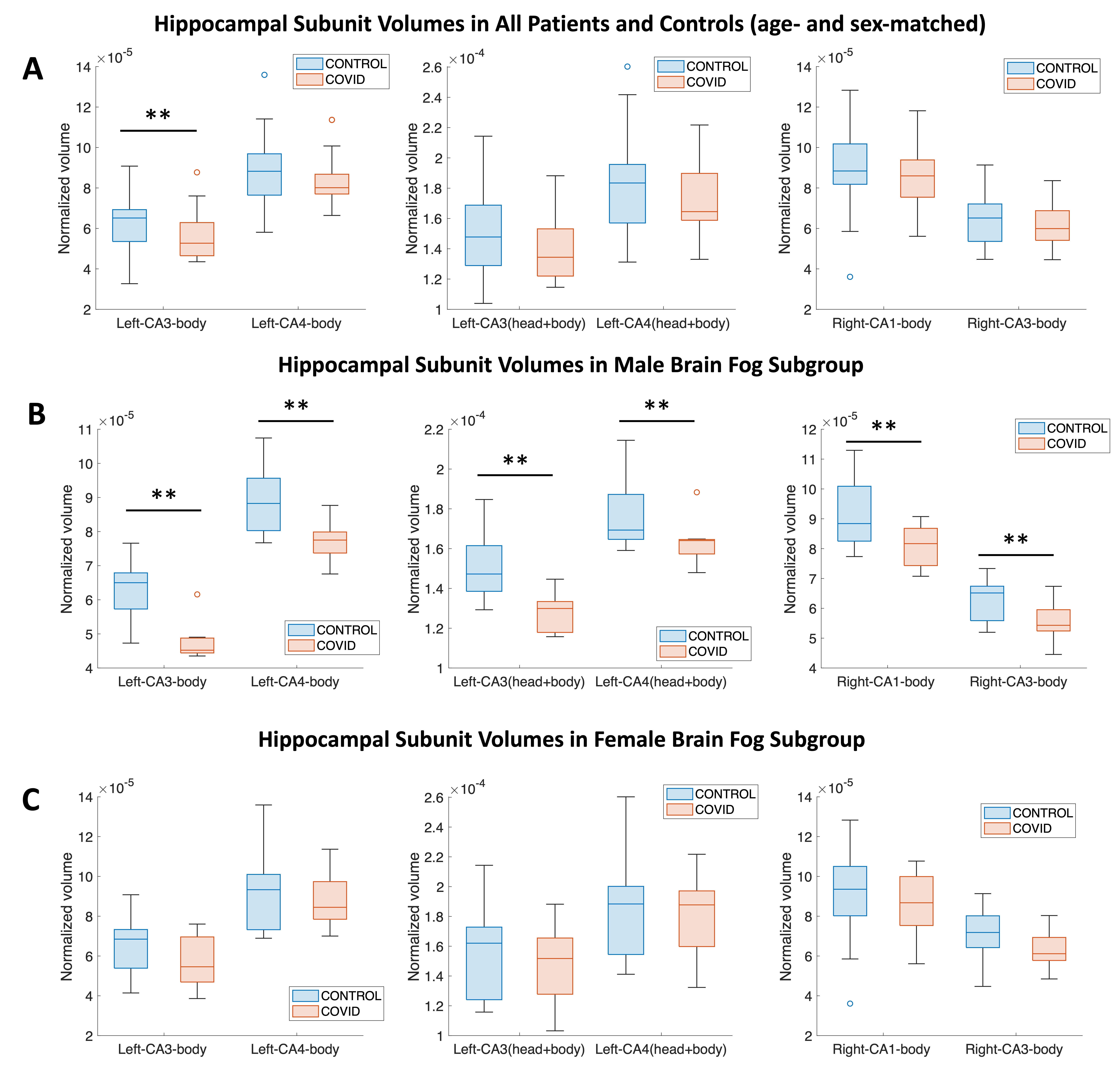
The frequency of the different neuroPASC categories in our COVID patient cohort is shown in Fig. 1.Analysis of hippocampal subunit segmentations showed that the volume of the left CA3 body was significantly decreased in all COVID patients (p = 0.036) (n=29) compared to age- and sex-matched controls (Fig. 2A). In the subgroup of COVID patients who reported the neuroPASC symptom of brain fog, several hippocampal subunit volumes were significantly decreased in male COVID patients (n=9) compared to age-matched male controls (Fig. 2B). There were no significant differences in hippocampal subunit volumes between female COVID patients reporting brain fog (n=12) and age-matched female controls (Fig. 2C). Similarly, in the subgroup of COVID patients who reported the neuroPASC symptom of anxiety, several hippocampal subunit volumes were significantly decreased in male COVID patients (n=7) compared to age-matched male controls, but not in female COVID patients (n=10) compared to age-matched female controls.
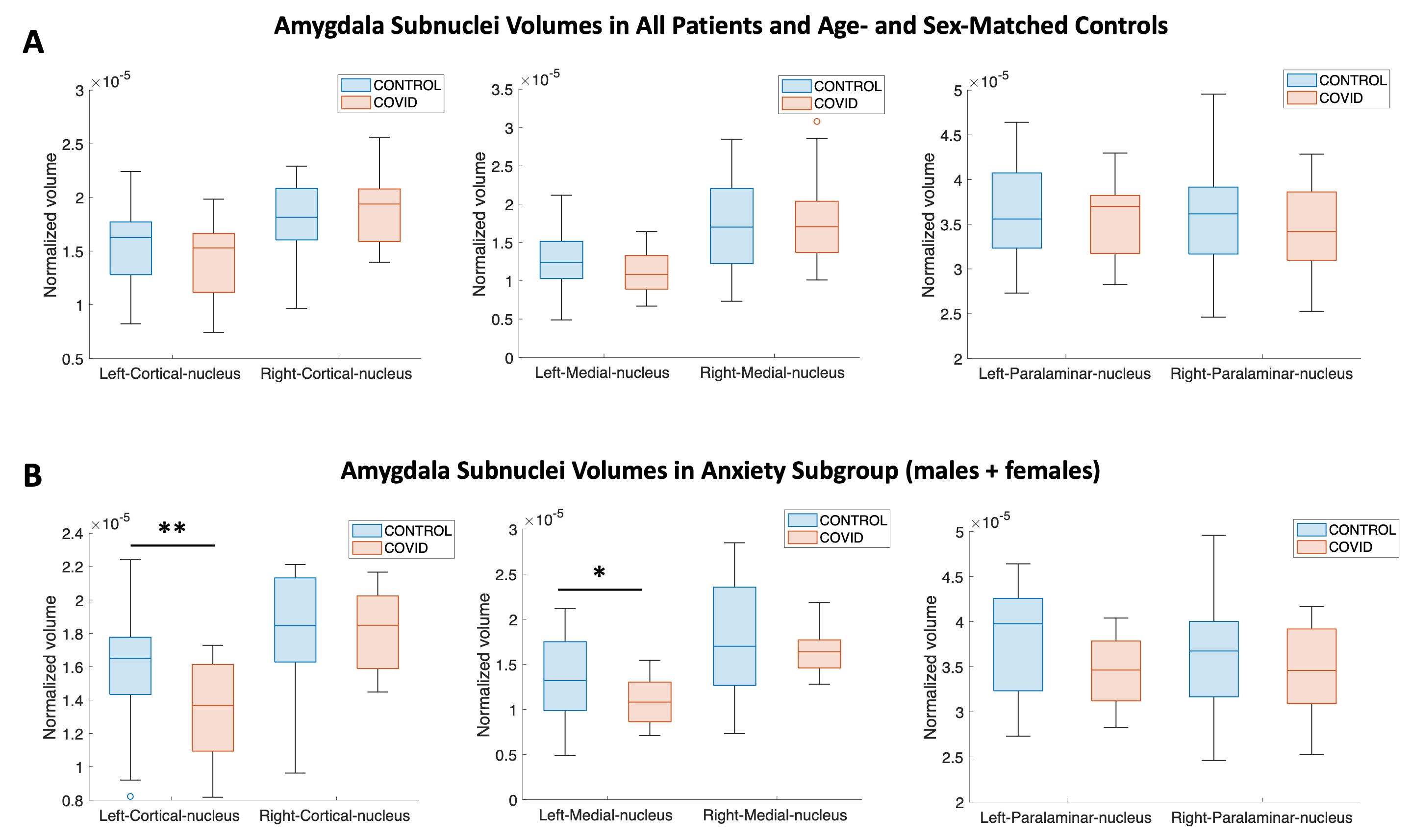
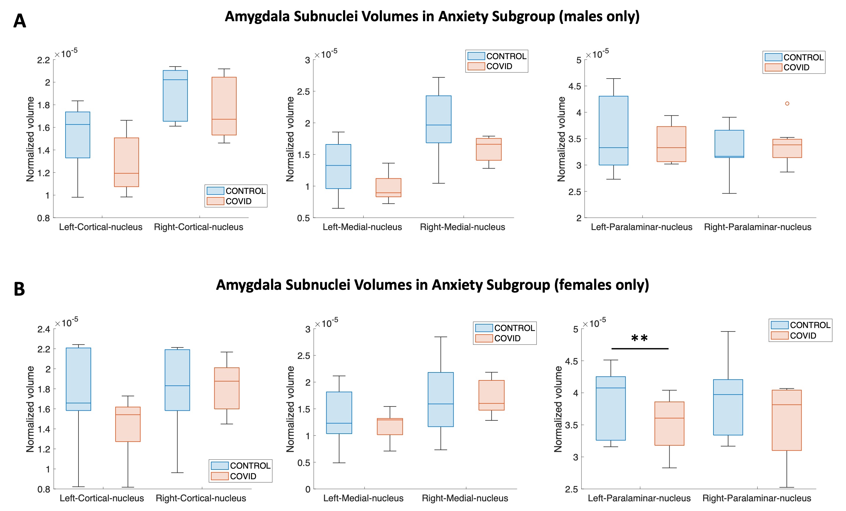
Analysis of amygdala subnuclei showed no significant difference COVID patients and controls when including all age- and sex-matched patients (n=29) (Fig. 3A). In the subgroup of patients (male+female) who reported the neuroPASC symptom of anxiety (n=16), volume of the left cortical nucleus was significantly decreased in COVID patients (p=0.049) compared to age- and sex-matched controls (Fig. 3B). The left medial nucleus had a trend towards being smaller in COVID patients compared to controls (p=0.096) (Fig. 3B). One amygdala subnucleus volume (left paralaminar nucleus) was significantly decreased in female COVID patients with anxiety (n=10) compared to age-matched female controls (p=0.045), but not in male COVID patients (n=7) compared to age-matched male controls (Fig. 4).
Discussion
Even though the COVID pandemic has ended, COVID-19 is still ongoing and remains a serious healthcare burden. The mechanisms of PASC/neuroPASC are still not well understood, causing limitations in treatment.Our results show that subtle but statistically significant alterations of volumes of the limbic structures may exist between COVID patients and healthy controls, especially for patients with the neuroPASC conditions of brain fog and anxiety. Decreased volumes of certain hippocampal subfields such as the CA3 subfield support the possibility of neuronal injury giving rise to brain fog, as suggested in other studies4,5. In addition, the neuroPASC condition of anxiety may be linked to changes in the amygdala, as suggested by our results. Finally, our results indicate that sex differences seen in COVID may be due to differences in how the limbic structures are altered between male and female patients.
The results presented here are preliminary and ongoing. Further analysis of exploring additional neuroPASC-based subgroups are planned.
Acknowledgements
The authors wish to acknowledge Brian Mathew for help with patient recruitment. The authors also wish to acknowledge Drs. David Putrino and Laura Tabacof for help with patient recruitment.
The authors acknowledge the National Institutes of Health (NIH) for the following sources of funding support for this study:
NIH NINDS 1R21NS122389-01
NIH NCI 5R01CA202911-05
References
1. Chen N, et al., Epidemiological and clinical characteristics of 99 cases of 2019 novel coronavirus pneumonia in Wuhan, China: a descriptive study. Lancet, 2020. 395(10223): p. 507-513.
2. Ladopoulos T, et al., “COVID-19: Neuroimaging Features of a Pandemic”. J Neuroimaging, 2021. 31(2): p. 228-243
3. Thaweethai T, et al., “Development of a Definition of Postacute Sequelae of SARS-CoV-2 Infection.” JAMA. 2023 Jun 13;329(22):1934-1946.
4. Nouraeinejad A, The functional and structural changes in the hippocampus of COVID-19 patients. Acta Neurol Belg. 2023 Aug;123(4):1247-1256.
5. Bayat AH, et al., COVID-19 causes neuronal degeneration and reduces neurogenesis in human hippocampus. Apoptosis. 2022 Dec;27(11-12):852-868. doi: 10.1007/s10495-022-01754-9. Epub 2022 Jul 25.
6. Fischl B. “FreeSurfer”. NeuroImage. 2012 Aug;62(2):774–781.
Figures