2078
Longitudinal neuroimaging combined with multi-omics reveals cerebral structural and functional changes in COVID-191radiology, Beijing Tiantan Hospital of Capital Medical University, Beijing, China, 2Beijing TianTan Hospital of Capital Medical University, Beijing, China, 3United Imaging Research Institute of Intelligent Imaging, Beijing, China
Synopsis
Keywords: Infectious Disease, COVID-19
Motivation: COVID-19's neurological effects remain ambiguous, with limited exploration of neuroimaging changes versus clinical symptoms, especially through pre-SARS-CoV-2 exposure longitudinal imaging, remains less explored.
Goal(s): Understand brain change patterns and identify therapeutic targets for post-COVID-19 brain injury and neuropsychiatric effects.
Approach: Utilized consistent neuroimaging on patients in acute recovery across two centers. Employed multiple clinical tools to gauge neuropsychiatric manifestations. Blood samples analyzed for protein profiles indicative of inflammation and neural damage.
Results: Found significant vulnerability in the limbic lobe, observed contrasting pathological changes in symmetrical brain regions, and highlighted potential genetic and neurotrophic factors influencing neuropsychiatric symptoms post-COVID-19.
Impact: This study offers clinicians a clearer path to target COVID-19-related neuropsychiatric symptoms. It prompts further inquiry into genetic influences on brain alterations, enabling tailored therapeutic strategies for affected patients.
Introduction
The neurological implications of COVID-19, caused by the SARS-CoV-2 virus, have been reported with accounts of structural and functional changes in the brain. Neuropsychiatric symptoms, supported by pathological evidence, show the virus's capability to infiltrate neural regions [1, 2]. However, a comprehensive understanding of how these changes on neuroimaging correlate with clinical presentations remains elusive. Very few studies offer insights through longitudinal imaging initiated pre-SARS-CoV-2 exposure, particularly within Asian populations [3]. This research aims to bridge this gap by using a longitudinal approach combined with diverse neuroimaging techniques, shedding light on brain alterations during the acute recovery phase. Additionally, proteomics serve as a robust tool for translating the clinical and neuroimaging observations, unraveling the intricate molecular dynamics at play.Methods
Sixity-six patients were evaluated during their acute recovery phase, termed SCAN-2, with the onset of COVID-19 controlled within a month of their baseline examination (SCAN-1). Inclusion criteria required available neuropsychiatric and neuroimaging data pre- and post-infection. Clinical attributes and neuropsychiatric manifestations were gauged through tools like the Montreal Cognitive Assessment (MoCA), Hamilton Depression and Anxiety Scale (HAMD and HAMA), Pittsburgh Sleep Quality Index (PSQI), Fatigue Severity Scale (FSS), and a self-reporting questionnaire. Neuroimaging was consistently executed across two centers, Beijing Tiantan and Hubei Shiyan Renmin Hospital, at the same 3.0 T system encompassing 3D T1WI, diffusion kurtosis imaging (DKI), and resting-state blood oxygenation level dependent functional MRI (bold-fMRI). Assessment parameters included the volume of cortex and deep gray matter and cortical thickness of gray matter (GM) the fractional anisotropy (FA) and the intracellular volume fraction (ICVF) abstracted from DKI and reflected microstructural disorders in white matter (WM), and the amplitude low-frequency fluctuation (ALFF) abstracted from bold-fMRI to reflect neural activity alterations. Concurrent with SCAN-2, blood samples were procured, testing for ApoE isoforms, and protein profiles indicative of inflammation and neural damage. Paired t-tests ascertained clinical and imaging changes, while correlation analyses sought associations between clinical changes, protein profiles, and genetic risks.Results
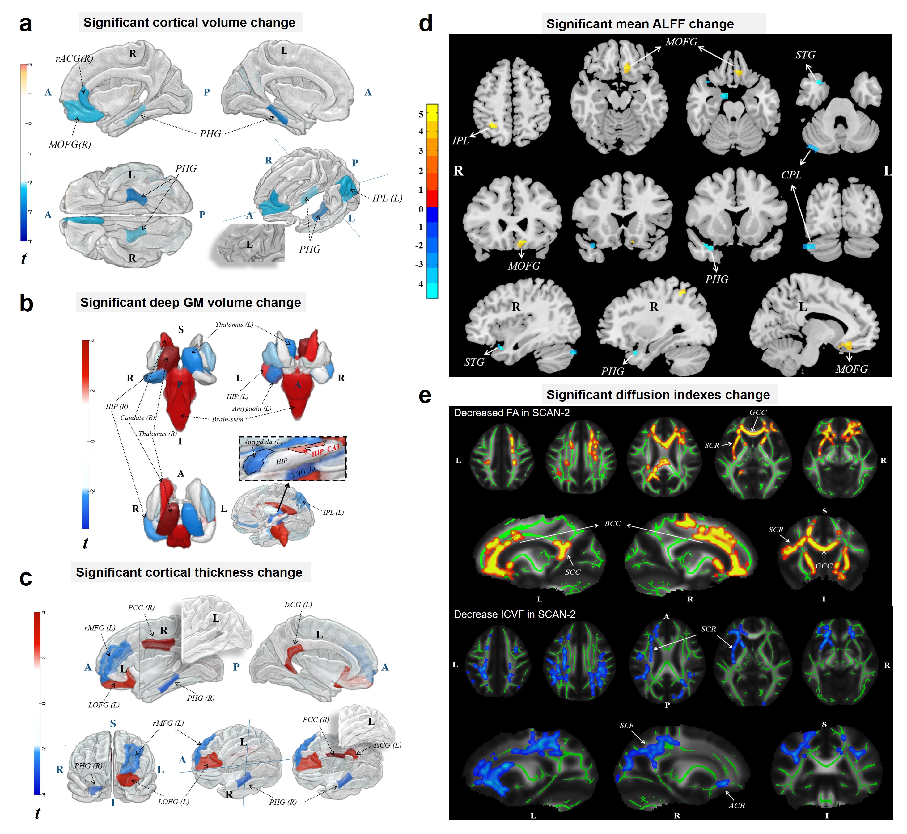
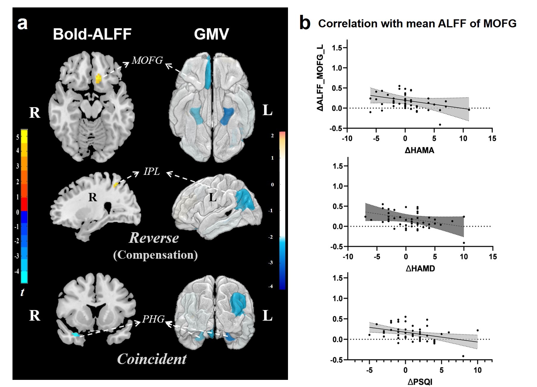
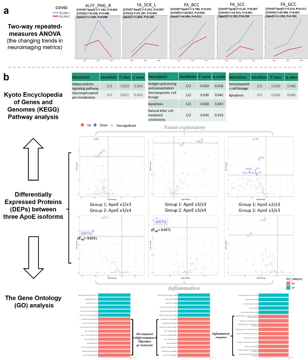
Our results underscored the limbic lobe's susceptibility in COVID-19, evidencing concurrent volume and thickness reduction in GM (all r < -2.0, all FDR corrected P < 0.05), WM diffusional index decreases (voxel significance at FDR corrected P <0.05), and hypoactivity in neuronal regions (voxel level P < 0.01 and cluster level P < 0.05 with GRF correction), which correlated with pro-inflammatory cytokines (all r < -0.30, all P < 0.05) and NEFL (r = -0.32, P = 0.028) (Figure 1). Interestingly, symmetrical brain regions or ipsilateral hemisphere function-related regions demonstrated morphological and functional changes with contrasting pathological implications (Figure 2). A noted lack of compensatory mechanisms in regions on contralateral hemisphere or functional related regions to the affected areas, especially the prefrontal lobe, was associated with neuropsychiatric issues. Additionally, older patients exhibited heightened neuroinflammation and brain injury biomarkers post-infection, represented by the hippocampal CA3 subfield volume. Lastly, ApoE polymorphisms potentially influenced hypo-neuronal activity (Figure 3), and sleep efficiency declines correlated with neurotrophic factors (NRTN and NT-3).Conclusion
Through the integration of longitudinal neuroimaging and proteomics, our study elucidates the structural and functional cerebral alterations resulting from COVID-19. Validating the pathophysiological underpinnings, we spotlight potential therapeutic targets for COVID-19-related neuropsychiatric symptoms, paving the way for protective strategies for vulnerable populations.Acknowledgements
No acknowledgement found.References
[1]. Stein, S.R., et al., SARS-CoV-2 infection and persistence in the human body and brain at autopsy. Nature, 2022. 612(7941): p. 758-763.
[2]. Crunfli, F., et al., Morphological, cellular, and molecular basis of brain infection in COVID-19 patients. Proc Natl Acad Sci U S A, 2022. 119(35): p. e2200960119.
[3]. Douaud, G., et al., SARS-CoV-2 is associated with changes in brain structure in UK Biobank. Nature, 2022. 604(7907): p. 697-707.
Figures