2077
Neurochemical Alterations Caused by Persistent COVID-19-Induced Anosmia: Probing Orbitofrontal Cortex Metabolites and Potential Biomarker1Guilan University of Medical Sciences, Rasht, Iran (Islamic Republic of), 2Center for Magnetic Resonance Research, University of Minnesota Medical School, Minneapolis, MN, United States
Synopsis
Keywords: Infectious Disease, COVID-19, Magnetic Resonance Spectroscopy, Anosmia, Orbitofrontal Cortex
Motivation: Olfactory impairments caused by COVID-19 have been addressed in neuroimaging findings over the past years. However, long-term complications on brain orbitofrontal cortex (OFC) needs to be further investigated.
Goal(s): Our goal was to assess metabolites level change in the brain OFC region in COVID-induced anosmia subjects in comparison to healthy population.
Approach: 54 subjects categorized into control, short- and long-term anosmia groups were studied using single voxel MRS. Metabolite level alterations of their brain OFC region were investigated.
Results: We found that NAA, Cho, and Cr levels in brain OFC region decreased as COVID-induced anosmia duration and severity increased.
Impact: The demonstrated results of alteration in brain metabolites level influenced by the severity of COVID-induced anosmia highlights MRS as a robust assessment tool for physicians to better investigate olfactory function impairment in the brain orbitofrontal cortex.
Introduction
COVID-19 symptoms can involve multiple organs and systems with short- and long-term complications1. A variety of neuropsychiatric manifestations including anosmia have been identified in COVID patients 2. Olfactory abnormalities are the most common neuroimaging findings 3 and evidence relates anosmia to olfactory processes and central nervous system (CNS) dysfunction. However, there has been less attention paid to COVID consequences on orbitofrontal cortex (OFC). Proton Magnetic Resonance Spectroscopy (1H-MRS) is a non-invasive technique to study chronic manifestations of CNS disorders. This study aims to use 1H-MRS to investigate alterations of brain metabolites impacted by persistent COVID-19-induced anosmia in regions associated with olfaction, particularly in OFC.Methods
27 subjects with COVID-19-induced anosmia and 27 healthy individuals were studied. In the first scenario, healthy group was compared to the entire anosmia group. Then, anosmia group was followed and categorized into two subgroups. Those who had anosmia after three months but improved after six months were included in Group 2, and cases who had still anosmia after six months were classified as Group 3, while healthy subjects were considered as Group 1. A national standard smell identification test (IR-SIT) was employed with scores ranging from 10 (severe) to 24 (normal) to diagnose anosmia.Proton single-voxel spectroscopy data using PRESS pulse sequence along with structural brain images from all subjects were acquired at a clinical 1.5 T scanner. A cubic voxel (2×2×1 cm3) was located on the right OFC in the brain while avoiding contact with the subcutaneous fat, skull, vasculature, arachnoid space, and cerebrospinal fluid as shown in Figure 1. 1H-MRS data was processed, and metabolic peak intensities were particularly measured for N-acetyl aspartate (NAA), choline (Cho), and Creatine (Cr) reported as integrals. Ratios of NAA/Cr, NAA/Cho, and Cho/Cr were calculated for normalization. One-way ANOVA test was used to compare MRS data among different groups (P<0.005). A linear regression was applied to the whole subject’s population data to investigate potential correlation between MRS data and IR-SIT scores.Results
Figure 1 shows the voxel placement on the right orbitofrontal cortex as well as spectrums associated with three groups in the right panel. Figure 2 shows the metabolites’ level comparison between the entire anosmia group (Group 1) and with control group (Group 2). All three metabolites exhibited lower levels in anosmia group, though, NAA difference was more pronounced (147±3 vs 301±2). In contrast, Cho/Cr was the only metabolic ratio that was higher in anosmia group (0.45 vs 0.36).In a differentiated way, Figure 3 shows the difference in metabolites' level and ratios among the three groups. The longer period anosmia group subjects belonged to, the lower their metabolite levels and ratios were, except for Cho/Cr, which increased for longer anosmia. Figure 4 shows the correlation between MRS-derived metabolite data and IR-SIT test scores. The lower test scores are associated with severe and longer anosmia. The regression line in NAA/Cho ratio had the highest R-squared (0.81) compared to the other five parameters.Discussion
A significant decrease was observed in NAA, Cho, and Cr levels and ratios such as NAA/Cho, and NAA/Cr in the brain orbitofrontal cortex of anosmia subjects. Severe and longer anosmia cases (group 3) had even further reduction in the metabolite levels. Among metabolic parameters employed to study OFC, NAA/Cho ratio had the highest linearity correlation with the smell identification test scores (SIT) for anosmia.Conclusion
Noticeable reduction in some of the vital metabolites’ levels impacted by COVID-19 induced anosmia in brain orbitofrontal cortex highlights the link between anosmia and function impairment in this cortex. The NAA/Cho ratio derived from MRS data can be a potential biomarker for anosmia severity and recovery examination. In addition, proton MRS is a robust and reliable approach for assessing anosmia and detecting COVID-19-related dysfunction in the brain olfactory region.Acknowledgements
No acknowledgement found.References
1. Davis, H. E., McCorkell, L., Vogel, J. M. & Topol, E. J. Long COVID: major findings, mechanisms and recommendations. Nat. Rev. Microbiol. 21, 133–146 (2023).
2. Ernst, T. et al. Neuronal and Glial Metabolite Abnormalities in Participants With Persistent Neuropsychiatric Symptoms After COVID-19: A Brain Proton Magnetic Resonance Spectroscopy Study. J. Infect. Dis. jiad309 (2023).
3. Afsahi, A. M. et al. Brain MRI findings in neurologically symptomatic COVID-19 patients: a systematic review and meta-analysis. J. Neurol. 270, 5131–5154 (2023).
Figures
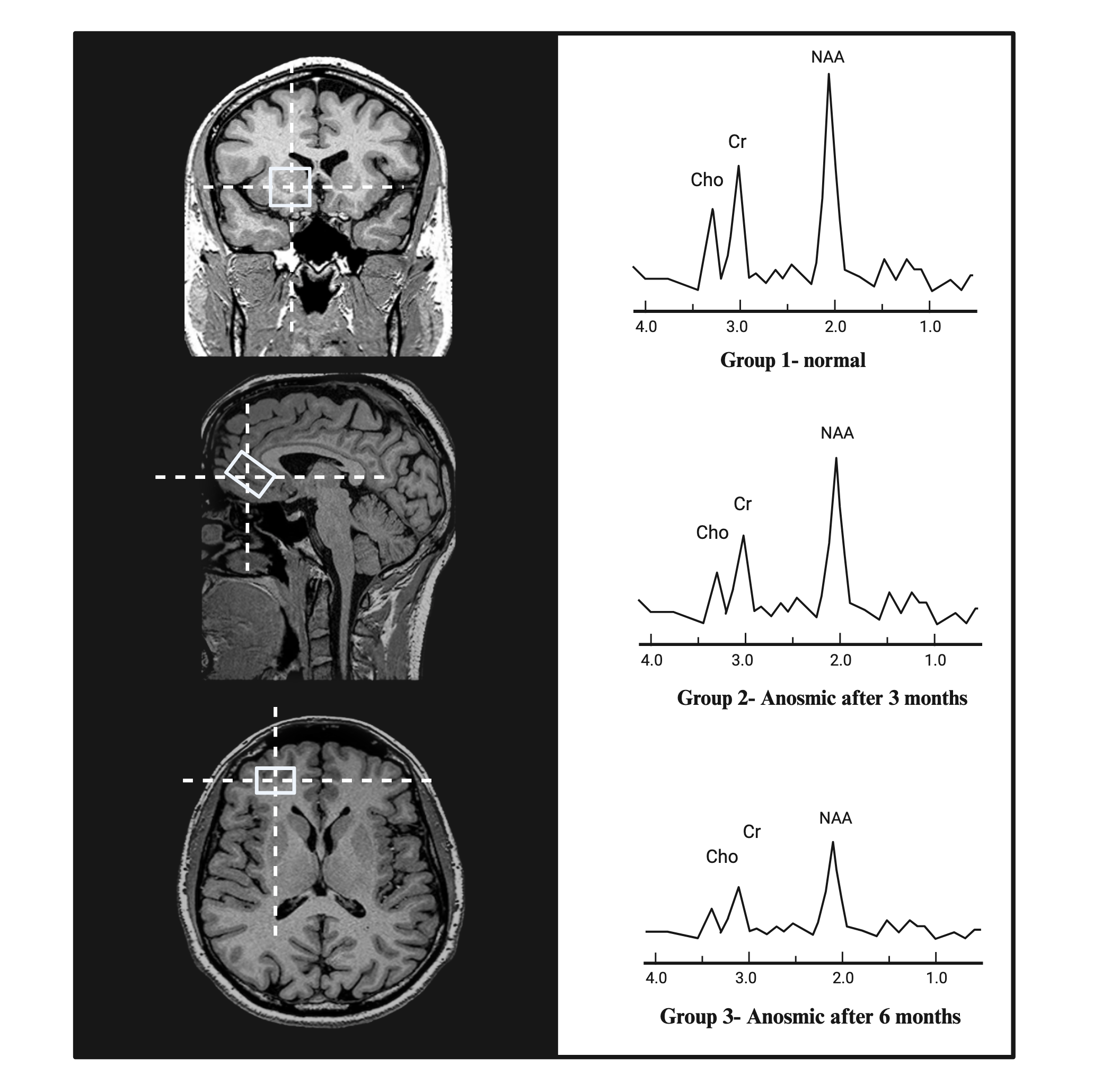
Figure 1: Voxel location on the brain right orbitofrontal cortex shown in three views (left panel). Spectrums of three groups involved in the study (right panel)
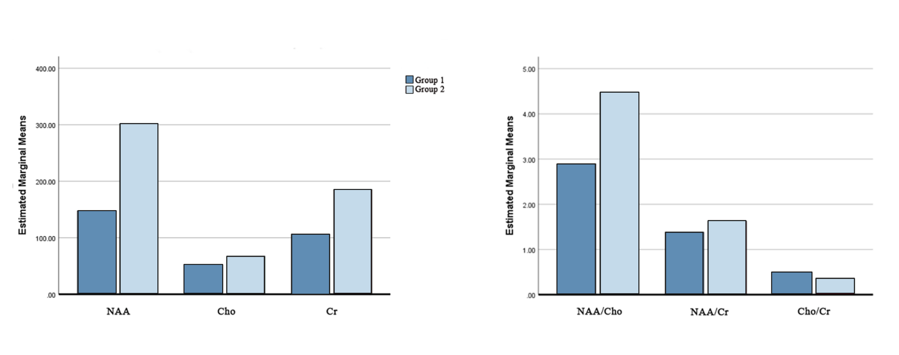
Figure 2: Metabolites level (left panel) and metabolic ratios (right panel) between the entire anosmia population (group 1) vs healthy population (group 2). Levels are reported as integral.
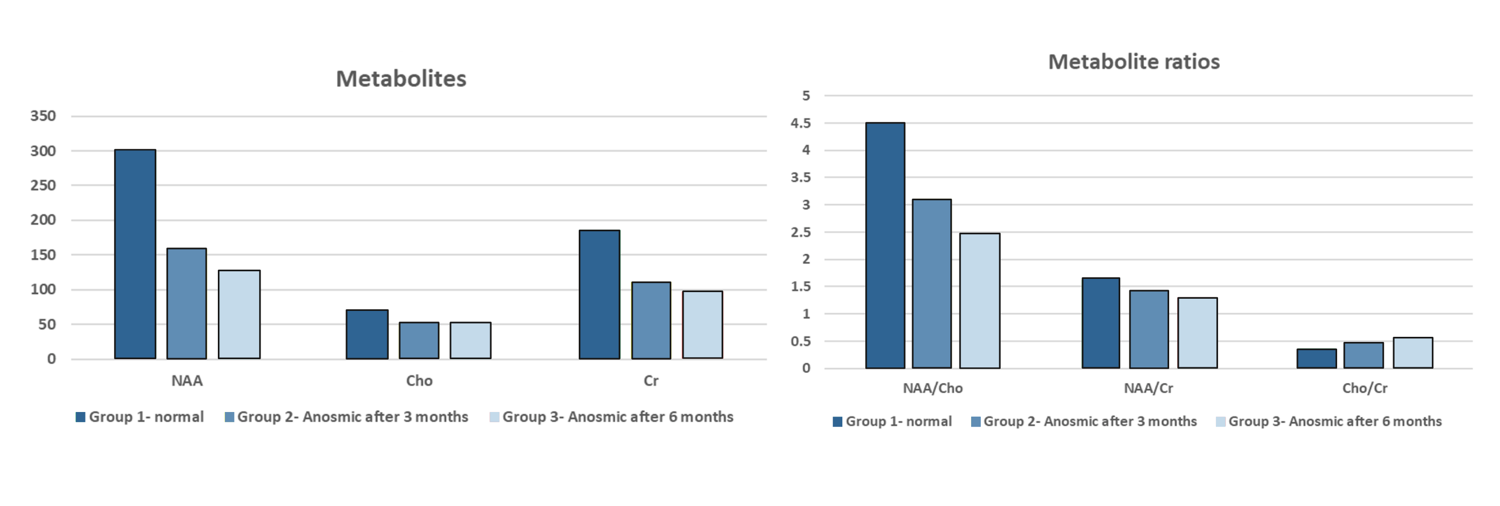
Figure 3: Comparison of metabolites level (left panel) as well as metabolic ratios (right panel) between three groups categorized as control (1), anosmic after three months (2), and anosmic after six months (3). Levels are reported as integral.
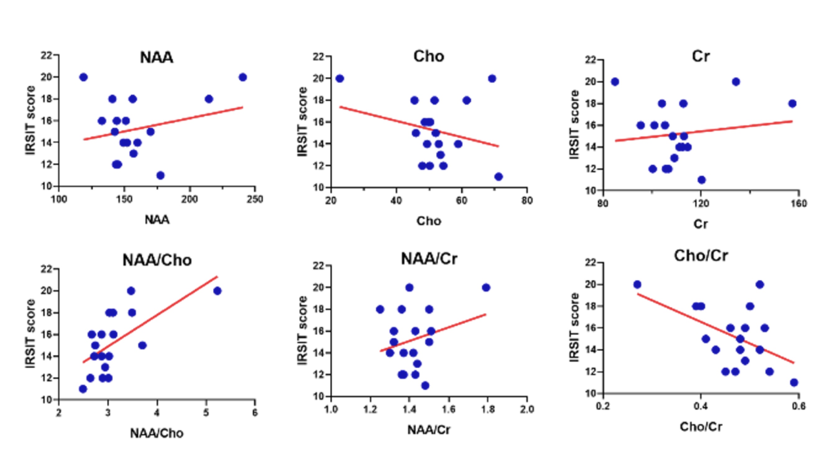
Figure 4: Correlation between MRS-derived metabolite data of the whole population and IR-SIT test scores along with fitted linear regression (red). The lower test scores signify severe anosmia.