2075
White matter alterations in brain fog: A Long-Covid study1Department of Clinical and Experimental Epilepsy, UCL Queen Square Institute of Neurology, Faculty of Brain Sciences, University College London, London, United Kingdom, 2NMR Research Unit, Queen Square MS Centre, Department of Neuroinflammation, UCL Queen Square Institute of Neurology, Faculty of Brain Sciences, University College London, London, United Kingdom, 3Department of Brain & Behavioural Sciences, University of Pavia, Pavia, Italy, 4Department of Medical Physics and Biomedical Engineering, Centre for Medical Image Computing (CMIC), University College London, London, United Kingdom, 5E-Health Center, Universitat Oberta de Catalunya, Barcelona, Spain, 6Radiomics Group, Vall d’Hebron Institute of Oncology, Vall d’Hebron Barcelona Hospital Campus, Barcelona, Spain, 7Neurology-Neuroimmunology Department Multiple Sclerosis Centre of Catalonia (Cemcat), Vall d’Hebron Barcelona Hospital Campus, Barcelona, Spain, 8Digital Neuroscience Center, IRCCS Mondino Foundation, Pavia, Italy
Synopsis
Keywords: Neuroinflammation, COVID-19
Motivation: The symptoms of brain fog include problems with concentration, memory, attention, which result in difficulties with communication. This can make it extremely difficult for an individual to carry out daily tasks and responsibilities as well as maintain relationships deteriorating their quality of life.
Goal(s): To investigate white matter alteration in Long-COVID.
Approach: Voxel-wise analysis of core white matter voxels using advanced MRI metrics and neuropsychological scores.
Results: Results highlight promising perspective for further investigations and potential clinical interpretation of Long-COVID syndromes
Impact: The addition of MRI enables a more thorough exploration of the diverse cognitive dimensions affected in individuals experiencing brain fog and perhaps the possibility to understanding the involvement of WM alterations as either a risk factor or consequence of Long-COVID.
Introduction
The majority of people who develop COVID-19 after contracting SARS-CoV-2 typically make a full recovery, yet some individuals (approx. 30%) continue to experience a variety of symptoms, ranging from neurological, respiratory to cardiovascular, weeks or months thereafter [1]. The persistence of such symptoms beyond 12 weeks is defined as Long COVID or post-COVID-19 syndrome [2].As previously demonstrated in various studies [3][4], ’brain fog' is commonly reported among the plethora of ongoing symptoms experienced. Brain fog is a lay term used to indicate a condition of confusion, forgetfulness, and a lack of focus and mental clarity, and is also referred to as mental fatigue. In addition, brain fog is commonly observed in other neurological diseases such as multiple sclerosis [5]. There are different theories behind which cognitive domains are involved and it is believed that working memory and executive function play a crucial part in the underlying mechanisms [6].
There are a multitude of possible, overlapping causes to Long-COVID that are still unknown or under debate [7]. Previous studies have shown brain structural alterations following COVID-19 [8]. In this study we used advanced MRI metrics to investigate white matter (WM) microstructural alterations at a voxel-wise level in Long-Covid participants to understand possible mechanisms associated with brain fog.
Methods
Cohort and ProtocolsThe dataset is composed of 72 subjects (25 healthy controls (HC) (15F, 42±11y), and 47 Long-COVID subjects (33F, 46±13y) scanned and clinically assessed between June 2020 and August 2023. The clinical and neuropsychological assessment included the Modified Fatigue Impact Scale (MFIS) and SDMT, to evaluate cognitive impairment.
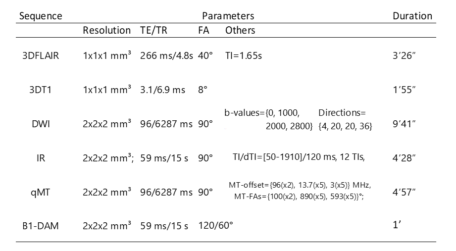
MRI data were acquired using a Philips Ingenia CX 3T scanner with the product 32-channel head coil. Additional details are reported in Table 1.
Image analysis
Several advanced MRI metrics were calculated through quantitative magnetization transfer, inversion recovery and diffusion-weighted imaging, respectively: bound pool fraction (BPF) and spin-spin relaxation time of the bound pool (T2B), longitudinal relaxation time (T1), and fractional anisotropy (FA), mean diffusivity (MD), orientation dispersion index (ODI) and neurite density index (NDI).
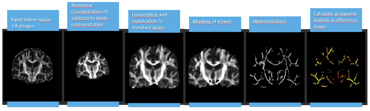
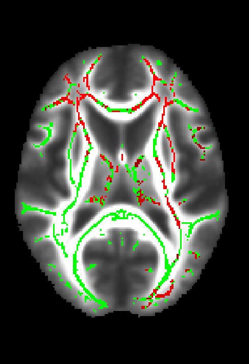
FSL tract-based spatial statistics [9] was used to extract the core of WM based on subject specific FA maximum values. Voxel-wise analysis of the core skeleton of white matter assessing changes of FA, MD, BPF, T1, T2B, ODI and NDI was performed (Figure2), using 5000 permutations and corrected for multiple comparisons using threshold-free cluster enhancement (TFCE). Age and gender were used as covariates. For all tests, a p-value < 0.01 was considered statistically significant.
Moreover, skeletonised maps for each metric were correlated at voxel level with SDMT z-score to assess the impact of MRI metrics on cognitive impairment.
Results
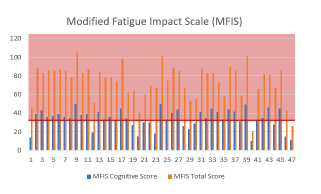
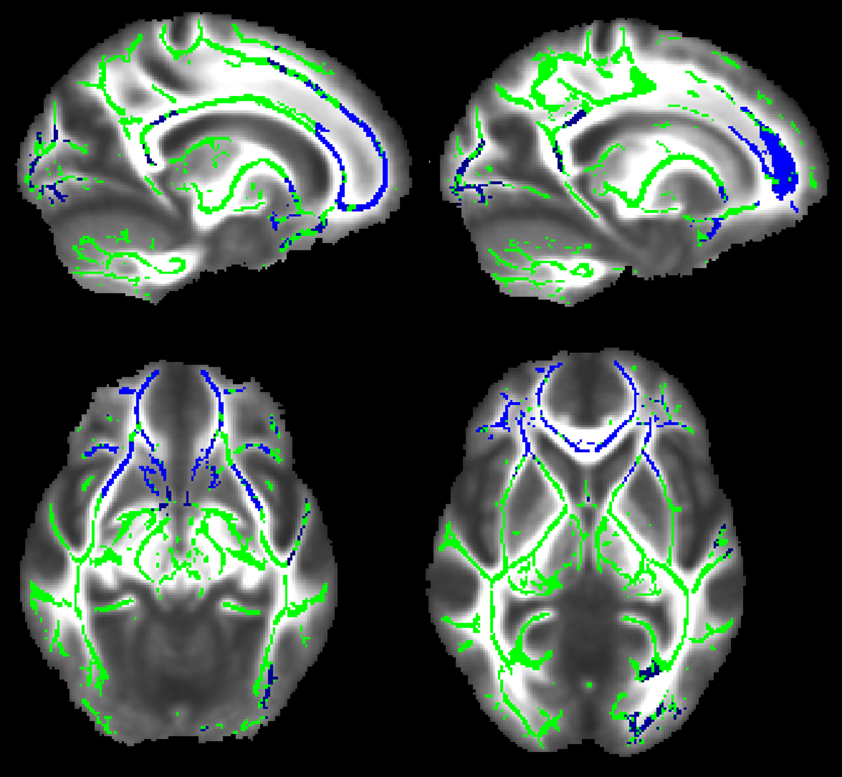
The MFIS scores showed that all Long-Covid subjects reported moderate to severe fatigue (Figure3). A total score of 38 is commonly used as a cut-off to discriminate between fatigue and non-fatigue individuals [10]. In most patients in our cohort, the cognitive subscale of the MFIS alone is enough to recognise patients with fatigue.BPF and T2B showed regional alterations in Long-COVID compared to HC, as shown in Figure 2 and 3.
T2B widespread alterations were observed in the frontal lobe, diencephalon and left occipital lobe, with increased T2B values being observed in Long-COVID compared to HC. Widespread alterations of BPF were found in the frontal lobe and more localised to the left temporal and occipital lobes, with reduced BPF being observed in Long-COVID.
No alterations were found using FA, MD, ODI and NDI. No correlation was found between SDMT and MRI metrics.
Discussion and conclusion
Our method demonstrates that WM is specifically affected in people with Long-COVID. Indeed, BPF and T2b maps detected widespread WM alterations, while microstructural metrics did not capture any loss of integrity. These results highlight the potential of BPF and T2b as valuable tools for detecting subtle myelin alterations in Long-COVID subjects, offering a promising perspective for further investigations and potential clinical interpretation of Long-COVID syndromes.As brain fog is not a specific symptom but rather a condition of generalized neuropsychological impairment, its causes may be attributed to deficits in various cognitive domains. Therefore, its assessment should be ascertained through the use of a series of complementary neuropsychological tests to better describe the multifaceted nature of this syndrome. The addition of MRI would enable a more thorough exploration of the diverse cognitive dimensions affected in individuals experiencing brain fog and perhaps the possibility to understanding the involvement of WM alterations as either a risk factor or consequence of Long COVID.
Acknowledgements
This study was funded by the Horizon2020 (Research and Innovation Action Grants Human Brain Project 945539 (SGA3)), the Biomedical Research Centre (BRC), and Rosetrees Trust. EG receives funding from TDC Technology Dedicated to Care. FPr received a Guarantors of Brain fellowship 2017–2020 and is supported by the National Institute for Health Research (NIHR), the Biomedical Research Centre initiative at University College London Hospitals (UCLH). FG receives the support of a fellowship from "la Caixa" Foundation (ID 100010434). The fellowship code is “LCF/BQ/PR22/11920010”.RS receives funding from the BRC (BRC1130/HEI/RS/11041). FPa receive funding from H2020 Research and Innovation Action Grants Human Brain Project (#785907, SGA2 and #945539, SGA3). H2020 Research and Innovation Action Grants Human Brain Project 785907 and 945539 (SGA2 and SGA3) fund ED'A. Moreover, the project was supported by the MNL Project “Local Neuronal Microcircuits” of the Centro Fermi (Rome, Italy) to ED'A. This work was also supported by #NEXTGENERATIONEU (NGEU) and funded by the Ministry of University and Research (MUR), National Recovery and Resilience Plan (NRRP), project MNESYS (PE0000006) - A Multiscale integrated approach to the study of the nervous system in health and disease (DN. 1553 11.10.2022).CGWK receives funding from Horizon2020 (Research and Innovation Action Grants Human Brain Project 945539 (SGA3)), BRC (#BRC704/CAP/CGW), MRC (#MR/S026088/1), Ataxia UK, Rosetrees Trust (#PGL22/100041 and #PGL21/10079). CGWK is a shareholder in Queen Square Analytics Ltd. JM receives funding from BRC.References
[1] H. E. Davis, L. McCorkell, J. M. Vogel, and E. J. Topol, “Long COVID: major findings, mechanisms and recommendations,” Nature Reviews Microbiology 2023 21:3, vol. 21, no. 3, pp. 133–146, Jan. 2023, doi: 10.1038/s41579-022-00846-2.
[2] A. Nalbandian et al., “Post-acute COVID-19 syndrome”, doi: 10.1038/s41591-021-01283-z.
[3] A. A. Asadi-Pooya et al., “Long COVID syndrome-associated brain fog,” J Med Virol, vol. 94, no. 3, pp. 979–984, Mar. 2022, doi: 10.1002/JMV.27404.
[4] F. Ceban et al., “Fatigue and cognitive impairment in Post-COVID-19 Syndrome: A systematic review and meta-analysis,” Brain Behav Immun, vol. 101, pp. 93–135, Mar. 2022, doi: 10.1016/J.BBI.2021.12.020.
[5] A. Chaudhuri and P. O. Behan, “Fatigue in neurological disorders,” Lancet, vol. 363, no. 9413, pp. 978–988, Mar. 2004, doi: 10.1016/S0140-6736(04)15794-2.
[6] A. B. Reiss et al., “Long COVID, the Brain, Nerves, and Cognitive Function,” Neurology International 2023, Vol. 15, Pages 821-841, vol. 15, no. 3, pp. 821–841, Jul. 2023, doi: 10.3390/NEUROLINT15030052.
[7] H. E. Davis, L. Mccorkell, J. M. Vogel, and E. J. Topol, “Long COVID: major findings, mechanisms and recommendations,” Nature Reviews Microbiology |, vol. 21, pp. 133–146, 2023, doi: 10.1038/s41579-022-00846-2.
[8] F. Crunfli et al., “Morphological, cellular, and molecular basis of brain infection in COVID-19 patients,” Proc Natl Acad Sci U S A, vol. 119, no. 35, Aug. 2022, doi: 10.1073/PNAS.2200960119.
[9] S. M. Smith et al., “Tract-based spatial statistics: voxelwise analysis of multi-subject diffusion data,” Neuroimage, vol. 31, no. 4, pp. 1487–1505, Jul. 2006, doi: 10.1016/J.NEUROIMAGE.2006.02.024.
[10] R. D. Larson, “Psychometric Properties of the Modified Fatigue Impact Scale,” Int J MS Care, vol. 15, no. 1, p. 15, 2013, doi: 10.7224/1537-2073.2012-019.
Figures