2072
Associations Between Microstructure, White Matter Hyperintensity Severity, and Cognitive Impairment: An Intravoxel Incoherent Motion Study1Shengli Clinical Medical College of Fujian Medical University; Radiology department of Fujian Provincial Hospital, Fuzhou, China, 2MR Research Collaboration Team, Siemens Healthineers Ltd., Shanghai, China
Synopsis
Keywords: White Matter, Aging, cognitive impairment
Motivation: It is still unclear what the mechanism of the effect of white matter hyperintensities (WMHs) and cognitive impairment is.
Goal(s): To use intravoxel incoherent motion (IVIM) to evaluate the microvascular and parenchymal microstructural changes of WMHs and to investigate their association with cognitive impairment.
Approach: The IVIM parameters were compared between patients and controls, different ROIs, and different WMH severity. Additionally, a multivariate linear regression analysis was conducted to explore the factors influencing cognitive scores.
Results: WMH patients had higher parenchymal diffusivity than the control group. Furthermore, increased parenchymal diffusivity was associated with an increased risk of cognitive decline.
Impact: The IVIM technique has the potential to provide a quantitative marker of parenchymal diffusivity for assessing the severity of WMH and may be useful in quantifying cognitive dysfunction in patients with WMH.
Introduction
White matter hyperintensities (WMHs) are a common age- and vascular risk factor-related disease that has been associated with cognitive impairment. However, the mechanism of this effect remains unclear. Previous studies have found hypoperfusion in WMHs using magnetic resonance imaging (MRI) Perfusion 1, while others have reported no significant difference in Cerebral Blood Volume (CBV) between WMH regions and Normal Appearing White Matter (NAWM)2. In some cases, increased CBV was even found in WMHs3. Intravoxel incoherent motion (IVIM) is a noninvasive diffusion-weighted imaging technique that can quantitatively measure microvasculature and parenchymal microstructural tissue properties4, and can separate microvascular and parenchymal MRI effects, unlike the conventional diffusion-weighted technique in which microvascular signals confound the parenchymal signal. The purpose of the present study was to investigate the correlation between changes in microcirculation and microstructure indicated by IVIM parameters and the severity of WMH and cognitive function.Methods
Forty-nine WMH patients and 31 healthy controls underwent a 3T MRI scanner (MAGNETOM Prisma, Siemens Healthcare, Erlangen, Germany) using a 20-element head coil. The sequences included T1-weighted (T1W), T2-weighted (T2W), T2W fluid-attenuated inversion recovery (FLAIR), and diffusion weighted imaging with 12 b-values (0, 50, 100, 150, 200, 250, 300, 500, 800, 1000, 1200, 1500 s/mm2) and the scanning parameters were: TR= 3600ms, TE= 80ms, slice thickness =5mm, FOV= 220mm × 220mm. The IVIM parametric maps were reconstructed using prototype software (Body Diffusion Toolbox 1.4; Siemens Healthcare). The IVIM dual exponential model parameters (parenchymal diffusivity D, intravascular diffusivity D*, and perfusion fraction f) were obtained in the regions of interest (ROIs). ROIs include deep white matter hyperintensities (DWMH), which is located apart from the cerebral ventricle in subcortical white matter (uniformly selected in the center of semioval); periventricular white matter hyperintensities (PWMH), which is attached to the ventricular system (selected in the contiguous with the margins of each lateral ventricle); and NAWM, generally chosen in the normal white matter adjacent to PWMH. ROIs were manually segmented on IVIM images using software (imagesRadiAnt DICOM Viewer 2021) by a neuroradiologist, who also identified and excluded infarcts and perivascular spaces (PVS). The Fazekas scale rates WMH severity in the periventricular and subcortical region combined, on a 0-3-point scale5. DWMH and PWMH two partial scores were added to obtain a total score (0-6 points). The severity of WMH patients was categorized as severe (3+3, 3+2, 3+1, 3+0, 2+2) or mild (0+1, 1+1, 1+2). Independent-samples t-test or Mann-Whitney U test was utilized to compare IVIM parameters between patients and controls. The Kruskal-Wallis test or one-way analysis of variance was used to compare IVIM parameters among DWMH, PWMH, and NAWM for patients. Wilcoxon two-sample test or independent-samples t-test was used to assess the differences in IVIM parameters based on the severity of WMH. Multivariate linear regression analysis was conducted to explore the factors influencing cognitive scores. Significance was inferred for p<0.05.Results
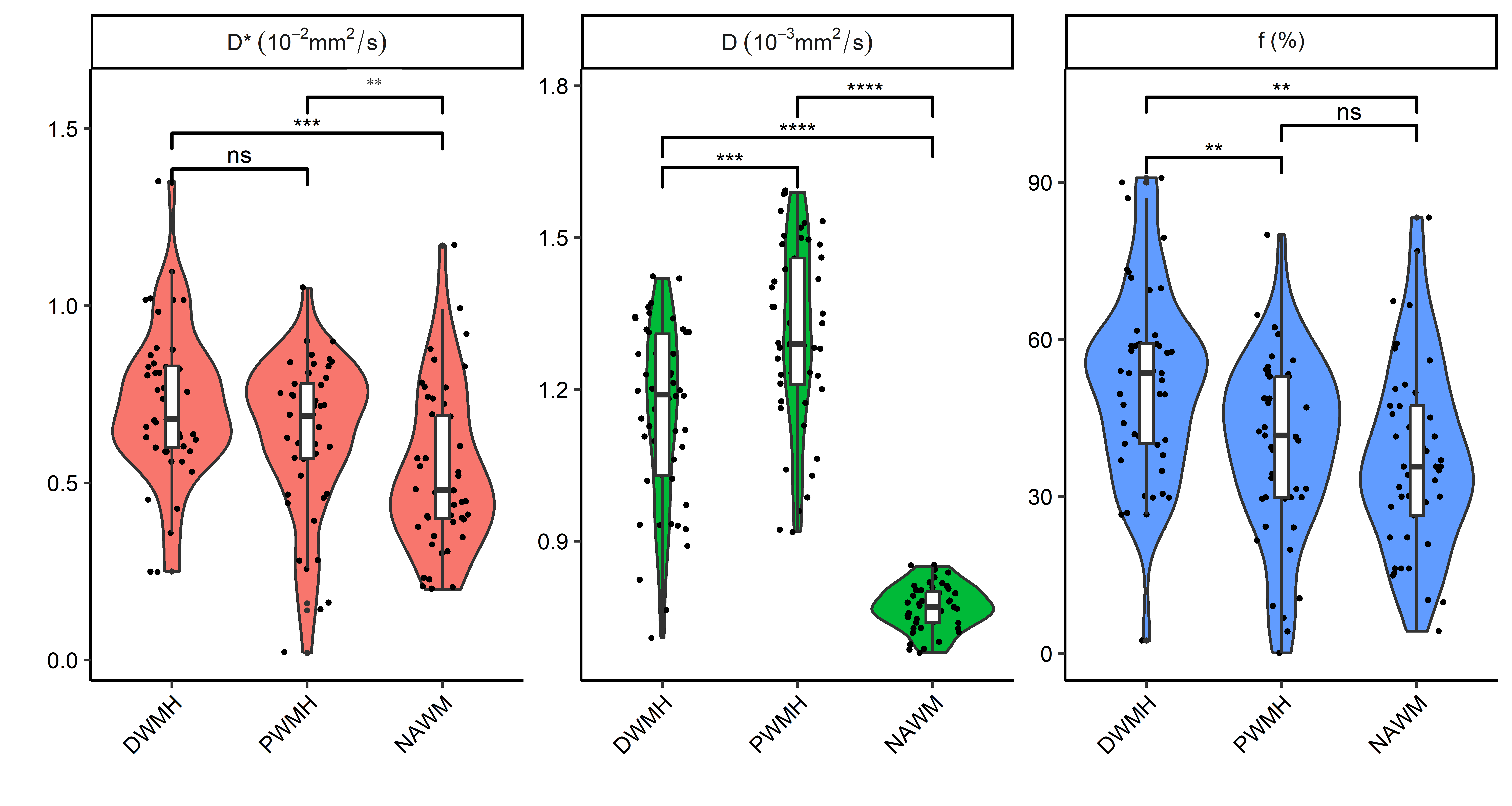
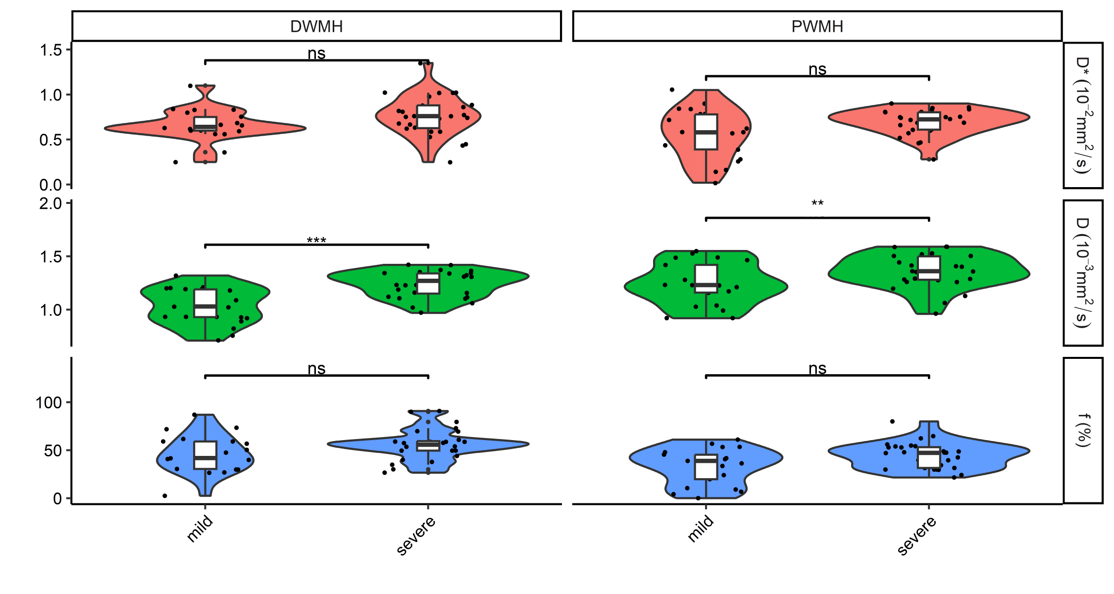
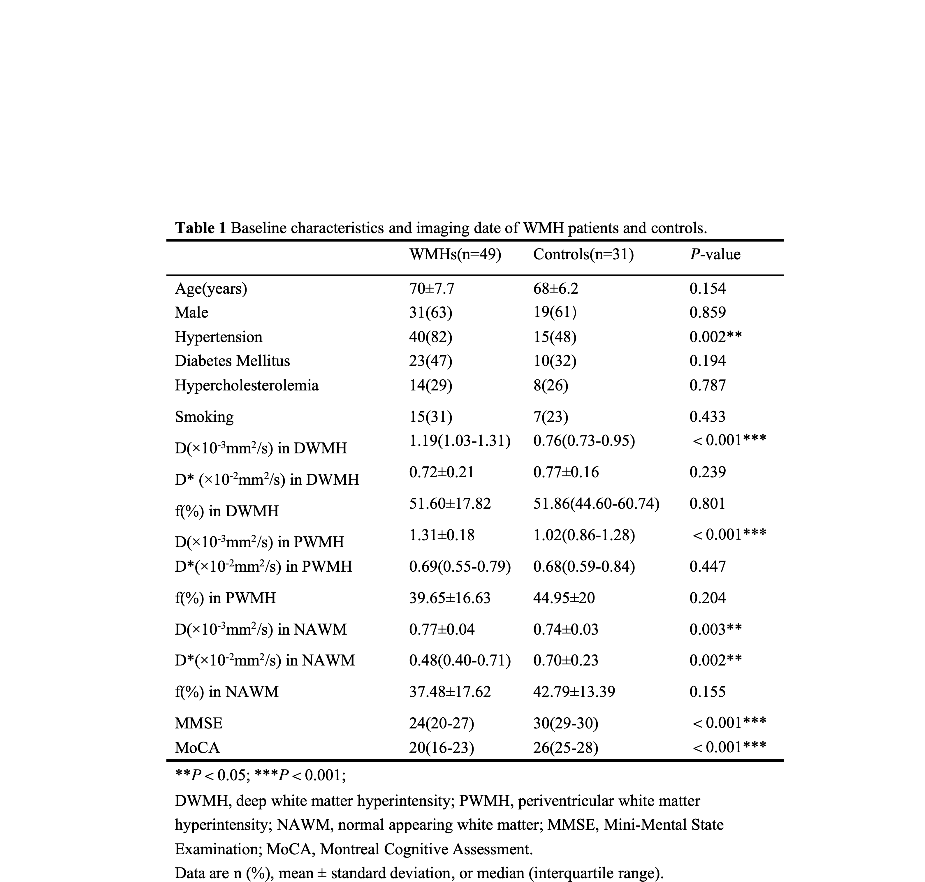
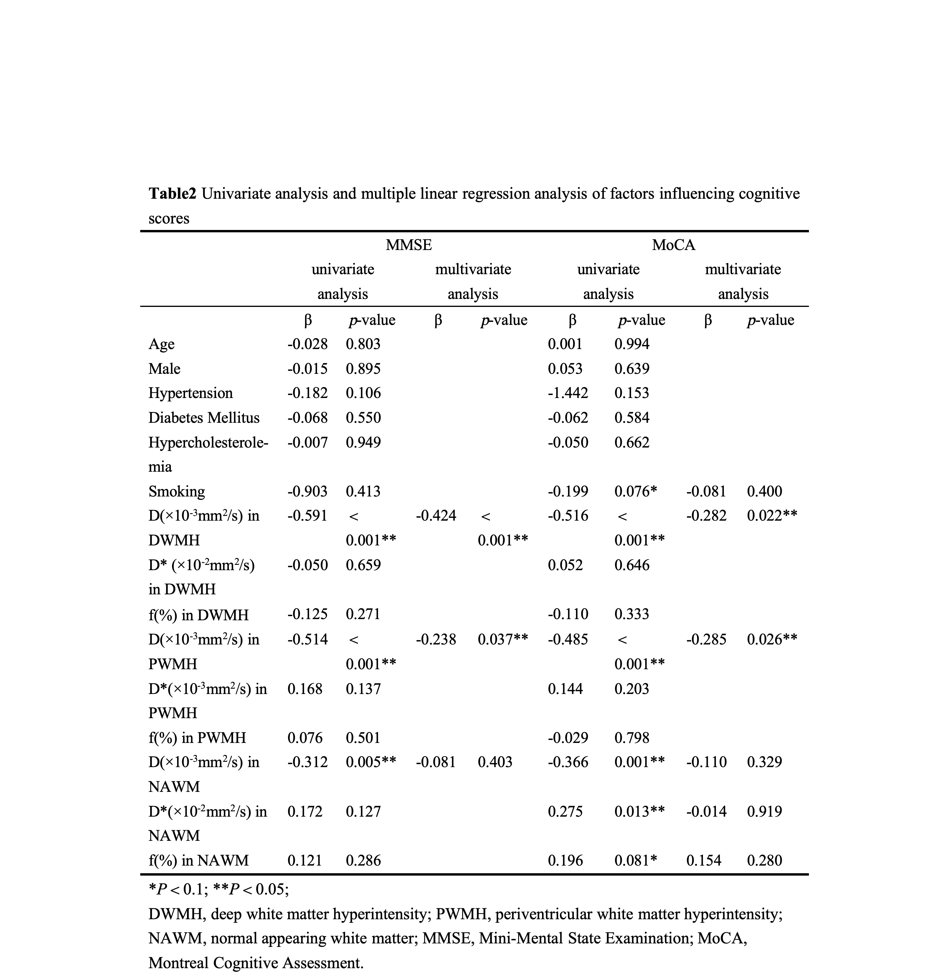
Patients with WMH exhibited significantly higher parenchymal diffusivity (D) than controls in DWMH, PWMH, and NAWM (all p< 0.05) (Table 1). IVIM parameters in the three groups (DWMH, PWMH, NAWM) were significantly different for patients (all p<0.001) (Fig. 1). The severe WMH group had significantly higher parenchymal diffusivity (DWMH and PWMH) than mild WMH (both p<0.05)(Fig. 2). Multiple linear regression analysis identified D in DWMH and PWMH as influencing cognitive function scores (all p< 0.05). Details of the univariate analysis and multiple linear regression models are listed in Table 2.Discussion
This study aimed to investigate the applicability of IVIM in WMHs by assessing the microvasculature and parenchymal microstructure. The results showed that patients with WMH had higher parenchymal diffusivity (D) than the control group, and an increasing parenchymal diffusivity was associated with increased risks of dementia or cognitive decline. Furthermore, parenchymal diffusivity measured with IVIM in the DWMH region showed the greatest relevance to cognitive score. Additionally, patients with severe WMH had higher parenchymal diffusivity D in both DWMH and PWMH than those with mild WMH.Conclusions
IVIM has the potential to provide a quantitative marker of parenchymal diffusivity for assessing the severity of WMH. This marker may also serve as a quantitative indicator of cognitive dysfunction in WMH patients.Acknowledgements
No acknowledgement found.References
[1] Markus H S, Lythgoe D J, Ostegaard L, et al. Reduced cerebral blood flow in white matter in ischaemic leukoaraiosis demonstrated using quantitative exogenous contrast based perfusion MRI. J Neurol Neurosurg Psychiatry. 2000, Jul; 69(1): 48-53.
[2] Marstrand J R, Garde E, Rostrup E, et al. Cerebral perfusion and cerebrovascular reactivity are reduced in white matter hyperintensities. Stroke. 2002, Apr; 33(4): 972-6.
[3] Yamada K, Sakai K, Owada K, et al. Cerebral white matter lesions may be partially reversible in patients with carotid artery stenosis. AJNR Am J Neuroradiol. 2010, Aug; 31(7): 1350-2.
[4] Le Bihan D, Breton E, Lallemand D, et al. Separation of diffusion and perfusion in intravoxel incoherent motion MR imaging. Radiology. 1988, Aug;168(2): 497-505.
[5] Fazekas F, Chawluk J B, Alavi A, et al. MR signal abnormalities at 1.5 T in Alzheimer's dementia and normal aging. AJR Am J Roentgenol. 1987, Aug; 149(2): 351-6.
Figures