2069
Alterations in the lateralization of the infant brain with white matter injury1First Affiliated Hospital of Xi'an Jiaotong University, XI'AN, China
Synopsis
Keywords: White Matter, Neonatal
Motivation: To investigation of cerebral asymmetry in full-term newborns and alterations in the lateralization of the infant brain with white matter injury
Goal(s): To explore brain volume laterality in normal full-term newborns and the effect of infant with WMI on laterality.
Approach: Calculated asymmetry index, used non-parametrically to test the significant, compared the difference of AI with the WMI group.
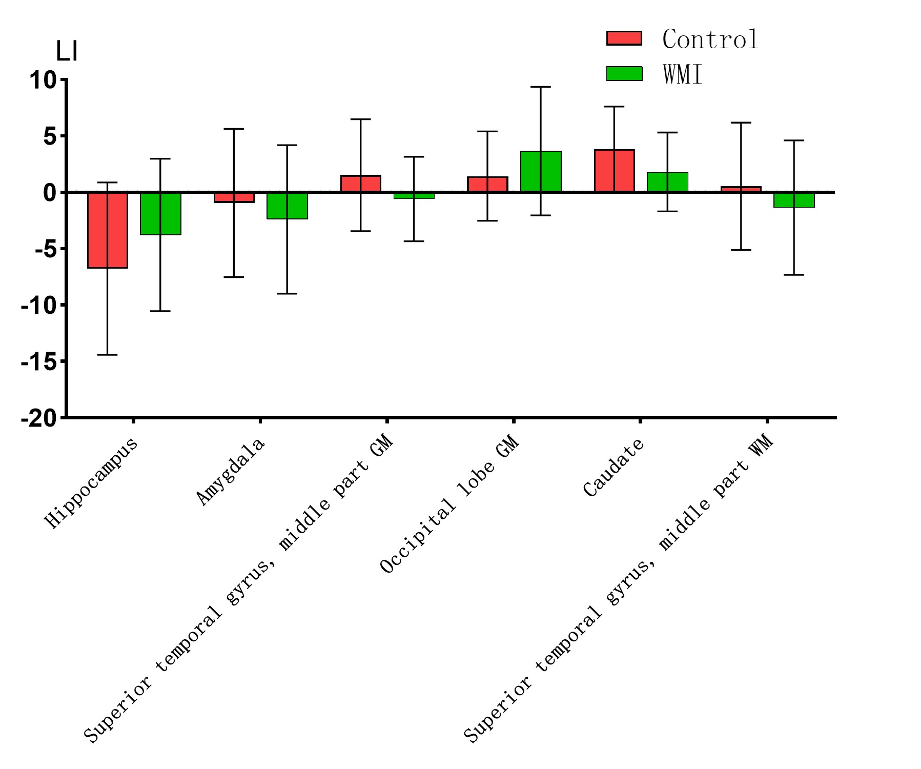
Results: The distribution of lateralized significance in brain regions between groups was near the same. Significant differences were found between the groups in the hippocampus, amygdala, superior temporal gyrus middle part GM, occipital lobe GM, superior temporal gyrus middle part WM, and caudate nucleus.
Impact: WMI can cause localized lateralized changes in brain regions, which may be associated with future adverse developmental outcomes and might be a biomarker for predicting prospective developmental outcomes of WMI.
Introduction
Cerebral hemispheric lateralization is the basic organization of the human brain1-3, and the phenomenon is not random but results from a unique pattern of structural design that confers evolutionary functional advantages4.The brain already has left-right asymmetry in the neonatal period and even in the fetal period5, which lays the foundation of basic lateralization differentiation for further postnatal adaptation to environmental change. White matter injury (WMI) are the most common type of brain injury in infancy (over 20% incidence), especially common in preterm infants6. WMI can lead to alterations in the localization of the injury and its distal white matter microstructure. Exploring changes in the lateralization of brain regions in infants with white matter injury may provide new insights into determining future developmental outcomes.Methods
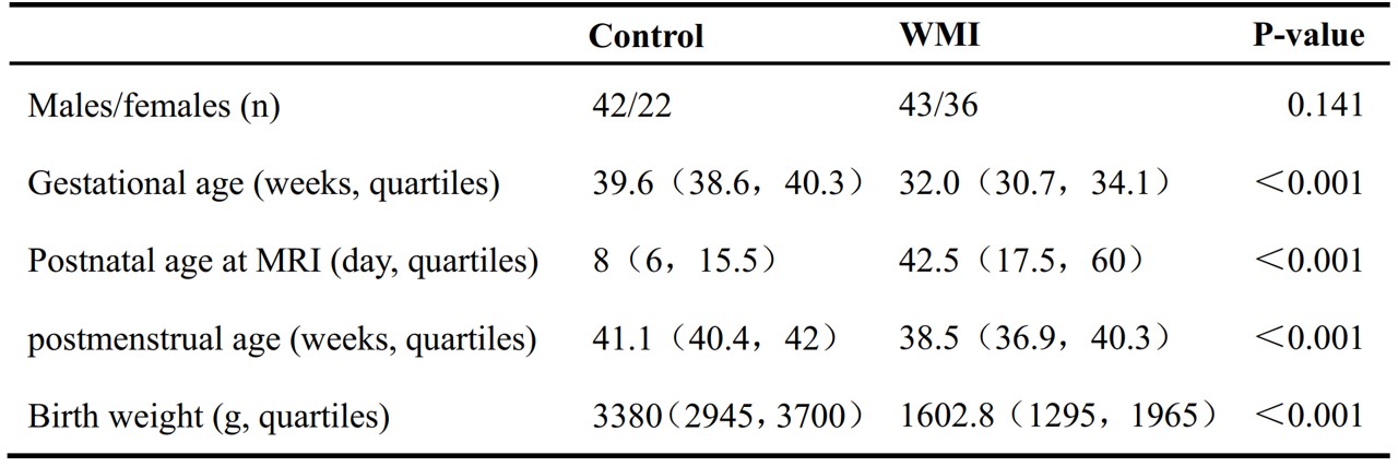
This study has been approved by the institutional IRB of the First Affiliated Hospital of Xi'an Jiaotong University and written informed consents were obtained from the children’s parents. From November 2010 to September 2021, we retrospectively collected 64 full-term newborns with no abnormalities in routine MRI(group control)and 79 infants with white matter injury (Table 1). Because preterm birth being high risk factor for WMI, the WMI group selected the population for MRI examinations from preterm birth corrected to nearly full term. The infants were given 10% chloral hydrate 0.5 ml/kg about 30 minutes before the MRI examination. After the scan, the doctor in charge and parents escorted the infants back to the neonatal care unit. All infants were scanned with GE Signa HDxt 3.0 T MRI equipment. The acquired MRI sequence and parameters are as follows: 3D-T1WI: TR=10.28 ms, TE=4.62 ms, slice thickness=1 mm, matrix acquisition=240×240, FOV=24 cm, imaging resolution=1×1×1 mm3. With the DHCP brain parcellation criteria, a deep learning model was trained to segment the brain into 87 regions by using the V-Net with bottleneck layer and the volume of each brain area was calculated. AI(Asymmetry Index)= 100 × (VL-VR)/(VL+VR). Nonparametric tests were used to analyze the significance of each brain region laterality, general linear models used to analyze between-group differences in brain region lateralization with correcting for whole-brain volume and left hemisphere-right hemisphere as covariates7. All analyses were performed using SPSS software (version 27). P<0.05 indicates that the correlation is statistically significant.Results
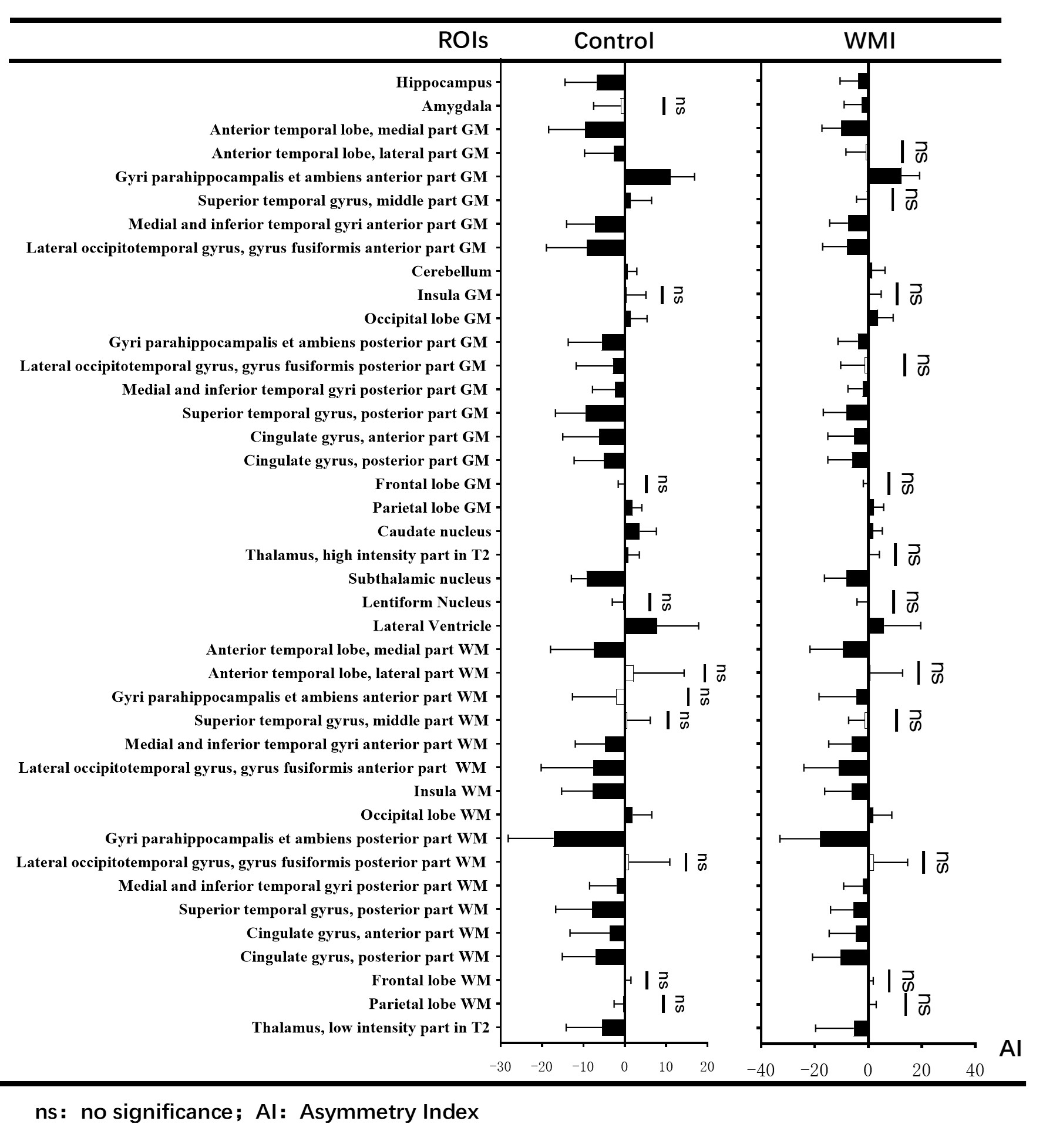
The distribution of lateralized significance in brain regions between the white matter injury group and the control group was near the same,which right lateralization predominates(Figure 1). Significant differences were found between the groups in the hippocampus, amygdala, superior temporal gyrus middle part GM, occipital lobe GM, superior temporal gyrus middle part WM, and caudate nucleus (Figure 2).Discussion
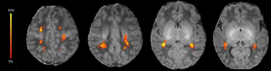
In terms of the results of Figure 1 in general, same between the two groups, brain areas of primary sensory, motor and essential functions have developed and show their own lateralization in the early postnatal period. Then we found that superior temporal gyrus middle part GM(gray matter) and WM(white matter) were more neutral of lateralization in white matter injury group than in the control, which is consistent with our previous results: The lesions are more likely to occur at the area(Figure 3). Compared with the control group, there were higher postnatal age at MRI in the WMI group. Based on previous literature, the overall developmental trajectory of the brain is from back to front8. The longer the postnatal age, the more exposed to the external visual environment in infants, consequently the more likely the visual cortex, occipital lobe GM, is to develop earlier compared with the control group, which is consistent with our results: there is higher AI in occipital lobe GM in WMI group. The results, caudate nucleus left lateralized, the hippocampus and amygdala right lateralized, are consistent with previous studies7,9-12, but there was lower degree of lateralization incaudate nucleus and hippocampus, higher in amygdala, which are related to learning, memory, cognition and the like, in the WMI group. In other words, the results may be associated with poor prognostic outcomes with white matter injury in the future.Conclusion
WMI can cause localized lateralized changes in brain regions, which may be associated with future adverse developmental outcomes and might be a biomarker for predicting prospective developmental outcomes of WMI.Acknowledgements
This work was supported by the National Natural Science Foundation of China (82272618, 81971581). Please address correspondence to Jian Yang, e-mail: yj1118@mail.xjtu.edu.cn and Xianjun Li, e-mail: xianj.li@mail.xjtu.edu.cn.References
1. Corballis, M.C. and I.S. Haberling, The Many Sides of Hemispheric Asymmetry: A Selective Review and Outlook. J Int Neuropsychol Soc, 2017. 23(9-10): p. 710-718.https://doi.org/10.1017/S1355617717000376
2.Beaton, A.A., The lateralized brain: the neuroscience and evolution of hemispheric asymmetries. Laterality, 2018: p. 1-4.https://doi.org/10.1080/1357650X.2018.1499749
3.Hirnstein, M., K. Hugdahl, and M. Hausmann, Cognitive sex differences and hemispheric asymmetry: A critical review of 40 years of research. Laterality, 2019. 24(2): p. 204-252.https://doi.org/10.1080/1357650X.2018.1497044
4.Kuo, F. and T.F. Massoud, Structural asymmetries in normal brain anatomy: A brief overview. Ann Anat, 2022. 241: p. 151894.https://doi.org/10.1016/j.aanat.2022.151894
5.Rajagopalan, V., et al., Mapping directionality specific volume changes using tensor based morphometry: an application to the study of gyrogenesis and lateralization of the human fetal brain. Neuroimage, 2012. 63(2): p. 947-58.https://doi.org/10.1016/j.neuroimage.2012.03.092
6.Kersbergen KJ, et al. Different patterns of punctate white matter lesions in serially scanned preterm infants. PLoS One. 2014 Oct 3;9(10):e108904. https://doi: 10.1371/journal.pone.0108904.
7.Williams CM, et al. Comparing brain asymmetries independently of brain size. Neuroimage. 2022 Jul 1;254:119118. https://doi: 10.1016/j.neuroimage.2022.119118.
8.Dubois, J., et al., THE EARLY DEVELOPMENT OF BRAIN WHITE MATTER: A REVIEW OF IMAGING STUDIES IN FETUSES, NEWBORNS AND INFANTS. Neuroscience, 2014. 276: p. 48-71.https://doi.org/10.1016/j.neuroscience.2013.12.044
9.Rogers, B.P., et al., Systematic Error in Hippocampal Volume Asymmetry Measurement is Minimal with a Manual Segmentation Protocol. Front Neurosci, 2012. 6: p. 179.https://doi.org/10.3389/fnins.2012.00179
10.Sotardi, S., et al., Voxelwise and Regional Brain Apparent Diffusion Coefficient Changes on MRI from Birth to 6 Years of Age. Radiology, 2021. 298(2): p. 415-424.https://doi.org/10.1148/radiol.2020202279
11.Ratnarajah, N., et al., Structural connectivity asymmetry in the neonatal brain. Neuroimage, 2013. 75: p. 187-194.https://doi.org/10.1016/j.neuroimage.2013.02.052
12.Machado-Rivas, F., et al., Normal Growth, Sexual Dimorphism, and Lateral Asymmetries at Fetal Brain MRI. Radiology, 2022. 303(1): p. 162-170.https://doi.org/10.1148/radiol.211222
Figures