2067
White matter changes in intrauterine fetal brain with mild to moderate isolated ventriculomegaly based on diffusion magnetic resonance imaging.1Shandong University, Jinan, China, 2Zhejiang University, Hangzhou, China, 3Beijing Hospital, Beijing, China
Synopsis
Keywords: White Matter, Fetus
Motivation: There is a controversial argue about the prognosis of fetuses with isolated ventriculomegaly in clinical practice.
Goal(s): Application of diffusion Magnetic Resonance Imaging (dMRI) in fetuses with isolated ventriculomegaly to explore structure changes of white matter fibers.
Approach: Collecting brain dMRI data in isolated ventriculomegaly and healthy fetuses, analyzing differences in white matter structure between the two groups using diffusion tensor imaging (DTI) and fixel based analysis (FBA) methods.
Results: Significant differences were found in DTI or FBA parameters of white matter fibers including the inferior longitudinal fasciculus, corpus callosum, fornix, sagittal stratum and corticospinal tract between the two groups.
Impact: There are changes in the brain white matter structure in intrauterine fetuses with isolated ventriculomegaly, which provides a certain objective basis for potential risks of neurodevelopmental disorders and prenatal counseling.
Introduction
Although it is currently widely believed that the prognosis of the vast majority of isolated ventriculomegaly (IVM) fetuses is good, especially those with mild to moderate lateral ventricular dilation[1], follow-up studies have found that mild to moderate IVM fetuses also have a risk of neurological dysplasia[2]. At present, there are few studies exploring the brain microstructural changes of IVM fetuses[3, 4], and there have been no reports on using dMRI methods to evaluate the changes in the white matter structure of intrauterine IVM fetuses. This study aims to analyze the changes in the structural integrity of white matter fibers in intrauterine fetuses with mild to moderate IVM using a combination of DTI and FBA parameters.Methods
Twenty fetuses diagnosed with mild to moderate IVM were collected prospectively, with gestational age ranging from 24 to 36 (29.9 ± 3.6) weeks, and 22 normal control fetuses with gestational age ranging from 24 to 36 (30.2 ± 3.7) weeks. All fetuses were subjected to routine cranial magnetic resonance imaging and dMRI data were collected. dMRI data was processed to compare the differences in four DTI parameters: fractional anisotropy (FA), mean diffusivity (MD), axial diffusivity (AD), and radial diffusivity (RD) of seven main fiber bundles, including the genu and splenium of the corpus callosum, middle cerebellar peduncle, the anterior thalamic radiation, cortico-spinal tract, inferior longitudinal fasciculus, and inferior fronto-occipital fasciculus, between the two groups; And differences in FBA parameters including fiber density (FD), fiber cross-sectional area (FC), and fiber density and cross-section (FDC) of the whole brain. The comparison of DTI metrics between the two groups was conducted using an independent sample t test. For fibers such as the anterior thalamic radiation, corticospinal tract, inferior longitudinal fasciculus, and inferior fronto-occipital fasciculus, the differences were compared by averaging the measured values in bilateral regions.Results
Compared with the normal control fetal group, there was a statistically significant increase in the FA value of the inferior longitudinal fasciculus in mild to moderate IVM fetuses (0.142 ± 0.012 vs 0.133 ± 0.015, P=0.033), and no differences were found between other DTI metrics of the resting fiber bundles. The whole brain FBA results showed that FD decreased while FC increased in the corpus callosum, fornix, and sagittal stratum in IVM fetuses, while FDC decreased in the cortico-spinal tract.Discussion
This study shows that changes in DTI or FBA parameters in IVM fetuses are located in the corpus callosum, fornix, sagittal stratum, and corticospinal tract. Therefore, the integrity of white matter fibers around the ventricles is affected by ventricular dilation. In fact, previous DTI studies[5-7] on IVM newborns have also found changes in the DTI metrics of these fibers. This study's dMRI analysis of intrauterine fetuses indicates that these vulnerable areas already have alternations during the fetal period. The change in the integrity of white matter fibers means that corresponding functions may be affected, such as the corticospinal tract, which serves as the largest projection fiber, affecting the voluntary movement of limbs[8]; The sagittal stratum contains important connecting fibers such as the inferior longitudinal fasciculus and optic radiation, which, together with the association fibers corpus callosum, play important roles in language, cognitive and attention skills, and vision[9, 10]. The changes in the fiber structure of the key white matter in this study may help explain potential developmental abnormalities in IVM fetuses in the future.Conclusion
There are changes in the brain white matter structure in intrauterine fetuses with mild to moderate IVM, which provides a certain objective basis for prognosis evaluation.Acknowledgements
We acknowledge for all pregnant women participated in this study and all authors who contributed to this study.References
[1] Scelsa, B., Rustico, M., Righini, A., Parazzini, C., Balestriero, M.A., Introvini, P., Spaccini, L., Mastrangelo, M., Lista, G., Zuccotti, G.V., Veggiotti, P., 2018. Mild ventriculomegaly from fetal consultation to neurodevelopmental assessment: A single center experience and review of the literature. Eur J Paediatr Neurol 22, 919-928.
[2] Pagani, G., Thilaganathan, B., Prefumo, F., 2014. Neurodevelopmental outcome in isolated mild fetal ventriculomegaly: systematic review and meta-analysis. Ultrasound in Obstetrics & Gynecology 44, 254-260.
[3] Vasung, L., Rollins, C.K., Zhang, J., Velasco-Annis, C., Yang, E., Lin, P.-Y., Sutin, J., Warfield, S.K., Soul, J., Estroff, J., Connolly, S., Barnewolt, C., Gholipour, A., Feldman, H.A., Grant, P.E., 2023. Abnormal development of transient fetal zones in mild isolated fetal ventriculomegaly. Cerebral Cortex 33, 1130-1139.
[4] Hahner, N., Benkarim, O.M., Aertsen, M., Perez-Cruz, M., Piella, G., Sanroma, G., Bargallo, N., Deprest, J., Gonzalez Ballester, M.A., Gratacos, E., Eixarch, E., 2019. Global and Regional Changes in Cortical Development Assessed by MRI in Fetuses with Isolated Nonsevere Ventriculomegaly Correlate with Neonatal Neurobehavior. American Journal of Neuroradiology.
[5] Gilmore, J.H., Smith, L.C., Wolfe, H.M., Hertzberg, B.S., Smith, J.K., Chescheir, N.C., Evans, D.D., Kang, C., Hamer, R.M., Lin, W., Gerig, G., 2008. Prenatal Mild Ventriculomegaly Predicts Abnormal Development of the Neonatal Brain. Biological Psychiatry 64, 1069-1076.
[6] Goodlett, C.B., Fletcher, P.T., Gilmore, J.H., Gerig, G., 2009. Group analysis of DTI fiber tract statistics with application to neurodevelopment. Neuroimage 45, S133-S142.
[7] Lockwood Estrin, G., Kyriakopoulou, V., Makropoulos, A., Ball, G., Kuhendran, L., Chew, A., Hagberg, B., Martinez-Biarge, M., Allsop, J., Fox, M., Counsell, S.J., Rutherford, M.A., 2016. Altered white matter and cortical structure in neonates with antenatally diagnosed isolated ventriculomegaly. NeuroImage: Clinical 11, 139-148.
[8] Fuelscher, I., Hyde, C., Efron, D., Silk, T.J., 2021. Manual dexterity in late childhood is associated with maturation of the corticospinal tract. Neuroimage 226, 117583.
[9] Zemmoura, I., Burkhardt, E., Herbet, G., 2021. The inferior longitudinal fasciculus: anatomy, function and surgical considerations. J Neurosurg Sci 65, 590-604.
[10] Blaauw, J., Meiners, L.C., 2020. The splenium of the corpus callosum: embryology, anatomy, function and imaging with pathophysiological hypothesis. Neuroradiology 62, 563-585.
Figures
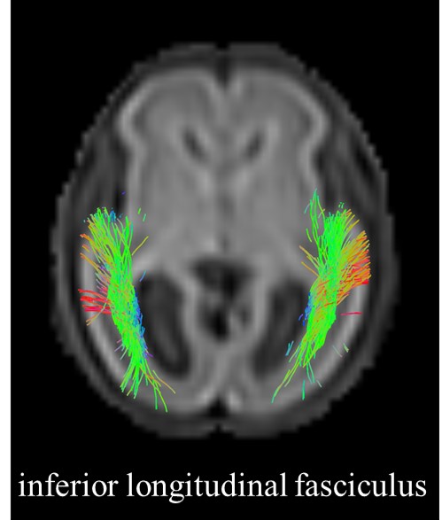
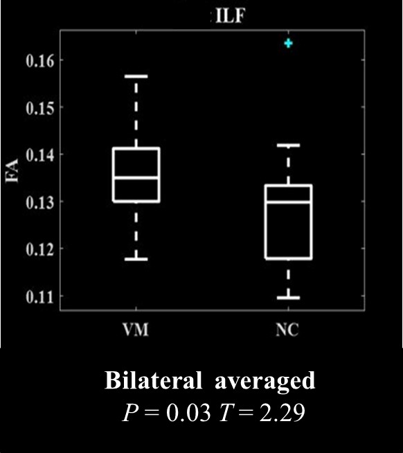
Independent sample t-test results for FA values of the inferior longitudinal fasciculus between the two groups. There was a statistically significant increase in the FA value of the inferior longitudinal fasciculus in mild to moderate IVM fetuses.
ILF, inferior longitudinal fasciculus; FA, fractional anisotropy; VM, ventriculomegaly; Bilateral averaged, average value of bilateral measured area
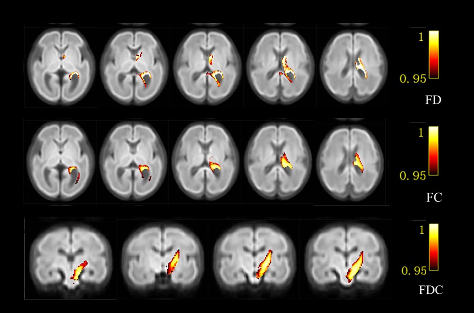
The whole brain FBA results showed the distribution of brain regions with significant differences in FD, FC, and FDC values between the two groups (P<0 05, FWE correction)
FD, fiber density; FC, fiber cross-section; FDC, fiber density and cross-section