2066
Abnormal white matter integrity revealed by Tracts Constrained by Underlying Anatomy (TRACULA) in obsessive-compulsive disorder1Department of Radiology and Huaxi MR Research Center (HMRRC), Functional and Molecular Imaging Key Laboratory of Sichuan Province, West China Hospital, Sichuan University, Chengdu, China, 2Mental Health Center, West China Hospital of Sichuan University, Sichuan Clinical Medical Research Center for Mental Disorders, Chengdu, China
Synopsis
Keywords: White Matter, White Matter, obsessive-compulsive disorder
Motivation: Diffusion tensor imaging (DTI) is a useful imaging technique to investigate white matter (WM) integrity and previous studies had revealed the disruption of WM related to neuropathology in obsessive-compulsive disorder (OCD). However, few study had reliably located the defect at track level.
Goal(s): To investigate the WM integrity in OCD on a whole brain level using newly developed tractography method.
Approach: We used Tracts Constrained by Underlying Anatomy (TRACULA) software to investigate the WM integrity in OCD.
Results: We found widespread disruption of WM integrity in OCD, and for the first time, we observed abnormality in bilateral acoustic radiation in OCD.
Impact: Using the innovated fiber tractography methodology, we were able to detect abnormality in bilateral acoustic radiation in OCD, which had been suggested by previous study using resting state fMRI.
Introduction
Obsessive-compulsive disorder (OCD) is a common psychiatric disorder with high prevalence, high disability, causing severe impaired social functioning in OCD patients. Diffusion tensor imaging (DTI) has revealed microstructural abnormality in white matter (WM) in OCD. Previous DTI studies had revealed fractional anisotropy (FA) alterations in OCD using voxel-based analysis at whole brain level, but results were blurred by methodologic drawbacks such as misalignment in fiber tracts. Tracts Constrained by Underlying Anatomy (TRACULA)[1] is a useful way to avoid those blemishes and thus in current study we tried to explore WM microstructural alterations in a group of non-morbidity OCD patients, and further associate the change with clinical characteristics.Methods
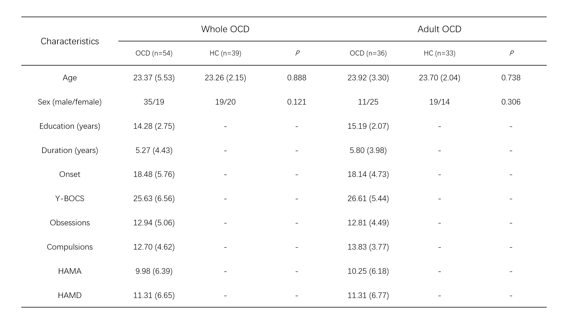
A total of fifty-four medication-free OCD patients without comorbidity (36 adults and 18 adolescents) and thirty-nine healthy controls (HC) were enrolled in the present study. T1-weighted structural MRI and DTI images were performed on a 3.0T MR scanner (Siemens TrioTim, Chengdu, China) equipped with a 32-channel head coil. Clinical symptoms were evaluated using Yale-Brown Obsessive Compulsive Scale (Y-BOCS), Hamilton Depression rating scale (HAMD) and Hamilton Anxiety Rating Scale (HAMA). 42 tracts were reconstructed automatically based on the anatomical neighborhood priors using TRACULA software. Statistical analyses were performed using SPSS version 26.0. FA, mean diffusivity (MD), radial diffusivity (RD) and axial diffusivity (AD) were extracted and compared using an analysis of covariance with age and sex. Additionally, we performed a partial correlation analysis between these metrics and clinical characteristics in OCD patients (covariates: age, sex and education years). We also performed these analyses in a subgroup of adult OCD patients alone. Multiple comparison correction used false discovery rate (FDR).Results
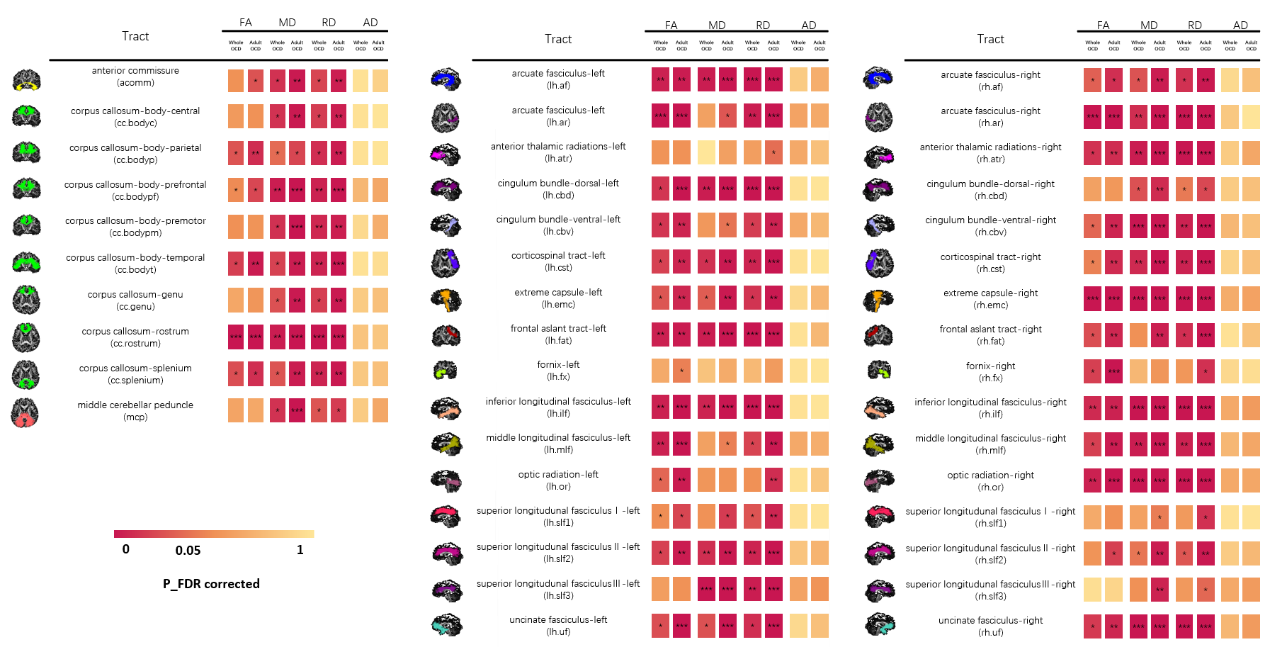
We found OCD patients showed lower FA in multiple WM tracts including corpus callosum, bilateral acoustic radiation, bilateral uncinate fasciculus et al, with adult OCD patients showed more obvious results of reduced FA in more WM tracts (Figure 1). MD and RD alterations were more extensive, and most of these alterations overlapped with FA alteration, but no AD alteration was observed. There is no significant correlation between diffusion parameters and clinical characteristics in whole OCD group. But in adult OCD group, FA in prefrontal body of corpus callosum (cc.bodypf) positively correlated with compulsive scores (p_FDR=0.042, r=0.56); and FA in left middle longitudinal fasciculus (lh.mlf) positively correlated with scores for HAMA (p_FDR=0, r=0.58).Discussion
Our finding of abnormalities in tracts including rostrum of corpus callosum, uncinate fasciculus were consistent with previous studies[2,3]. The abnormality in bilateral acoustic radiation, as first reported in OCD, represented the microstructural impairment in a primary sensory pathway, which conveys auditory information from the medial geniculate nucleus (MGN) of the thalamus to the auditory cortex on the transverse temporal gyrus of Heschl (HG)[4]. The rostrum of corpus callosum connects the orbital surfaces of the frontal lobes, therefore the results support that the orbital prefrontal region is involved in the pathophysiology of OCD[5]. Uncinate fasciculus is an important structural connect of fronto-limbic circuit, involved in the regulation of social-emotional processing and apparent memory[6].Conclusion
We use a newly innovated tractography method to detect WM alteration in OCD. Widespread WM integrity disruption is detected in whole OCD group with adult patients showed more prominent. And for the first time, we find abnormality in bilateral acoustic radiation which may underly the neural mechanism for impairment in primary sensory pathways.Acknowledgements
This study was supported by National Key R&D Program of China (Grant No. 2022YFF1202400) and National Natural Science Foundation of China (Grant No.82372080).References
1. Maffei, C et al. “Using diffusion MRI data acquired with ultra-high gradient strength to improve tractography in routine-quality data.” NeuroImage vol. 245 (2021): 118706. doi:10.1016/j.neuroimage.2021.118706
2. Zhang, Zeng et al. “Microstructural white matter abnormalities in obsessive-compulsive disorder: A coordinate-based meta-analysis of diffusion tensor imaging studies.” Asian journal of psychiatry vol. 55 (2021): 102467. doi:10.1016/j.ajp.2020.102467
3. Jenkins, Lisanne Michelle et al. “Shared white matter alterations across emotional disorders: A voxel-based meta-analysis of fractional anisotropy.” NeuroImage. Clinical vol. 12 1022-1034. 28 Sep. 2016, doi:10.1016/j.nicl.2016.09.001
4. Maffei C, Sarubbo S, Jovicich J. Diffusion-based tractography atlas of the human acoustic radiation. Sci Rep. 2019 Mar 11;9(1):4046. doi: 10.1038/s41598-019-40666-8. PMID: 30858451; PMCID: PMC6411970
5. Saito, Yukiko et al. “Corpus callosum in patients with obsessive-compulsive disorder: diffusion-tensor imaging study.” Radiology vol. 246,2 (2008): 536-42. doi:10.1148/radiol.2462061469
6. Jayarajan, Rajan Nishanth et al. “White matter abnormalities in children and adolescents with obsessive-compulsive disorder: a diffusion tensor imaging study.” Depression and anxiety vol. 29,9 (2012): 780-8. doi:10.1002/da.21890
Figures