2058
Mapping structural disconnection and morphometric similarity in Multiple Sclerosis: a longitudinal study1University of Naples "Federico II", Naples, Italy, 2Sapienza University of Rome, Rome, Italy, 3National Research Council, Naples, Italy
Synopsis
Keywords: Multiple Sclerosis, Multiple Sclerosis, Network analysis, Structural disconnection, Morphometric similarity
Motivation: Multiple sclerosis can be modelled as a network disorder. Progressive demyelination and neurodegeneration lead to structural disconnection and disruption of the morphometric similarity between gray matter regions.
Goal(s): To obtain measures of structural disconnection and morphometric similarity networks from conventional MRI sequences and test whether they are sensitive to disease status and clinical disability.
Approach: 461 patients were imaged. Using publicly available software, we computed structural disconnection using white matter lesions masks and normative tractography atlases. Likewise, morphometric similarity was computed from standard FreeSurfer outputs.
Results: Structural disconnection and morphometric similarity networks are sensitive to disease status and explain clinical disability.
Impact: Measures of structural disconnection and morphometric similarity networks obtained from conventional MRI sequences are sensitive to multiple sclerosis and its related physical and cognitive disability. Our approach could represent a way to overcome the limitations of the standard network analyses.
Introduction
Multiple sclerosis (MS) can be conceptualized as a network disorder. The accumulation of white matter (WM) demyelinating lesions leads to progressive structural disconnection between gray matter (GM) regions,1 adding to other pathological processes that directly and indirectly damage the GM (i.e., microglial/macrophagic activation and cortical demyelination).2 The resulting progressive neurodegeneration3 disrupts the patterns of morphometric similarity between cortical regions, ultimately subverting the hierarchical organization of the brain.4 Network-based approaches could represent a tool to overcome the so-called “clinico-radiological paradox”, that is the gap between the clinical status and the radiological severity assessed through conventional MRI biomarkers, such as WM lesion load and brain atrophy.5 Nonetheless, these methods require advanced MRI sequences that are not routinely acquired and long processing times, hampering their application in clinical practice.Here, using conventional MRI and publicly available software, we assessed cross-sectional and longitudinal modifications of structural disconnection and morphometric similarity networks in MS, and tested whether they are sensitive to disease status and progression over time, and whether they could explain disease-related physical and cognitive disability.
Methods
We retrospectively collected 3T structural brain MRIs of 461 MS patients (age = 37.2 ± 10.6 years, F:M = 324:137), corresponding to 1235 visits (mean follow-up time = 1.9 ± 2.0 years, range = 0.1-13.3 years), and 55 healthy controls (age = 42.4 ± 15.7 years; F:M = 25:30). From 3D-T1w and FLAIR-T2w scans, WM lesions were automatically segmented and the brain was parcellated into 100 cortical (Schaefer atlas) and14 subcortical (Aseg atlas) regions. For MS patients, subject-level WM masks were registered to the MNI space and used to compute networks of structural disconnection: using the Lesion Quantification Toolkit,6 based on the HCP842 tractography atlas, disconnection between pairs of regions was estimated as the proportion of connecting streamlines passing through WM lesions. Likewise, with the Morphometric Inverse Divergence (MIND) method,4 we computed networks of morphometric similarity between cortical regions from 3D-T1w derived FreeSurfer outputs for both groups. Physical and cognitive disability were assessed with the expanded disability status scale (EDSS) and the symbol digit modalities test (SDMT), respectively.Using network-based statistics, the effect of time and clinical disability (and group, for MIND networks) were tested with linear mixed-effects models. Five-thousands permutations were used, with a statistical significance level set at p < 0.05 (FWER-corrected). Statistical analyses were carried out using R (version 4.1.2).Results
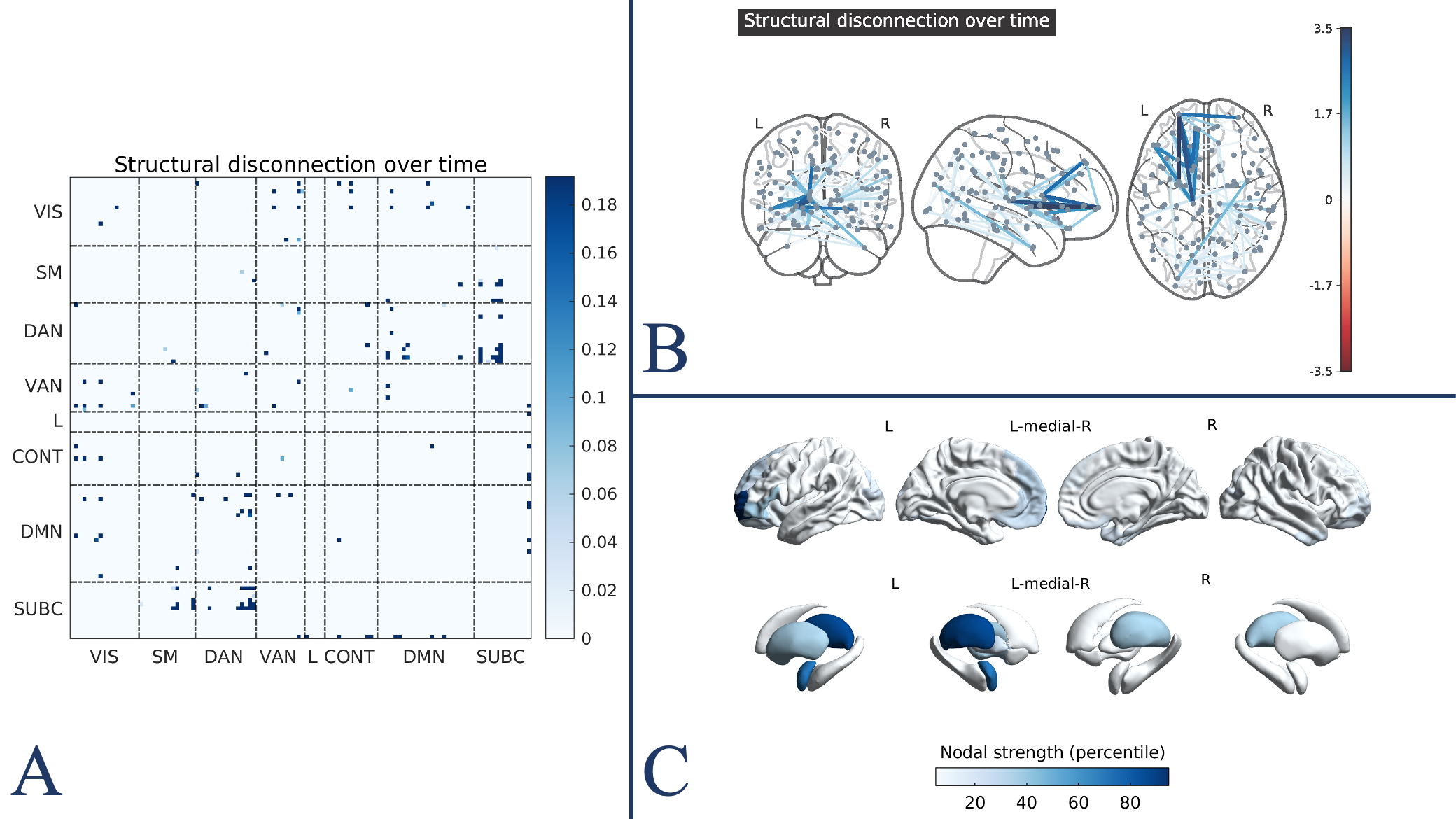
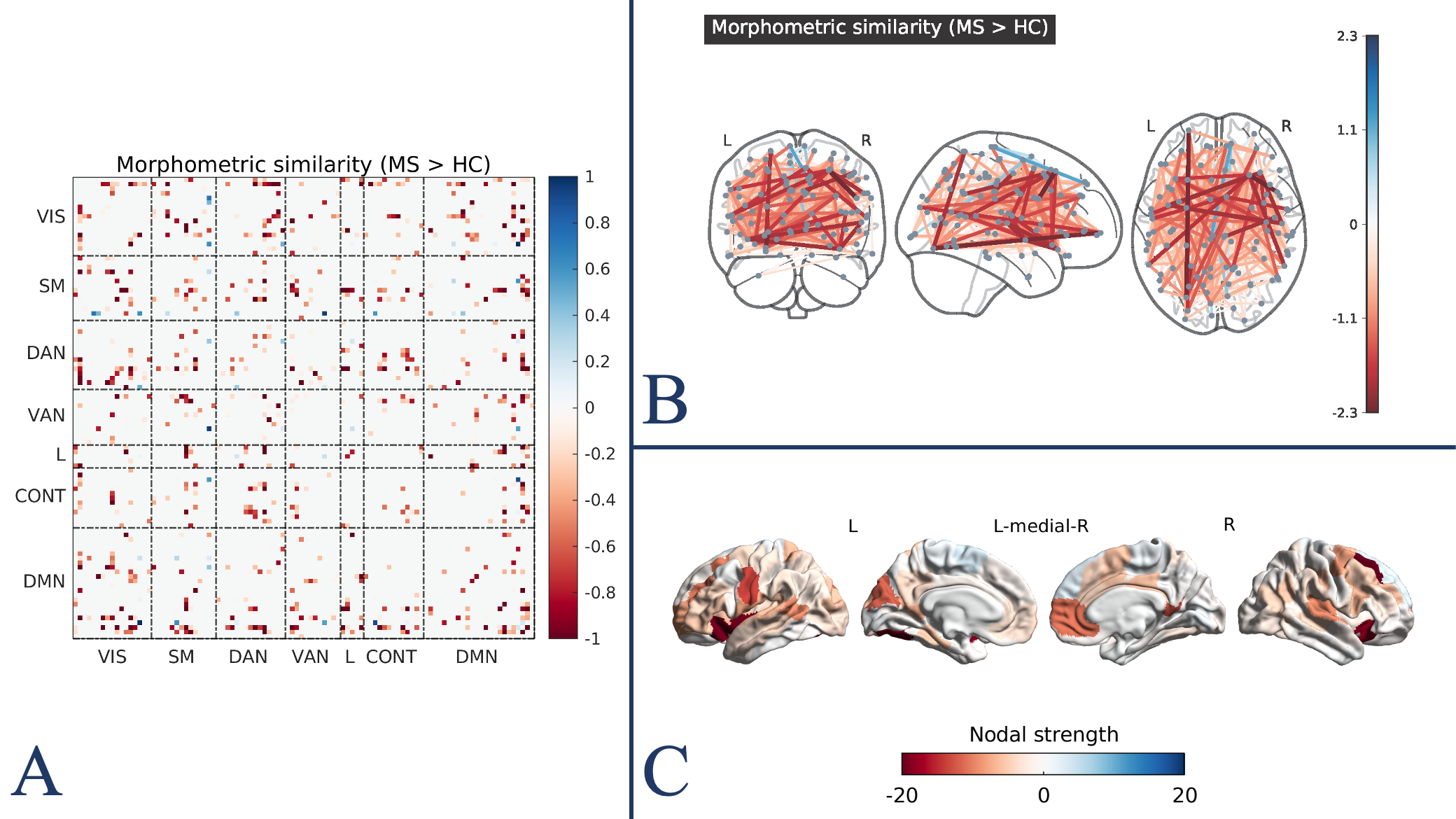
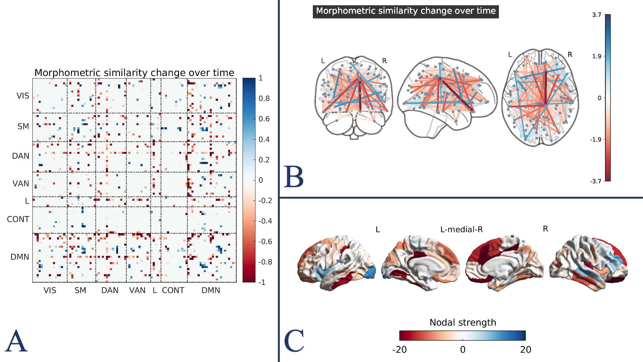
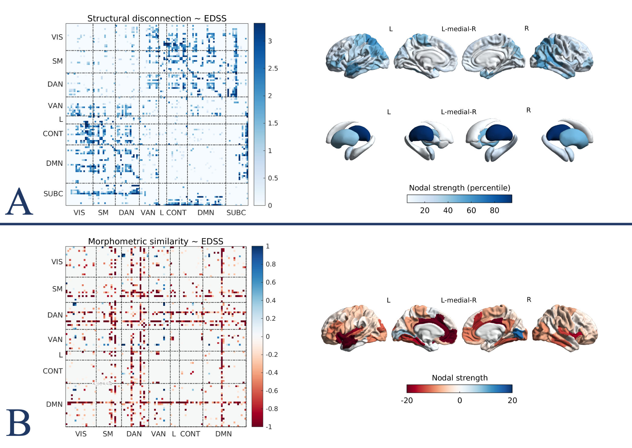
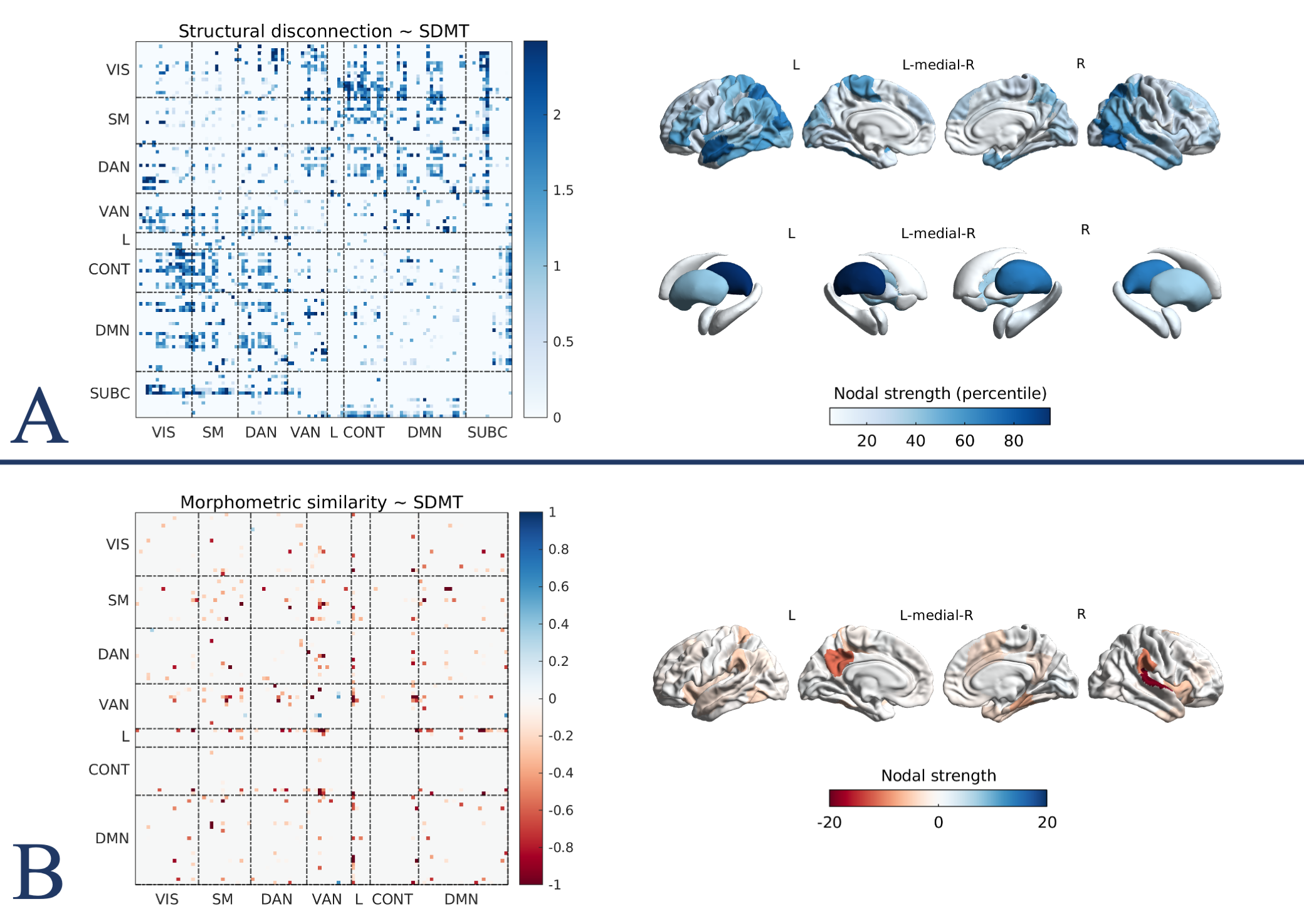
We identified a relatively small subnetwork of significant progressive structural disconnection (82 edges, pFWE = 0.04), mainly encompassing cortico-subcortical tracts (Figure 1). MIND networks were sensitive to disease status and progression over time, with distributed effects of decreased morphometric similarity in large subnetworks of 431 and 509 edges, respectively (pFWE < 0.01, Figures 2 and 3).Both structural disconnection and morphometric similarity alterations significantly explained clinical disability. In particular, we identified significant associations of EDSS with structural disconnection and MIND subnetworks of 960 and 670 edges, respectively (pFWE < 0.01, Figure 4). Similarly, SDMT was significantly associated with structural disconnection and MIND subnetworks of 988 and 202 edges, respectively (pFWE < 0.01, Figure 5).
Conclusions
We demonstrated that networks of structural disconnection and morphometric similarity, as assessed through conventional MRI, are sensitive to MS-related brain damage and its evolution over time. Moreover, they proved to be sensitive to physical and cognitive disability, potentially adding to established conventional MRI-derived measures as biomarkers of disease severity and progression. Extracting network measures from conventional MRI scans holds the potential for driving brain connectomics towards applicability in everyday clinical practice.Acknowledgements
No acknowledgement found.References
- Rise HH, Brune S, Chien C, et al. Brain disconnectome mapping derived from white matter lesions and serum neurofilament light levels in multiple sclerosis: A longitudinal multicenter study. NeuroImage: Clinical. 2022;35:103099. doi:10.1016/j.nicl.2022.1030992.
- Calabrese M, Magliozzi R, Ciccarelli O, Geurts JJG, Reynolds R, Martin R. Exploring the origins of grey matter damage in multiple sclerosis. Nat Rev Neurosci. 2015;16(3):147-158. doi:10.1038/nrn39003.
- Tsagkas C, Chakravarty MM, Gaetano L, et al. Longitudinal patterns of cortical thinning in multiple sclerosis. Human Brain Mapping. 2020;41(8):2198-2215. doi:10.1002/hbm.249404.
- Sebenius I, Seidlitz J, Warrier V, et al. Robust estimation of cortical similarity networks from brain MRI. Nat Neurosci. 2023;26(8):1461-1471. doi:10.1038/s41593-023-01376-75.
- Barkhof F. The clinico-radiological paradox in multiple sclerosis revisited: Current Opinion in Neurology. 2002;15(3):239-245. doi:10.1097/00019052-200206000-000036.
- Griffis JC, Metcalf NV, Corbetta M, Shulman GL. Lesion Quantification Toolkit: A MATLAB software tool for estimating grey matter damage and white matter disconnections in patients with focal brain lesions. NeuroImage: Clinical. 2021;30:102639. doi:10.1016/j.nicl.2021.102639
Figures