2057
Fixel-based analysis of multi-shell diffusion MRI reveals white matter changes after long-duration spaceflight1Lab for Equilibrium Investigations and Aerospace, Dept. of Physics, University of Antwerp, Antwerp, Belgium, 2imec-Vision Lab, Dept. of Physics, University of Antwerp, Antwerp, Belgium, 3Translational Neurosciences, University of Antwerp, Antwerp, Belgium, 4SSC RF – Institute of Biomedical Problems, Russian Academy of Sciences, Moscow, Russian Federation, 5Laboratory for Cognitive Research, National Research University Higher School of Economics, Moscow, Russia, Moscow, Russian Federation, 6Radiology Dept., National Medical Research Treatment and Rehabilitation Centre of the Ministry of Health of Russia, Moscow, Russian Federation, 7Institute for Neuroradiology, Ludwig-Maximilians-University, Munich, Germany
Synopsis
Keywords: White Matter, Tractography & Fibre Modelling, Spaceflight
Motivation: The effects of spaceflight on the central nervous system, and in particular the brain’s white matter (WM), are poorly understood.
Goal(s): To gain knowledge about the effect of long-duration spaceflight on the brain's WM.
Approach: We performed the first fixel-based analysis of diffusion MRI scans of 18 cosmonauts before and after long-duration spaceflight.
Results: We show widespread changes in the WM after spaceflight, which are predominantly macroscopic rather than microscopic. Moreover, we detect a net increase in the amount of WM fibers in the left superior and left middle cerebellar peduncles, providing evidence for neuroplasticity in the brain induced by long-duration spaceflight.
Impact: Better understanding and monitoring of the effect of space flight on the brain is crucial to ensure the health of space crews and their performance during long-duration space missions.
Introduction
The effects of spaceflight on the central nervous system, and in particular the brain’s white matter (WM), remain poorly understood [1]. Previous work using voxel-based analysis of diffusion MRI has shown a net increase of WM in the cerebellum after spaceflight, potentially reflecting neuroplasticity [2]. Another study using differential tractography has reported widespread changes in WM connections [3], but it remains unclear whether these reflect a true connectivity change or are merely the result of fluid and tissue displacements [1,2,4]. To elucidate these findings, we performed an advanced fixel-based analysis of diffusion MRI [5] of cosmonauts before and after long-duration spaceflight.Material and methods
Study design: 18 Roscosmos cosmonauts were scanned using diffusion-weighted MRI, before (pre-flight: avg 81 (SD 31) days) and shortly after (post-flight: 9 (3) days) a mission to the ISS (avg 195 (SD 60) days in space). Data acquisition: All data were acquired on a 3T MRI system equipped with a 16-channel receiver head coil using a twice-refocused pulsed gradient spin-echo echo-planar imaging sequence. A multi-shell diffusion-weighted acquisition scheme was prescribed, containing diffusion weightings of b = 0, 700, 1200, and 2800 s/mm2, applied in 8, 25, 45, and 75 directions, respectively. Other imaging parameters were: repetition/echo time of 7800/100 ms, voxel size of 2.4mm × 2.4mm × 2.4mm, matrix size of 100 × 100, 58 slices, and 1 excitation. Preprocessing: The raw MRI data were processed and analyzed using a state-of-the-art pipeline combining tools from MRtrix (www.mrtrix.org; version 3.0.3), FSL (http://fsl.fmrib.ox.ac.uk; version 6.0.5), and ANTs (http://stnava.github.io/ANTs/; version 2.4.0). First, the images were denoised to increase their signal-to-noise ratio [6]. Second, the Gibbs ringing artifact was suppressed to avoid spurious oscillations in the vicinity of sharp tissue boundaries [7]. Next, susceptibility-induced distortions, as well as motion and Eddy current-induced distortions, were corrected using an integrated approach [8]. Then, the low-frequency intensity nonuniformity, also known as the bias field, was corrected [9]. Last, images were upsampled spatially in all three dimensions using cubic b-spline interpolation to a voxel size of 1.25mm × 1.25mm × 1.25mm to improve the accuracy of downstream spatial normalization. Modeling: From the preprocessed data, the WM fiber orientation distribution function (fODF) was estimated in each voxel using multi-tissue constrained spherical deconvolution [10]. The fODF fields of all subjects and time points were subsequently warped to a study-specific template [11,12]. From the fODF fields and their spatial warps, we extracted the following fixel-based metrics: Fiber Density (FD): a microscopic measure that reflects changes in the amount of fibers within a fixed cross-section; Fiber cross-section (FC): a macroscopic measure (resulting from image warping) that reflects changes in the cross-section of a WM bundle; and Fiber density modulated by cross-section (FDC = FD ⨉ FC): a combined measure which takes into account both micro- and macroscopic effects, reflecting a net change in the capacity of the WM fibers to relay information [5]. Statistical analysis: nonparametric permutation testing with FWE correction and a significance threshold of P < 0.05 was performed using the default settings for fixel-based analysis within the MRtrix software package.Results and discussion
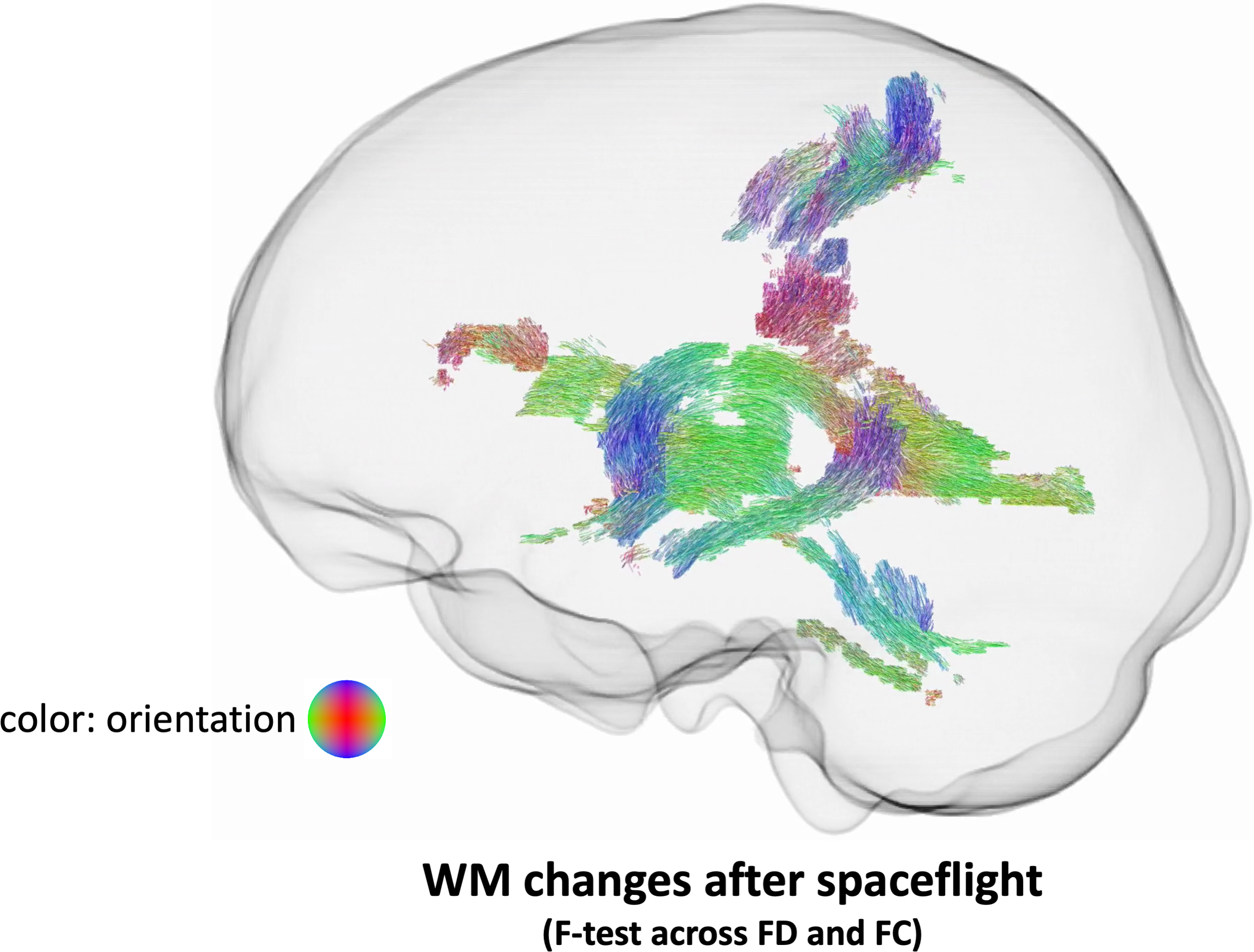
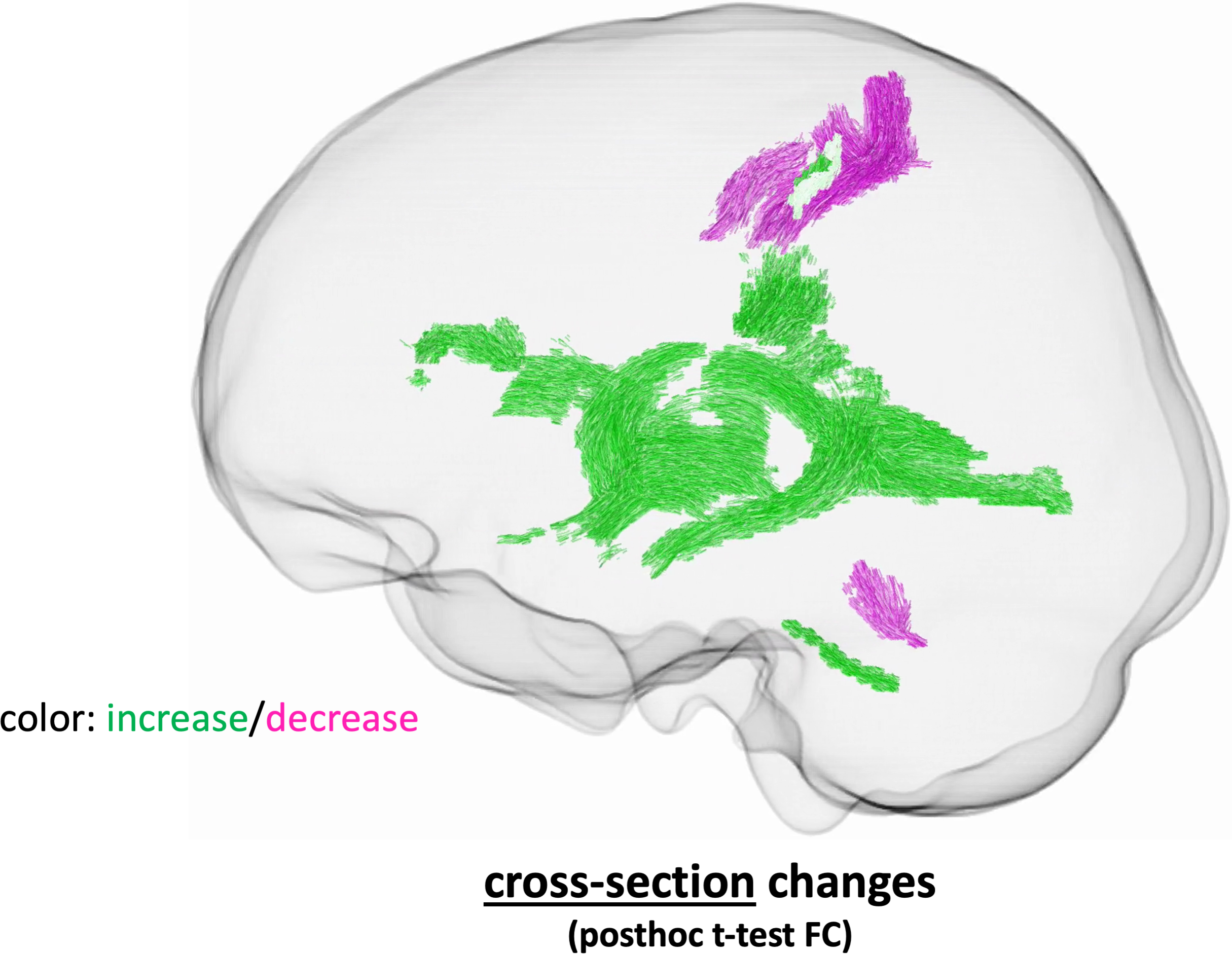
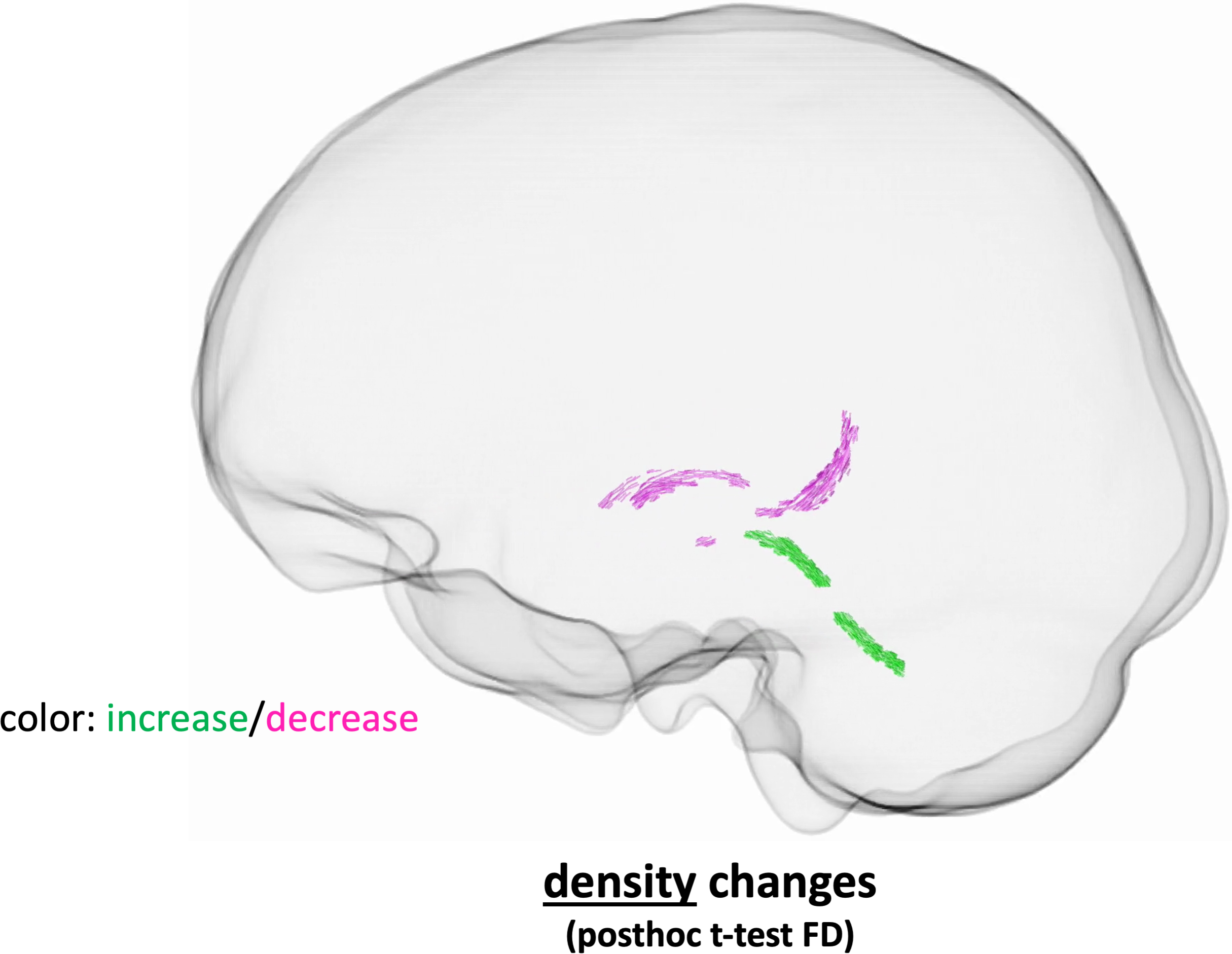
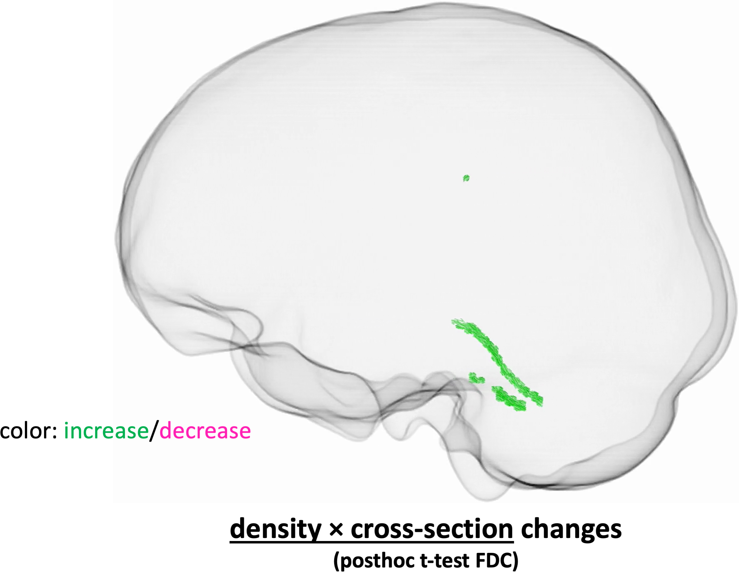
An omnibus F-test across both FD and FC revealed widespread changes in the WM as a result of long-duration spaceflight (Fig. 1), corroborating the overall findings of Doroshin et al. [3]. Subsequent posthoc paired t-tests show that the bulk of these changes are macroscopic in nature, i.e. changes in FC (Fig. 2), rather than microscopic, i.e. changes in FD (Fig. 3), and are thus unlikely to reflect widespread rewiring. Moreover, a close inspection of Fig. 3 reveals that any reductions in FD occur directly at the interface with CSF, meaning that they are likely the result of macroscopic fluid and tissue relocation, rather than true microscopic changes. When both FD and FC changes are taken into account simultaneously (FDC, Fig. 4), there appear to be no net decreases in WM, corroborating the hypothesis that most FD changes are the result of macroscopic fluid changes, rather than reflecting a true change in WM connectivity. Interestingly, FDC analysis did reveal a net increase of WM in the left superior and left middle cerebellar peduncles [13], which further supports the first evidence of neuroplasticity presented by Jillings et al. [2].Conclusion
This first fixel-based analysis of diffusion MRI data in cosmonauts shows widespread changes in the WM after spaceflight, which are predominantly macroscopic rather than microscopic. However, we did detect a net increase in the amount of WM fibers in the left superior and left middle cerebellar peduncles, providing additional evidence for neuroplasticity in the brain induced by long-duration spaceflight.Acknowledgements
No acknowledgement found.References
1. Roy-O’Reilly M, Mulavara A, Williams T. A review of alterations to the brain during spaceflight and the potential relevance to crew in long-duration space exploration. NPJ Microgravity. 2021;7: 5.
2. Jillings S, Van Ombergen A, Tomilovskaya E, Rumshiskaya A, Litvinova L, Nosikova I, Pechenkova E, Rukavishnikov I, Kozlovskaya IB, Manko O, Danilichev S, Sunaert S, Parizel PM, Sinitsyn V, Petrovichev V, Laureys S, Zu Eulenburg P, Sijbers J, Wuyts FL, Jeurissen B. Macro- and microstructural changes in cosmonauts’ brains after long-duration spaceflight. Sci Adv. 2020;6. doi:10.1126/sciadv.aaz9488
3. Doroshin A, Jillings S, Jeurissen B, Tomilovskaya E, Pechenkova E, Nosikova I, Rumshiskaya A, Litvinova L, Rukavishnikov I, De Laet C, Schoenmaekers C, Sijbers J, Laureys S, Petrovichev V, Van Ombergen A, Annen J, Sunaert S, Parizel PM, Sinitsyn V, Zu Eulenburg P, Osipowicz K, Wuyts FL. Brain Connectometry Changes in Space Travelers After Long-Duration Spaceflight. Front Neural Circuits. 2022;16: 815838.
4. Barisano G, Sepehrband F, Collins HR, Jillings S, Jeurissen B, Taylor JA, Schoenmaekers C, De Laet C, Rukavishnikov I, Nosikova I, Litvinova L, Rumshiskaya A, Annen J, Sijbers J, Laureys S, Van Ombergen A, Petrovichev V, Sinitsyn V, Pechenkova E, Grishin A, Zu Eulenburg P, Law M, Sunaert S, Parizel PM, Tomilovskaya E, Roberts DR, Wuyts FL. The effect of prolonged spaceflight on cerebrospinal fluid and perivascular spaces of astronauts and cosmonauts. Proc Natl Acad Sci U S A. 2022;119: e2120439119.
5. Raffelt DA, Tournier J-D, Smith RE, Vaughan DN, Jackson G, Ridgway GR, Connelly A. Investigating white matter fibre density and morphology using fixel-based analysis. Neuroimage. 2017;144: 58–73.
6. Veraart J, Novikov DS, Christiaens D, Ades-Aron B, Sijbers J, Fieremans E. Denoising of diffusion MRI using random matrix theory. Neuroimage. 2016;142: 394–406.
7. Kellner E, Dhital B, Kiselev VG, Reisert M. Gibbs-ringing artifact removal based on local subvoxel-shifts. Magn Reson Med. 2016;76: 1574–1581.
8. Andersson JLR, Sotiropoulos SN. An integrated approach to correction for off-resonance effects and subject movement in diffusion MR imaging. Neuroimage. 2016;125: 1063–1078.
9. Tustison NJ, Avants BB, Cook PA, Zheng Y, Egan A, Yushkevich PA, Gee JC. N4ITK: improved N3 bias correction. IEEE Trans Med Imaging. 2010;29: 1310–1320.
10. Jeurissen B, Tournier J-D, Dhollander T, Connelly A, Sijbers J. Multi-tissue constrained spherical deconvolution for improved analysis of multi-shell diffusion MRI data. Neuroimage. 2014;103: 411–426.
11. Raffelt D, Tournier J-D, Fripp J, Crozier S, Connelly A, Salvado O. Symmetric diffeomorphic registration of fibre orientation distributions. Neuroimage. 2011;56: 1171–1180.
12. Pietsch M, Christiaens D, Hutter J, Cordero-Grande L, Price AN, Hughes E, Edwards AD, Hajnal JV, Counsell SJ, Tournier J-D. A framework for multi-component analysis of diffusion MRI data over the neonatal period. Neuroimage. 2019;186: 321–337.
13. van Baarsen KM, Kleinnijenhuis M, Jbabdi S, Sotiropoulos SN, Grotenhuis JA, van Cappellen van Walsum AM. A probabilistic atlas of the cerebellar white matter. Neuroimage. 2016;124: 724–732.
Figures