2055
A Deep Learning model for segmentation of the core components mediating the Papez circuit in brain 7T-MRI1Department of Medical Imaging, Guizhou Provincial People’s Hospital, Guiyang, Guizhou, China, 2Department of Radiology, Southwest Hospital of Army Medical University, Chongqin, China
Synopsis
Keywords: White Matter, Neuro, 7T MRI; Papez circuit; segmentation; deep learning
Motivation: The core components mediating the Papez circuit are important targets or localization indicators for the clinical development of deep brain stimulation (DBS) in treating Alzheimer's disease (AD), refractory epilepsy, and other diseases.
Goal(s): Exploring deep learning model for segmentation of the core components mediating the Papez circuit in brain 7T-MRI
Approach: Fifty-five healthy volunteers were enrolled, and SegResVAE Net was used to segment core components.
Results: The SegResVAE Net exhibited the best Dice scores for the mammillary body, mammillothalamic tract, and the average of all components.
Impact: Segmentation of the core components mediating the Papez circuit would be beneficial for treating Alzheimer's disease (AD), refractory epilepsy, and other diseases.
Introduction
The mammillary body (MB), mammillothalamic tract (MTT), and post-commissural fornix (PF) are the core components that mediate the Papez circuit, they are involved in the regulation of mood, stress, anxiety, and memory functions1. Simultaneously, they are also important targets or localization indicators for the clinical development of deep brain stimulation (DBS) in treating Alzheimer's disease (AD), refractory epilepsy, and other diseases2, 3. The changes in MB volume and fornix are also used as biomarkers for early AD diagnosis4, 5. Thus, it is critical to quantify these core components accurately. The 7.0T MR scanner allows these core components (MB, MTT, and PF) to be visualized more clearly than 1.5T and 3.0T. Recently, the deep learning approach has shown promising performance in brain segmentation. However, no study tried to quantify or segment these core components. In this study, we aimed to segment MB, MTT, and PF in 7T-MR images for the first time using deep learning models.Materials and Methods
ParticipantsFifty-five healthy volunteers, older than 20 years of age, with no history of neurological disorders and no history of hypertension or diabetes, were enrolled.
MRI Acquisition
All patients underwent standard MRI examination on a 7.0T MR scanner (Terra, SIEMENS, Germany) with a 32-channel head orthogonal coil. T1WI was acquired with TR/TE: 5000/2 ms, FOV: 208×208 mm, Slice thickness: 0.65 mm, Pixel spacing: 0.68 mm, Matrix size: 320×300, Number of slices: 240, Flip angle: 5.0/3.0 deg.
Preprocessing
A radiologist with more than 8 years of neuroimaging experience manually drew the core components mediating the Papez circuit. Then, the original images were cropped into patches with the size of 64×64×64 centered on all the components. The inputs were normalized using the Z-score method. In the training phase, data augmentation techniques (random flipping, random translation, and random scaling) were used.
Segmentation Model
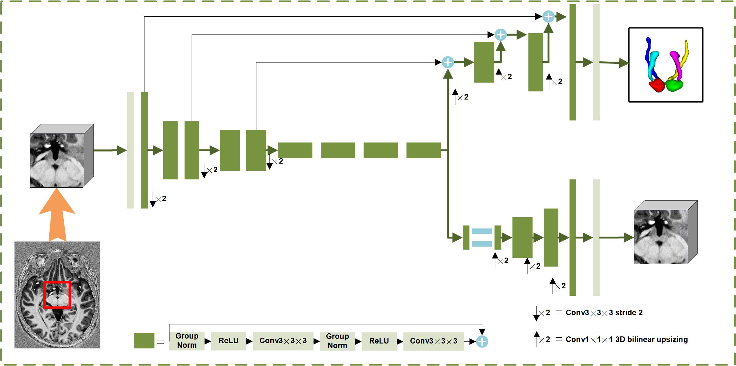
The SegResVAE Net6 was used to segment the core components mediating the Papez circuit. As shown in Figure 1, the SegResVAE Net contains three parts (the encoder part, the decoder part, and the variational auto-encoder (VAE) part). The encoder part extracted the higher-order semantic information from the input images. The decoder part with inter-level skip connections predicted the segment results. The VAE part reconstructed without inter-level skip connections reconstructed the input image dimensions to add more constraints. The overall loss function contains Dice loss (based on labels and predicted segmentation results), L2 loss (based on the input image and VAE output), and KL divergence (based on the input image and VAE output). The Adam optimizer7 with an initial learning rate of 1e−4 was used.
Results
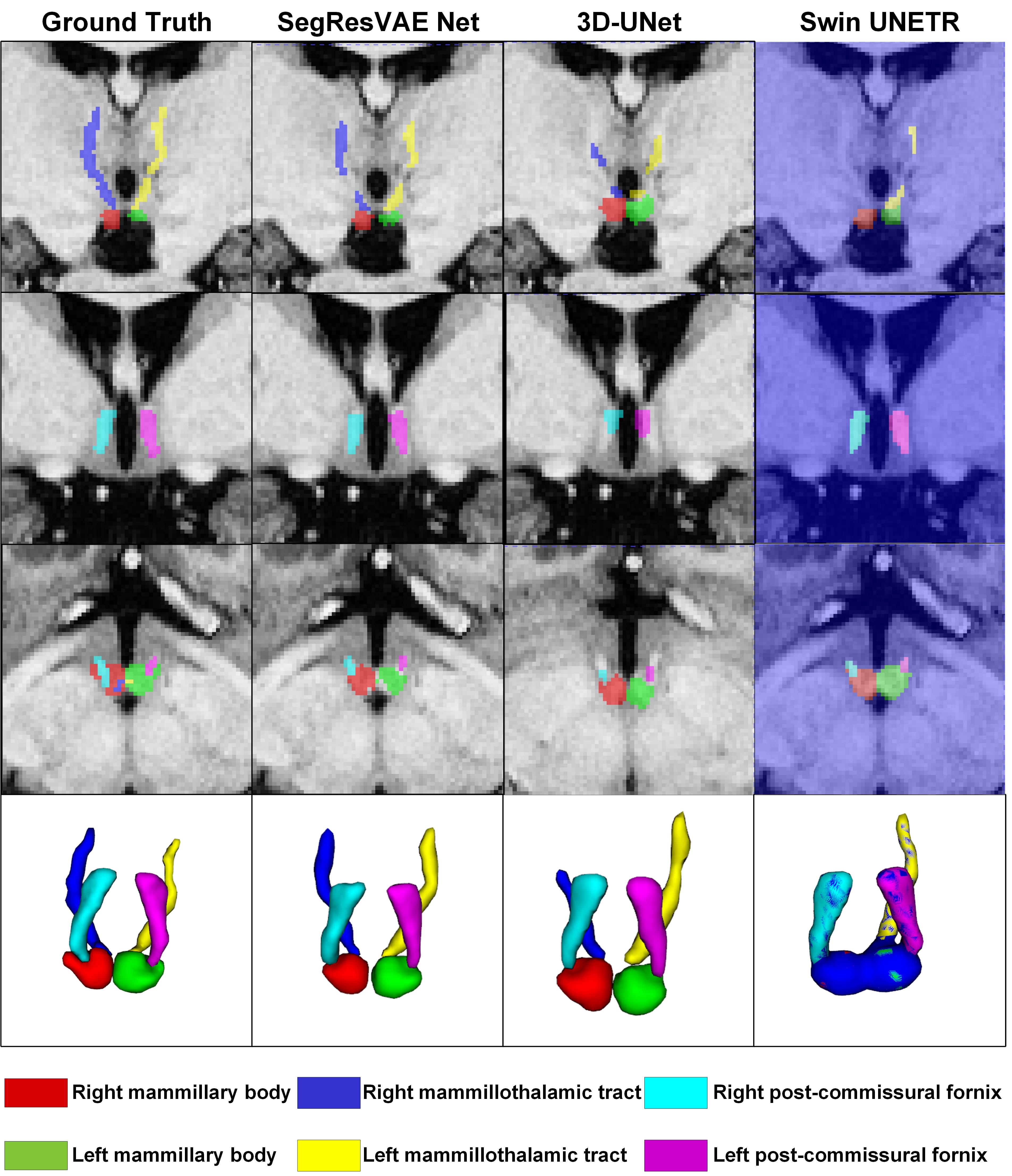
The model's performance was evaluated using Dice scores with a five-fold cross-validation method. We compared the SegResVAE Net with 3D-UNet8 and Swin UNETR9.The SegResVAE Net achieved the Dice scores for left MB, right MB, left MTT, right MTT, left PF, right PF, and the average of all components were 0.811, 0.807, 0.704, 0.705, 0.747, 0.796, and 0.761, respectively. The 3D-UNet achieved the Dice scores for left MB, right MB, left MTT, right MTT, left PF, right PF, and the average of all components were 0.801, 0.803, 0.687, 0.691, 0.782, 0.777, and 0.757, respectively. The Swin UNETR achieved the Dice scores for left MB, right MB, left MTT, right MTT, left PF, right PF, and the average of all components were 0.803, 0.004, 0.703, 0.426, 0.803, 0.801, and 0.590, respectively.
The SegResVAE Net exhibited the best Dice scores for MB, MTT, and the average of all components. The Swin UNETER exhibited the best Dice scores for PF but failed to segment the right MB and the right MTT. Figure 2 shows the comparison of segmented images of the three models.
Discussion and Conclusion
In this pilot study, we first segment the mammillary body, mammillothalamic tract, and post-commissural fornix in the brain based on the 7T-MR scanner using a deep learning model. The results of model segmentation showed great potential for quantitative analysis of the core components that mediate the Papez circuit.The SegResVAE network demonstrated the best segmentation performance, which may be due to the collaboration of the model's dual task of segmentation and reconstruction, and is more suitable for the small sample dataset. In this dataset with a small sample size, Swin UNETR with larger model parameters may not be able to segment the MB and MTT.
In conclusion, deep learning-based segmentation to the core components mediating the Papez circuit in 7T MRI may be potentially helpful for patients with AD, intractable epilepsy, and other diseases.
Acknowledgements
This work was supported by the Guizhou Provincial People’s Hospital Talent Fund (Peng Yunsong) under the Grant Hospital Talent Project [2022]-5.References
1. Dillingham CM, Milczarek MM, Perry JC and Vann SD. Time to put the mammillothalamic pathway into context. Neuroscience & Biobehavioral Reviews. 2021;121:60-74.
2. Yu D, Yan H, Zhou J, Yang X, Lu Y and Han Y. A circuit view of deep brain stimulation in Alzheimer’s disease and the possible mechanisms. Molecular Neurodegeneration. 2019;14:1-12.
3. Schaper FL, Plantinga BR, Colon AJ, Wagner GL, Boon P, Blom N, Gommer ED, Hoogland G, Ackermans L and Rouhl RP. Deep brain stimulation in epilepsy: a role for modulation of the mammillothalamic tract in seizure control? Neurosurgery. 2020;87:602.
4. Copenhaver BR, Rabin LA, Saykin AJ, Roth RM, Wishart HA, Flashman LA, Santulli RB, McHugh TL and Mamourian AC. The fornix and mammillary bodies in older adults with Alzheimer's disease, mild cognitive impairment, and cognitive complaints: a volumetric MRI study. Psychiatry Research: Neuroimaging. 2006;147:93-103.
5. Fletcher E, Raman M, Huebner P, Liu A, Mungas D, Carmichael O and DeCarli C. Loss of fornix white matter volume as a predictor of cognitive impairment in cognitively normal elderly individuals. JAMA neurology. 2013;70:1389-1395.
6. Myronenko A. 3D MRI brain tumor segmentation using autoencoder regularization. Brainlesion: Glioma, Multiple Sclerosis, Stroke and Traumatic Brain Injuries: 4th International Workshop, BrainLes 2018, Held in Conjunction with MICCAI 2018, Granada, Spain, September 16, 2018, Revised Selected Papers, Part II 4. 2019:311-320.
7. Kingma DP and Ba J. Adam: A method for stochastic optimization. arXiv preprint arXiv:14126980. 2014.
8. Kerfoot E, Clough J, Oksuz I, Lee J, King AP and Schnabel JA. Left-ventricle quantification using residual U-Net. Statistical Atlases and Computational Models of the Heart Atrial Segmentation and LV Quantification Challenges: 9th International Workshop, STACOM 2018, Held in Conjunction with MICCAI 2018, Granada, Spain, September 16, 2018, Revised Selected Papers 9. 2019:371-380.
9. Hatamizadeh A, Nath V, Tang Y, Yang D, Roth HR and Xu D. Swin unetr: Swin transformers for semantic segmentation of brain tumors in mri images. International MICCAI Brainlesion Workshop. 2021:272-284.
Figures