2053
Assessing Visual Pathway White Matter Degeneration in Primary Open-angle Glaucoma Using Diffusion Spectrum Imaging1Fudan University, Eye & ENT Hospital, Shanghai, China
Synopsis
Keywords: Head & Neck/ENT, Head & Neck/ENT
Motivation: The underlying structural pathophysiology of visual white matter degeneration in primary open-angle glaucoma (POAG) remains incompletely understood.
Goal(s): To characterize macro- and micro-structural abnormalities in the optic tract and optic radiation of POAG by multiple morphology and diffusion metrics derived from diffusion spectrum imaging(DSI)
Approach: DSI was performed on 34 bilateral POAG patients and 25 healthy controls (HCs), and multiple morphology and diffusion metrics were calculated and compared.
Results: Significant differences emerged in morphology and diffusion metrics between POAG patients and HCs.
Impact: Our demonstration of multiple morphology and diffusion changes in POAG utilizing DS suggests the macro- and micro-structural white matter abnormalities, which could be used for assessing POAG degenetation
Introduction
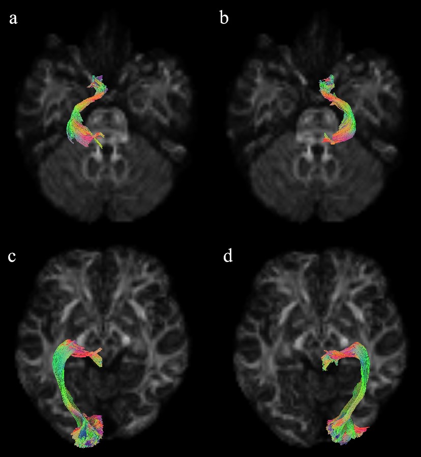
Primary open-angle glaucoma (POAG) has been recognized as a neurodegenerative disease with structural abnormalities in extraretinal visual white matter, particularly in the optic tract (OT) and the optic radiation (OR), but the underlying structural pathophysiology is not fully understood [1]. Diffusion MRI is the primary non-invasive modality for quantifying the structural neurodegeneration of glaucoma visual white matter and diffusion spectrum imaging (DSI) is a high angular resolution dMRI technique using probabilistic tractography, which can accurately depict the complex architecture of visual pathway and derive DTI-, NODDI- and MAP metrics through one single acquisition [2-4]. However, these metrics have not been applied on POAG. The objective of the current study was to comprehensively evaluate the he macro- and micro-structural abnormalities of OR and OT in POAG by multiple DSI-derived morphology and diffusion metrics.Methods
We conducted a prospective DSI study involving 34 bilateral POAG patients and 25 healthy controls (HCs). We derived and compared multiple morphology metrics, including volume, area, length, and shape metrics, as well as diffusion metrics such as diffusion tensor imaging (DTI), mean apparent propagator (MAP), and neurite orientation dispersion and density imaging (NODDI) metrics of the OT and OR. Statistical comparisons employed t-test or Mann-Whitney U test. Logistic regression models were constructed to differentiate between POAG patients and HCs using these MRI metrics.Results
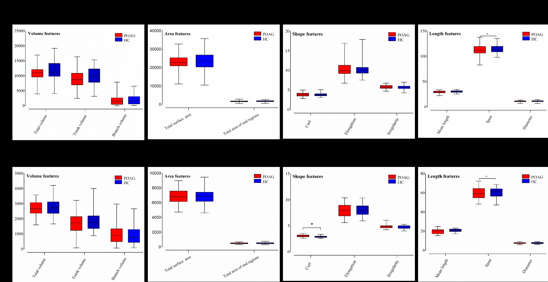
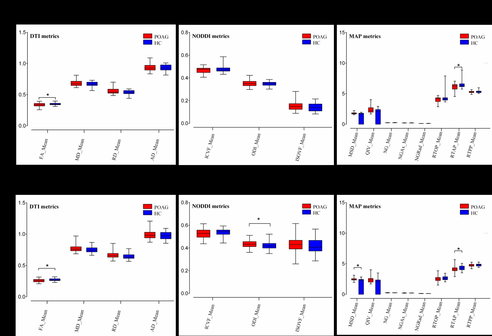
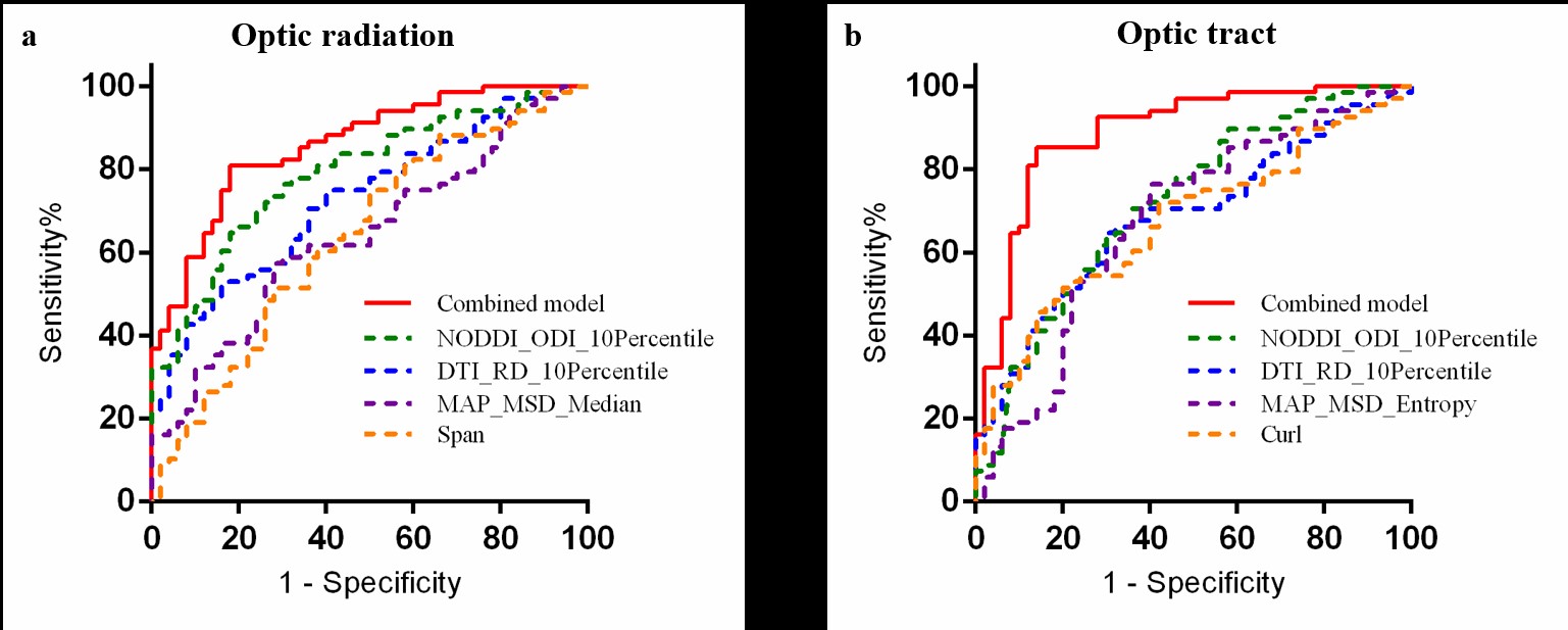
Significant differences emerged in morphology and diffusion metrics. In morphology, POAG patients exhibited length and shape changes, including a reduced span in the OR and a decreased span, along with a higher curl, in the OT compared to HCs (all p < 0.05). Regarding diffusion, POAG patients demonstrated decreased DTI_FA_Mean and MAP_RTAP_Mean in the OR, while showing lower DTI_FA_Mean and higher DTI_RD_Mean, MAP_RTAP_Mean, MAP_MSD_Mean, and NODDI_ODI_Mean in the OT (all p < 0.05). The logistic regression models, incorporating these MRI metrics, effectively discriminated between POAG patients and HCs, achieving AUCs of 0.860 for the OR and 0.888 for the OT.Discussion
Our findings revealed distinctive changes in the shape and length features, indicative of macro-structural degeneration in the OT and OR, which, to our knowledge, have not been previously reported. Moreover, we explored multiple diffusion metrics to examine micro-structural pathology, with the inclusion of MAP metrics for the first time, which demonstrated superior sensitivity compared to conventional DTI metrics. Furthermore, the study indicated that OR appeared to be less severely affected than OT. These results enhance our understanding of the visual white matter degeneration in POAG patients.Conclusions
DSI-derived morphology and diffusion metrics offer valuable insights into the macro- and micro-structural white matter degeneration in the OT and OR of POAG. Furthermore, the OR may exhibit less severe degeneration compared to the OT.Acknowledgements
This work was supported by the National Natural Science Foundation of China (Grant No. 81871341, 82271945, 81430007).References
1 Lawlor M, Danesh-Meyer H, Levin LA, Davagnanam I, De Vita E, Plant GT (2018) Glaucoma and the brain: Trans-synaptic degeneration, structural change, and implications for neuroprotection. Surv Ophthalmol 63:296-306
2 Yeh FC (2020) Shape analysis of the human association pathways. Neuroimage 223:117329
3 Mao C, Jiang W, Huang J et al (2022) Quantitative Parameters of Diffusion Spectrum Imaging: HER2 Status Prediction in Patients With Breast Cancer. Front Oncol 12:817070
4 She D, Huang H, Guo W, Jiang D, Zhao X, Kang Y, Cao D (2023) Grading meningiomas with diffusion metrics: a comparison between diffusion kurtosis, mean apparent propagator, neurite orientation dispersion and density, and diffusion tensor imaging. Eur Radiol 33:3671-3681
Figures