2049
Multimodal MRI Evaluation of Brain Plasticity Following 8 Weeks of Structured Functional and Strength Training1Core Technology Platform, New York University Abu Dhabi, Abu Dhabi, United Arab Emirates, 2New York University Abu Dhabi, Abu Dhabi, United Arab Emirates
Synopsis
Keywords: White Matter, Diffusion Tensor Imaging, exercise
Motivation: The impact of longer exercise durations and its correlation with comprehensive behavioral changes, such as improvements in strength and working memory, remains relatively underexplored.
Goal(s): In this study, we explore the impact of an 8-week strength training program on brain function and structure through multimodal MRI.
Approach: We recruited 21 participants, who had been leading a sedentary lifestyle before enrolling. Utilizing a Siemens Prisma scanner and Human Connectome Project protocols, we obtained multimodal MRI data, covering anatomical scans, diffusion imaging, resting state fMRI, and ASL scans.
Results: Preliminary findings highlight the importance of microstructural measurements in detecting exercise-induced brain changes.
Impact: This research highlights the potential for using multimodal MRI to characterize exercise-induced white matter plasticity in the brain, particularly in motor-related areas.
Background
Exercise has a well-documented positive impact on brain function, promoting neurogenesis, angiogenesis, and mitigating age-related effects [1]. However, the impact of longer exercise durations and its correlation with comprehensive behavioral changes, such as improvements in strength and working memory, remains relatively underexplored. In this pilot study, we aimed to evaluate the effects of an 8-week formal strength training program on brain structure and function using multimodal MRI [2]. Our focus was on longitudinal structural and white matter assessments, along with behavioral and working memory evaluations, aiming to characterize the effect sizes of various neuroimaging measurements. These measurements encompassed cortical thickness, white matter (WM) plasticity measures derived from standard DTI as well as advanced techniques generalized q-space imaging (GQI). Our findings hold significance for designing future longitudinal studies and wellness intervention research focused on quantifying the effects of strength and functional exercise on neuroimaging parameters.Methods
We recruited 21 participants, aged on average 20 years, with an equal gender distribution, all of whom had maintained a sedentary lifestyle in the year prior to their participation. We utilized a 3T Siemens Prisma scanner with a 64-channel head coil and followed established Human Connectome Project (HCP) protocols for acquiring multimodal MRI data [3], [4]. This included anatomical scans, diffusion imaging with Diffusion EPI sequence, and resting state fMRI and ASL scans. Anatomical scans comprised T1-weighted 3D MPRAGE and T2-weighted 3D SPACE sequences in the sagittal plane, with isotropic spatial resolution 0.8mm, a field of view of 256mm, and a GRAPPA of 2. The T1 sequence featured TR of 2400ms, and TE of 2.22ms, while the T2 sequence had a TR of 3200ms, and TE of 563ms (11 minutes acquisition time). We applied minimal preprocessing pipelines from the HCP to both anatomical and diffusion data . Subsequently, we constructed an unbiased template and conducted surface/volume parcellation of cortical and subcortical structures to identify subtle exercise-induced changes. Diffusion data was acquired using an SS-EPI sequence with multiband acceleration, yielding a spatial resolution of 1.5 x 1.5mm, slice thickness of 1.5mm, 92 slices, TR of 3230ms, TE of 89.20ms, and a multiband factor of 4. We analyzed thirteen white matter tracts associated with sensorimotor ability and working memory, including the Parahippocampal Cingulum, Superior Longitudinal Fasciculus (SLF I, II, & III), Corpus Callosum (Body, Major, and Minor Forceps), and the Corticospinal tract. Mean diffusivity, fractional anisotropy, and quantitative anisotropy were measured for each tract. Cohen’s d effect size was calculated for each structural parcellation in the FreeSurfer Desikan-Killiany atlas and for each WM tract based on the diffusion microstructure metrics to estimate the required sample size for future studies. G*Power software was then used to determine the sample size needed for designing larger prospective cohorts to investigate the effects of wellness interventions on multimodal MRI datasets.Results and Discussion
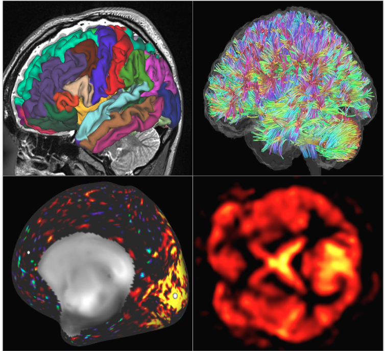
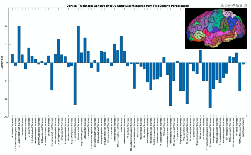
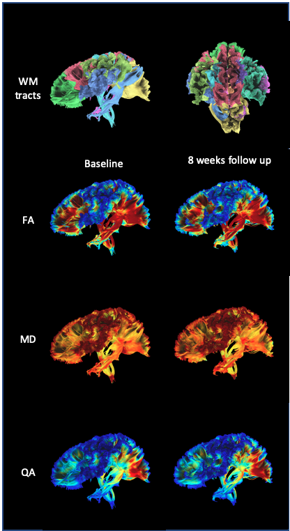
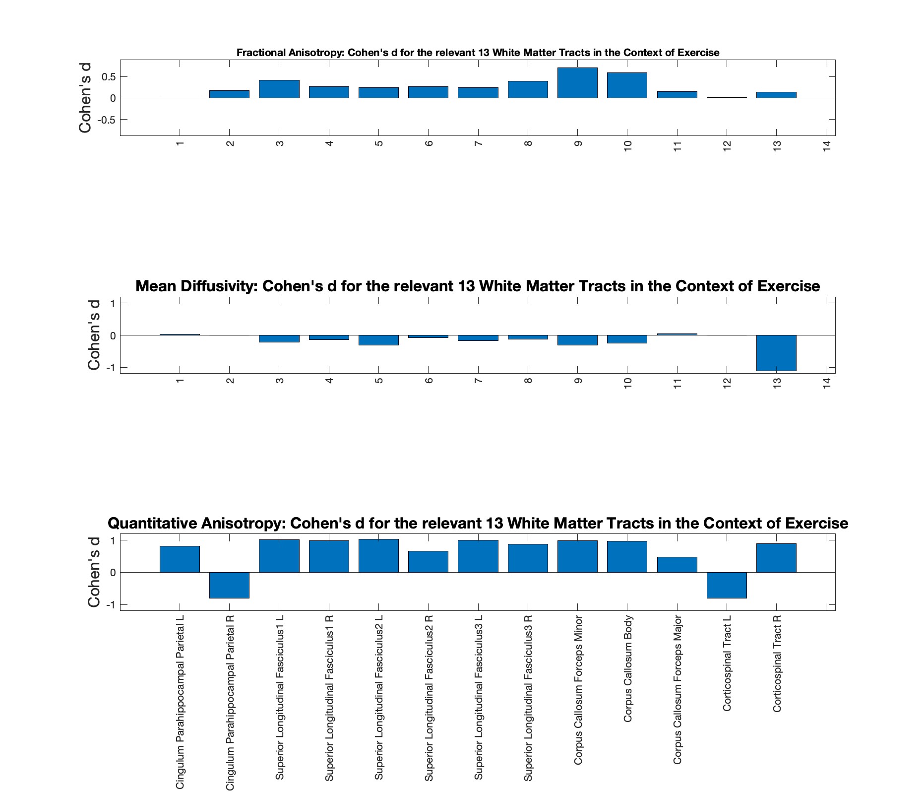
In Figure 1, we provide a selection of sample images obtained through multimodal MRI, covering structural segmentation, WM fiber tracking, resting state functional connectivity, and blood perfusion measures (with processing pending for the latter two modalities). These visualizations offer a preliminary glimpse into the dataset acquired during the study. Figure 2 showcases our analysis of Cohen’s d measures for the 72 cortical thickness parcellation measurements, revealing predominantly weak effects (Cohen’s d < 0.2). With a significance level of 0.05 and a power of 0.8, a sample size of 200 subjects would be required, making such a sample size infeasible. This outcome aligns with expectations, given the typically subtle structural changes within an 8-week timeframe. In contrast, our examination of WM plasticity, involving 5 subjects, yielded more promising results. The analysis suggests a moderate-to-strong effect size (Cohen’s d > 0.4), particularly for fractional anisotropy (FA) in areas relevant to motor function and coordination, such as the corpus callosum and corticospinal tract. These findings support the feasibility of employing sample sizes of fewer than 40 subjects to effectively detect exercise-induced effects on white matter fiber tracts, as illustrated in Figures 3 and 4. Our ongoing work includes the finalization of preprocessing for the remaining diffusion datasets and the incorporation of effect sizes from ASL and rsfMRI into the power analysis. In conclusion, our study offers preliminary evidence concerning the effect sizes of exercise-induced changes within HCP-style neuroimaging dataset. Furthermore, our findings emphasize the utility of sensitive microstructural measurements, such as diffusion imaging, over traditional anatomical sequences, in detecting the subtle yet significant physiological changes associated with the positive effects of exercise on the human brain.Acknowledgements
The experiments described herein were conducted using the facilities of the NYUAD Brain Imaging Core Technology Platform.References
[1] J. J. Steventon et al., “Changes in white matter microstructure and MRI-derived cerebral blood flow after 1-week of exercise training,” Sci. Rep., vol. 11, no. 1, pp. 1–12, 2021.
[2] S. Y. Bookheimer et al., “The Lifespan Human Connectome Project in Aging: An overview,” Neuroimage, vol. 185, no. September 2018, pp. 335–348, 2019.
[3] M. F. Glasser et al., “The Human Connectome Project’s neuroimaging approach,” Nature Neuroscience, vol. 19, no. 9. Nature Publishing Group, pp. 1175–1187, 01-Sep-2016.
[4] M. F. Glasser et al., “The minimal preprocessing pipelines for the Human Connectome Project,” Neuroimage, vol. 80, pp. 105–124, 2013.
Figures