2047
Disrupted structural brain networks and structural-functional decoupling in adolescent major depressive disorder1Department of Radiology, West China Hospital of Sichuan University, Chengdu, China
Synopsis
Keywords: Structural Connectivity, Diffusion Tensor Imaging, Major depressive disorder; Structural connectome; Graph theory
Motivation: Previous studies have demonstrated brain abnormalities in adolescents with major depressive disorder (MDD); however, how the topological organization of brain networks is affected is still unclear.
Goal(s): This study aimed to investigate the white matter (WM) structural topology in adolescent MDD.
Approach: The topological metrics of WM structural brain networks were analyzed using graph theory method.
Results: Adolescent MDD patients showed disrupted topological organization at the global, rich-club and modular levels, exhibited abnormal nodal centralities in multiple regions, and had a decrease in the coupling between structural and functional connectivity.
Impact: Our findings suggest widespread disruption of structural brain networks and structural-functional decoupling in adolescent MDD, which may provide new insights into the neurobiology of adolescent MDD.
Introduction
Major depressive disorder (MDD) is a multifactorial disorder with clinically heterogeneous features involving disturbances of mood and cognitive function 1, and tends to emerge during adolescence 2. Our understanding of the neurobiological mechanism of adolescent MDD lags behind that of adult MDD. Previous studies have demonstrated regional structural and functional abnormalities in adolescents with MDD; however, how brain networks are affected in adolescent MDD patients remains poorly understood. The present study aimed to explore the topological organization of white matter structural networks and the coupling strength between structural connectivity (SC) and functional connectivity (FC) in adolescent MDD.Methods
Ninety-four adolescents with MDD and 78 healthy controls (HCs) were recruited. Structural and functional MR imaging data were acquired using a 3.0-T scanner (GE Discovery MR750). A 90 × 90 structural and functional networks were constructed for each subject. The global and nodal topological properties, rich-club organization, and modular organization were analyzed using graph theory method. For each subject, we calculated the Pearson correlation coefficient between the SC and FC. Between-group differences in network topological metrics and SC-FC coupling were estimated using nonparametric permutation tests (10,000 iterations). The Bonferroni method was used to correct for multiple comparisons. Partial correlation analysis was used to assess the relationships between these altered metrics and clinical variables. Age, sex and BMI were set as covariates.Results
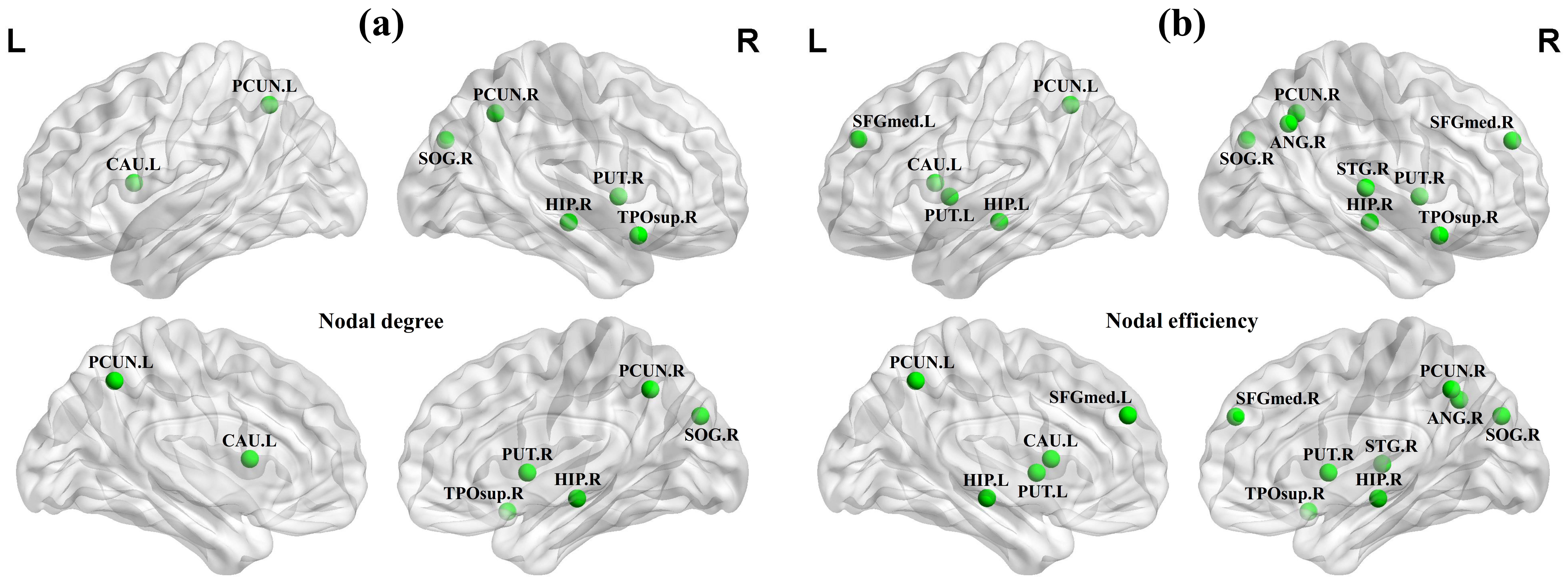
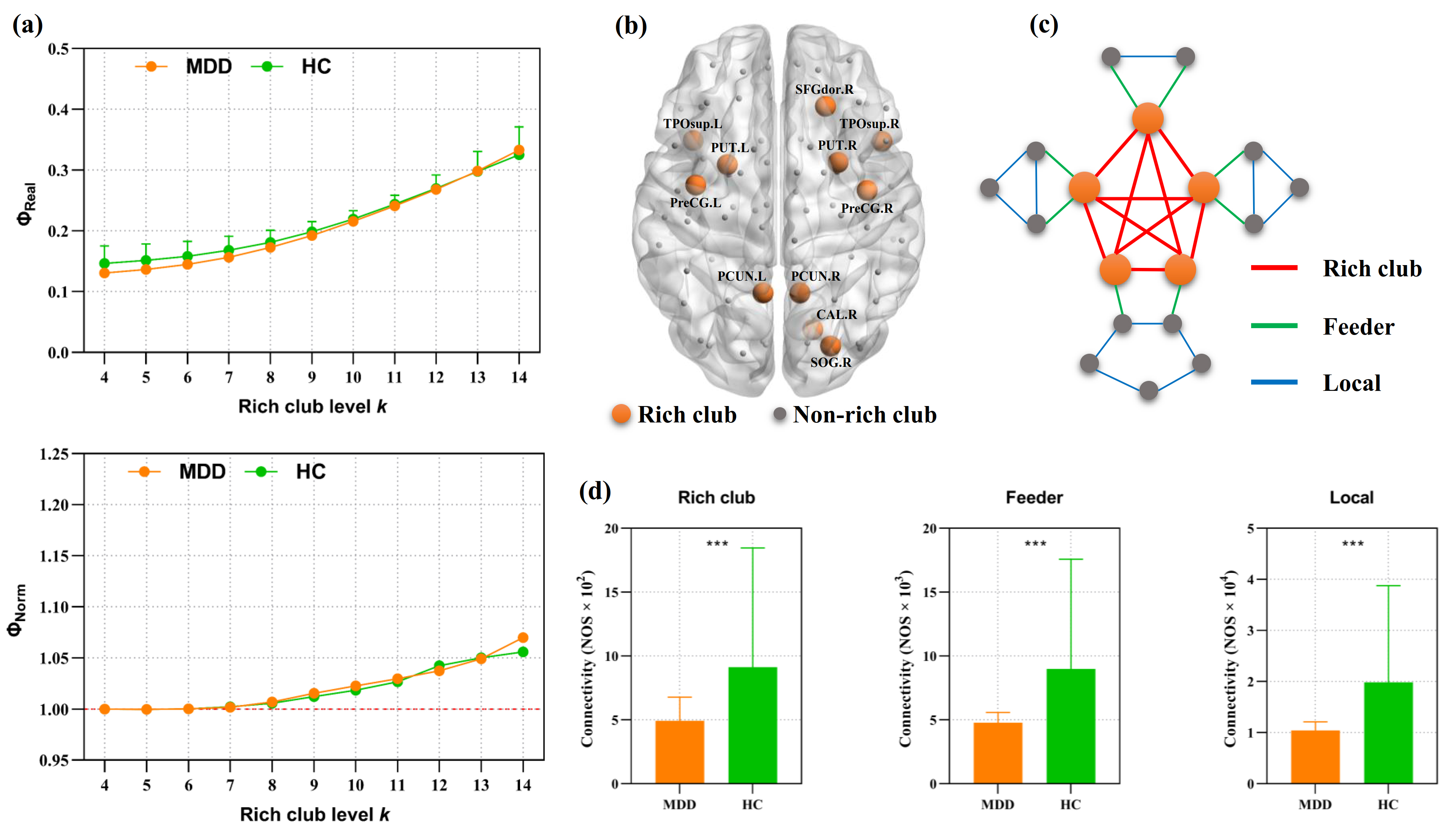

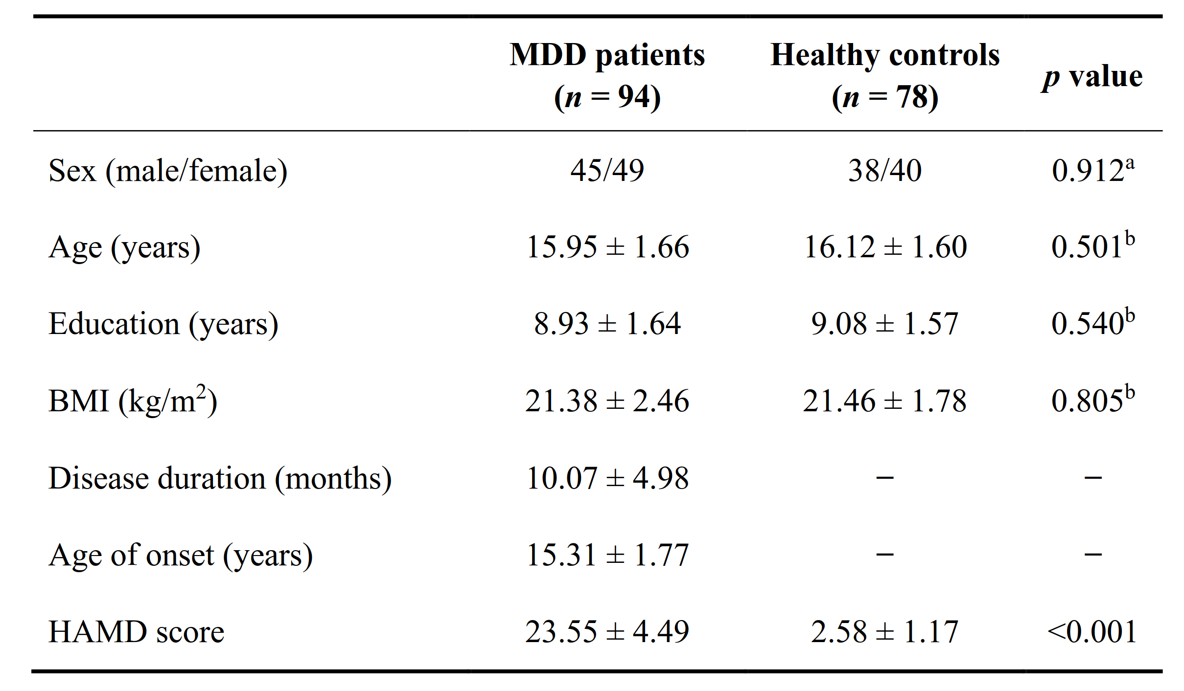
Detailed demographic and clinical characteristics of the two groups are shown in Figure 1. Adolescent MDD patients, relative to HCs, showed significantly lower Eglob, Eloc and Cp, and had significantly higher Lp and λ (all p < 0.05) (Figure 2). Adolescent MDD patients showed decreased nodal centralities in multiple regions (Figure 3). Rich-club organization was demonstrated in both groups (Figure 4a). Eleven regions were selected as rich-club nodes (Figure 4b). The edges of structural networks were classified into rich-club connections, feeder connections and local connections based on the categorization of the 90 nodes as rich-club and non-rich-club regions (Figure 4c). Compared to the HC group, adolescent MDD patients showed a significant decrease in rich-club connections, feeder connections and local connections (all p < 0.001) (Figure 4d). Five modules were identified at the group level: left prefrontal-subcortical module (module Ⅰ), right prefrontal-subcortical module (module Ⅱ), posterior default-mode and sensorimotor module (module Ⅲ), right default-mode and visual module (module Ⅳ), and left default-mode and visual module (module Ⅴ). Adolescent MDD patients showed significant decreased structural connections within all modules and between any pair of modules compared to healthy adolescents (all p < 0.05) (Figure 5). Adolescent MDD patients showed a significant reduction in strength of SC-FC coupling compared to controls (p = 0.012).Discussion
To our knowledge, the present study is the first to investigate the structural topological organization and the relationship between SC and FC in first-episode drug-naïve adolescent MDD patients. At the global level, our findings of decreased segregation and integration suggest that the brain structural networks of adolescent MDD patients are closer to a “weaker small-worldization” configuration 3. Additional modular and rich-club analysis revealed impaired rich-club organization and disrupted inter- and intra-modular connectivity of the structural brain networks in adolescent MDD patients. At the regional nodal level, the adolescent MDD-related decreases in nodal centralities were found in the medial prefrontal and lateral temporal regions, hippocampus, angular gyrus, and precuneus, most of which are components of the DMN 4. Similarly, decreased nodal centralities in the DMN was also found in the functional brain networks in patients with MDD 5. The DMN has been linked to depressive rumination 6, self-referential processing 7 and emotional appraisal 8. The adolescent MDD-related decreases in nodal centralities were also observed in the subcortical regions, including the left caudate nucleus and bilateral putamen that are involved in reward processing. Another interesting finding of this study was the decoupling between SC and FC in adolescents with MDD. A decrease in SC-FC coupling strength may suggest a loss of coherence of structural and functional connectomes. Prior study suggested that the coupling strength between SC and FC increased with age in the late developing human brain 9. A decreased level of SC-FC coupling may suggest that the functional brain networks in adolescent MDD patients are more dynamic and less constrained by the underlying anatomical connections.Conclusion
In summary, this study characterizes the structural connectome in adolescent MDD from different topological levels. These findings may provide new insights into the neurobiology of adolescent MDD.Acknowledgements
This study was supported by the National Natural Science Foundation of China (82271947, 81971595, 81820108018 and 81621003), the National Key R&D Program of China (2022YFC2009900), the Key Program of Natural Science Foundation of Sichuan Province (2022NSFSC0047), and the 1·3·5 Project for Disciplines of Excellence–Clinical Research Incubation Project, West China Hospital, Sichuan University (2020HXFH005).References
1. Malhi GS, Mann JJ. Depression. Lancet (London, England). 2018;392(10161):2299-2312.
2. Kessler RC, Avenevoli S, Costello EJ, et al. Prevalence, persistence, and sociodemographic correlates of DSM-IV disorders in the National Comorbidity Survey Replication Adolescent Supplement. Archives of general psychiatry. 2012;69(4):372-380.
3. Suo XS, Lei DL, Li LL, et al. Psychoradiological patterns of small-world properties and a systematic review of connectome studies of patients with 6 major psychiatric disorders. Journal of psychiatry & neuroscience : JPN. 2018;43(6):427.
4. Buckner RL, Andrews-Hanna JR, Schacter DL. The brain's default network: anatomy, function, and relevance to disease. Annals of the New York Academy of Sciences. 2008;1124:1-38.
5. Yang H, Chen X, Chen ZB, et al. Disrupted intrinsic functional brain topology in patients with major depressive disorder. Molecular psychiatry. 2021;26(12):7363-7371.
6. Hamilton JP, Farmer M, Fogelman P, Gotlib IH. Depressive Rumination, the Default-Mode Network, and the Dark Matter of Clinical Neuroscience. Biological psychiatry. 2015;78(4):224-230.
7. Qin P, Northoff G. How is our self related to midline regions and the default-mode network? NeuroImage. 2011;57(3):1221-1233.
8. Vanderhasselt MA, Baeken C, Van Schuerbeek P, Luypaert R, De Raedt R. Inter-individual differences in the habitual use of cognitive reappraisal and expressive suppression are associated with variations in prefrontal cognitive control for emotional information: an event related fMRI study. Biological psychology. 2013;92(3):433-439.
9. Hagmann P, Sporns O, Madan N, et al. White matter maturation reshapes structural connectivity in the late developing human brain. Proceedings of the National Academy of Sciences of the United States of America. 2010;107(44):19067-19072.
Figures
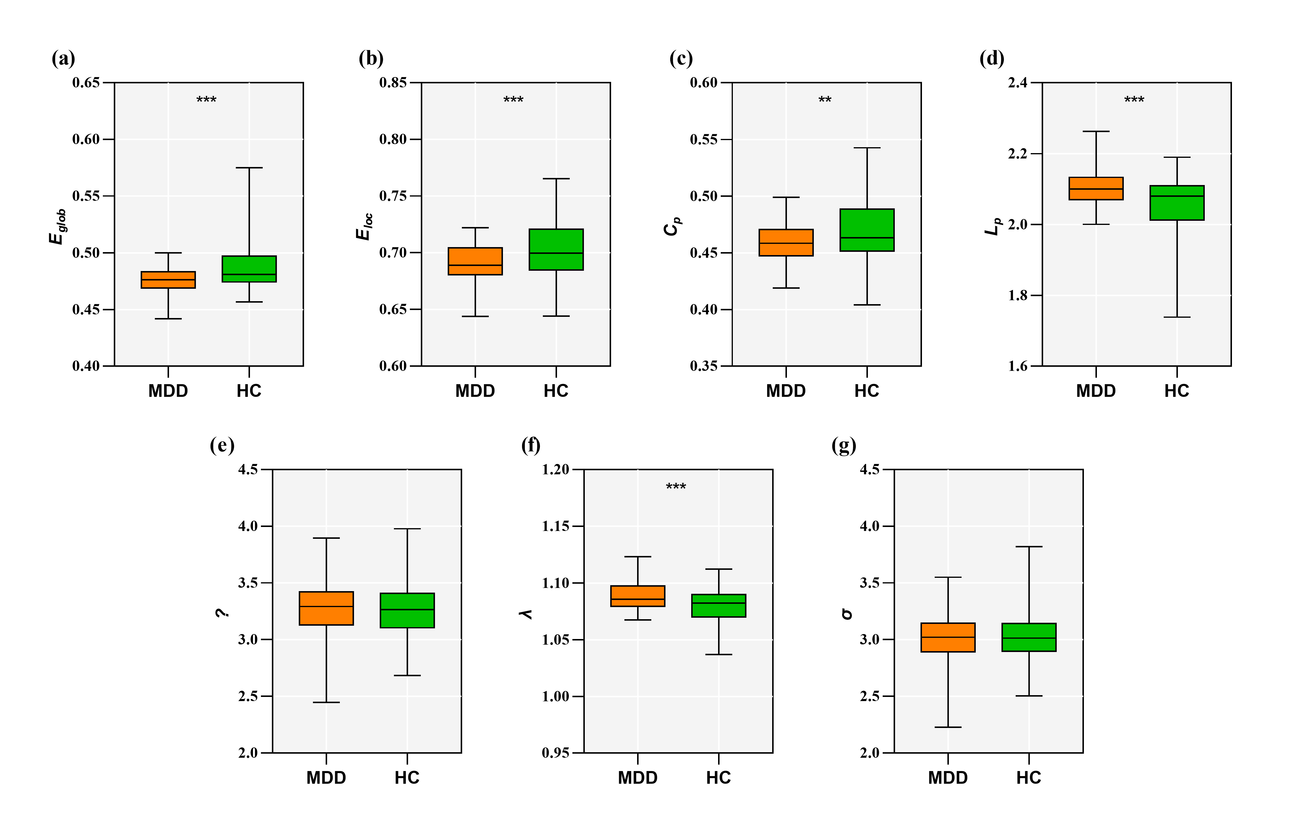
(λ) (f), and small-worldness (σ) (g) are shown using boxplots for the adolescent major depressive disorder (MDD) group (orange) and healthy control (HC) group (green). ** p < 0.01, *** p < 0.001.