2044
The involvement of the cerebellum in structural connectome changes in episodic migraine without aura1Department of Bioengineering, Instituto Superior Técnico, Universidade de Lisboa, Institute for Systems and Robotics - Lisboa, Lisbon, Portugal, 2Learning Health, Hospital da Luz, Lisbon, Portugal, Lisbon, Portugal, 3Imaging Department, Hospital da Luz, Lisbon, Portugal, Lisbon, Portugal, 4Neurology Department, Hospital da Luz, Lisbon, Portugal, Lisbon, Portugal, 5Center for Interdisciplinary Research in Health, Universidade Católica Portuguesa, Lisbon, Portugal, Lisbon, Portugal
Synopsis
Keywords: Structural Connectivity, Brain Connectivity, Migraine
Motivation: While the pathophysiology of migraine remains incompletely understood, several studies reported connectivity disruptions across large-scale brain networks.
Goal(s): To study changes in the structural connectome of migraine patients including cortical and subcortical regions as well as the cerebellum, often disregarded.
Approach: We performed tractography on diffusion MRI data and applied graph theory metrics to study connectome changes in episodic migraine patients and their healthy controls, using two different whole-brain parcellations.
Results: Patients show increased global efficiency and decreased characteristic path length, as well as increased connectivity of cerebellar regions with a greater node degree in the posterior lobe of the cerebellum.
Impact: This study sheds light on the importance of including regions other than the cortex in the structural connectome studies of migraine. Indeed, the cerebellum seems to play an important role in migraine, presenting increased connectivity with other regions.
Introduction
Migraine is one of the most prevalent neurological disorders worldwide (17% prevalence1). While its pathophysiology remains incompletely understood, several studies detected functional and structural disruptions across large-scale brain networks2-5. Structural connectivity has been studied using diffusion MRI (dMRI) tractography. Grey matter parcelations often define whole-brain network nodes for obtaining structural connectomes. However, the most commonly used atlases, Desikan or Schaffer6,7, include the cortex but miss important brain regions in migraine pathophysiology like the thalamus and cerebellum. We investigate structural connectome changes in migraine patients, including both cortical, subcortical and cerebellar regions.Methods
Population: 14 patients with low-frequency episodic menstrual migraine without aura (M) (36±9yrs), scanned during the interictal phase, and a control group of 15 healthy women (HC) (31±7yrs), in the corresponding menstrual cycle phase (post-ovulation).Acquisition and preprocessing: dMRI images were acquired from a 3T Siemens Vida MRI system with a 64-channel RF-receive head coil using: TR/TE=6800/89ms, 66 slices, GRAPPA factor 2, SMS factor 3, 2mm isotropic resolution. Sampling scheme: b=400,1000,2000s/mm2 shells along 32, 32, 60 directions, respectively, and 8 non-diffusion-weighted volumes. Data preprocessing followed the DESIGNER pipeline10.
Tractography and connectomics: Multi-shell multi-tissue constrained spherical deconvolution was performed in MRtrix11 using the anatomically constrained tractography framework. Probabilistic tractography was performed, with 10 million streamlines per subject, maximum length of 25cm and 1mm step size. Spherical-deconvolution Informed Filtering of Tractograms algorithm was applied. The connectivity matrix was built from individual subject streamlines, enforcing symmetry, using two different parcellations: cortical Schaefer atlas regions (100 regions) + 12 subcortical regions (SC) and 26 cerebellum regions (CB) of the AAL116 atlas12; and ii) the 116 cortical, SC and CB of the AAL116 atlas.
Statistical Analysis: Network-based Statistics (NBS) was used to compare groups (M>HC or HC>M), using the NBS toolbox13 (p<0.05). The regions (nodes) of each parcellation were then grouped into node aggregates according to: i) the 7 canonical resting-state networks identified by Yeo14+ subcortical regions + crus + posterior lobe of the cerebellum (PLC) + vermis; and ii) 4 lobes (occipital, parietal, frontal and temporal) + subcortical regions + crus + PLC + vermis. For all but the vermis, left and right regions were considered. Both global and local graph metrics were computed for each connectome using the Brain Connectivity (BCT) Toolbox15: characteristic path length (L), clustering coefficient (C), average degree (D) and global efficiency (GE) (global); and the degree of the node aggregates (local). Their correlation with clinical data was investigated.
Results
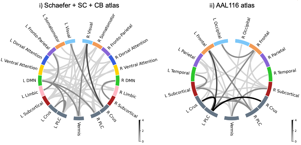
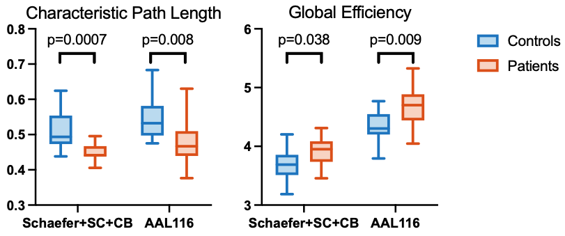
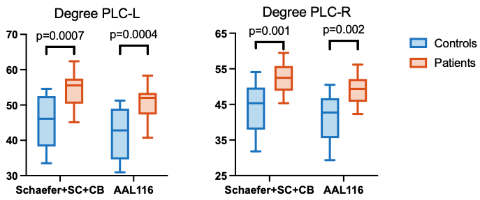
Significant connectome differences were found for the NBS contrast M>HC using both parcellations (Figure 1). Despite some differences, the two parcellations produced some common patterns, namely the increased connectivity between the left crus and the left PLC, between the occipital lobe/visual network and the vermis, and between cerebellar regions and frontal and parietal regions. Overall, a distinctive pattern of increased cerebellar connectivity was observed in patients.Concerning graph metrics, in both parcellations, L was decreased and GE was increased in migraine patients (Figure 2), while the node degree of both the right and left PLC was also increased (Figure 3). In AAL116, C and D were increased in migraine patients.
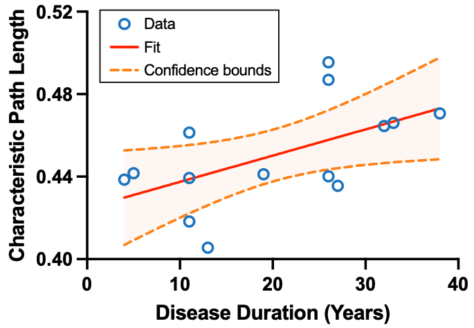
A significant positive correlation between L and the migraine disease duration was found (Figure 4).
Discussion and Conclusions
We found structural connectivity disruptions in migraine patients, especially involving the cerebellum. The cerebellum plays an inhibitory role in pain processing5, having several connections to the prefrontal cortex (via thalamus)14. The crus is thought to have cognitive and emotional representations, displaying activity during painful stimuli15. Therefore, the increased connectivity between the cerebellum and other regions may indicate a dysfunctional negative feedback loop where the inhibitory signal is not sensed by the thalamus5. The crus and the posterior lobe of the cerebellum are also involved in cognitive functions, thus changes in their connectivity potentially contribute to cognitive deficits common in migraine. Moreover, we found decreased L (and increased GE), consistent with the literature2,3,16,17 and hypothesized as leading to higher and more effective pain information dissemination in the brain circuitry. This integration increase was also present in the cerebellum as the PLC node degree was also increased. Interestingly, a positive correlation was found between L and disease duration potentially implying a plastic adaptation over time to migraine attacks. Importantly, the involvement of the cerebellum was consistently observed using two different brain parcellations, which supports the robustness of the findings. Overall, our results showcase the importance of the cerebellum in migraine pathophysiology, highlighting the importance of including it in connectome studies.Acknowledgements
We acknowledge the Portuguese Science Foundation through grants SFRH/BD/139561/2018, PTDC/EMD-EMD/29675/2017, LISBOA-01-0145-FEDER-029675 and UIDB/50009/2020.References
1. Hoffmann, J., Baca, S. M., & Akerman, S. (2019). Neurovascular mechanisms of migraine and cluster headache. Journal of Cerebral Blood Flow & Metabolism, 39(4), 573–594. https://doi.org/10.1177/0271678X17733655
2. Li, K., Liu, L., Yin, Q., Dun, W., Xu, X., Liu, J., & Zhang, M. (2017). Abnormal rich club organization and impaired correlation between structural and functional connectivity in migraine sufferers. Brain imaging and behavior, 11, 526-540.
3. Silvestro, M., Tessitore, A., Caiazzo, G., Scotto di Clemente, F., Trojsi, F., Cirillo, M., ... & Russo, A. (2021). Disconnectome of the migraine brain: a “connectopathy” model. The Journal of Headache and Pain, 22(1), 1-11.
4. Planchuelo-Gomez, A., Garcia-Azorin, D., Guerrero, A. L., Aja-Fernandez, S., Rodriguez, M., & de Luis-Garcia, R. (2020). Structural connectivity alterations in chronic and episodic migraine: A diffusion magnetic resonance imaging connectomics study. Cephalalgia, 40(4), 367-383.
5. Mehnert, J., & May, A. (2019). Functional and structural alterations in the migraine cerebellum. Journal of Cerebral Blood Flow & Metabolism, 39(4), 730-739.
6. Desikan, R. S., Ségonne, F., Fischl, B., Quinn, B. T., Dickerson, B. C., Blacker, D., Buckner, R. L., Dale, A. M., Maguire, R. P., Hyman, B. T., & others. (2006). An automated labeling system for subdividing the human cerebral cortex on MRI scans into gyral based regions of interest. Neuroimage, 31(3), 968–980.
7. Schaefer, A., Kong, R., Gordon, E. M., Laumann, T. O., Zuo, X.-N., Holmes, A. J., Eickhoff, S. B., & Yeo, B. T. T. (2018). Local-Global Parcellation of the Human Cerebral Cortex from Intrinsic Functional Connectivity MRI. Cerebral Cortex, 28(9), 3095–3114. https://doi.org/10.1093/cercor/bhx179
8. Ades-Aron, B., Veraart, J., Kochunov, P., McGuire, S., Sherman, P., Kellner, E., Novikov, D. S., & Fieremans, E. (2018). Evaluation of the accuracy and precision of the diffusion parameter EStImation with Gibbs and NoisE removal pipeline. NeuroImage, 183, 532–543. https://doi.org/10.1016/j.neuroimage.2018.07.066
9. Tournier, J. D., Smith, R., Raffelt, D., Tabbara, R., Dhollander, T., Pietsch, M., ... & Connelly, A. (2019). MRtrix3: A fast, flexible and open software framework for medical image processing and visualisation. Neuroimage, 202, 116137.
10. Tzourio-Mazoyer, N., Landeau, B., Papathanassiou, D., Crivello, F., Etard, O., Delcroix, N., Mazoyer, B., & Joliot, M. (2002). Automated Anatomical Labeling of Activations in SPM Using a Macroscopic Anatomical Parcellation of the MNI MRI Single-Subject Brain. NeuroImage, 15(1), 273–289. https://doi.org/10.1006/nimg.2001.0978
11. Zalesky, A., Fornito, A., & Bullmore, E. T. (2010). Network-based statistic: Identifying differences in brain networks. NeuroImage, 53(4), 1197–1207. https://doi.org/10.1016/j.neuroimage.2010.06.041
12. Yeo, B. T., Krienen, F. M., Sepulcre, J., Sabuncu, M. R., Lashkari, D., Hollinshead, M., Roffman, J. L., Smoller, J. W., Zöllei, L., Polimeni, J. R., Fischl, B., Liu, H., & Buckner, R. L. (2011). The organization of the human cerebral cortex estimated by intrinsic functional connectivity. Journal of neurophysiology, 106(3), 1125–1165. https://doi.org/10.1152/jn.00338.2011
13. Rubinov, M., & Sporns, O. (2010). Complex network measures of brain connectivity: Uses and interpretations. NeuroImage, 52(3), 1059–1069. https://doi.org/10.1016/j.neuroimage.2009.10.003
14. Middleton, F. A., & Strick, P. L. (2001). Cerebellar projections to the prefrontal cortex of the primate. Journal of neuroscience, 21(2), 700-712.
15. Moulton, E. A., Elman, I., Pendse, G., Schmahmann, J., Becerra, L., & Borsook, D. (2011). Aversion-related circuitry in the cerebellum: responses to noxious heat and unpleasant images. Journal of neuroscience, 31(10), 3795-3804.
16. Liu, J., Ma, S., Mu, J., Chen, T., Xu, Q., Dun, W., ... & Zhang, M. (2017). Integration of white matter network is associated with interindividual differences in psychologically mediated placebo response in migraine patients. Human Brain Mapping, 38(10), 5250-5259.
17. Dai, L., Zheng, Q., Chen, X., Wang, J., Peng, Y., Hu, C., ... & Li, Y. (2021). Altered brain structural topological properties and its correlations with clinical characteristics in episodic migraine without aura. Neuroradiology, 63, 2099-2109.
Figures