2043
Habenula functional connectivity predicts prior night’s sleep in healthy adults: A 5.0 T fMRI study1Department of Radiology, Zhongnan Hospital of Wuhan University, Wuhan, China
Synopsis
Keywords: Structural Connectivity, High-Field MRI, sleep
Motivation: The habenula is evolutionarily conserved brain region which play a crucial role in sleep regulation
Goal(s): To analysis the structural and functional relationship between habenula and individual sleep by 5T ultrahigh-resolution MRI
Approach: Using ultrahigh-resolution structural and functional MRI, we parcellated lateral and medial portions of the habenula and calculated structural and functional connectivity on the basis of these parcellations
Results: We parcellated lateral and medial portions of the habenula and identified connectivity with salience, limbic and part of somatomotor networks, which led to the loss in average sleep and cognitive processes
Impact: For the first time we identified the correlation between habenula and individual sleep using 5T ultrahigh-resolution MRI. By analyzing 300 samples high -resolution data, we revealed the neurological basis of individual sleep regulation, and provided new opinions for the mechanism of sleep diseases.
Background and purpose
Sleep is an evolutionarily conserved behavior in animals and humans, which plays crucial role in strength recovery, memory consolidation, synaptic homeostasis, and waste clearance from the nervous system.(Verweij et al., 2014) Brain connectivity encoding individual sleep regulation may be rooted in small, evolutionally conservative nuclei, for example, the habenula (Hb).(Namboodiri et al., 2016) (Killgore, 2013)The habenula (Hb) is a phylogenetically brain region in a small structure with a volume of 31-36 mm3 and its accurate outlining has been a challenge on conventional functional imaging.(Aizawa et al., 2013) Despite in small size, the habenula acts as a critical neuroanatomical hub that regulates behavioral responses to pain, stress, anxiety, reward and sleep. (Boulos et al., 2017)It has been proven that intersubject variability in sleep predicts resting fMRI functional connectivity.(Li et al., 2020) Herein, we investigate the potential role of Hb in sleep-related attention and memory loss.Methods
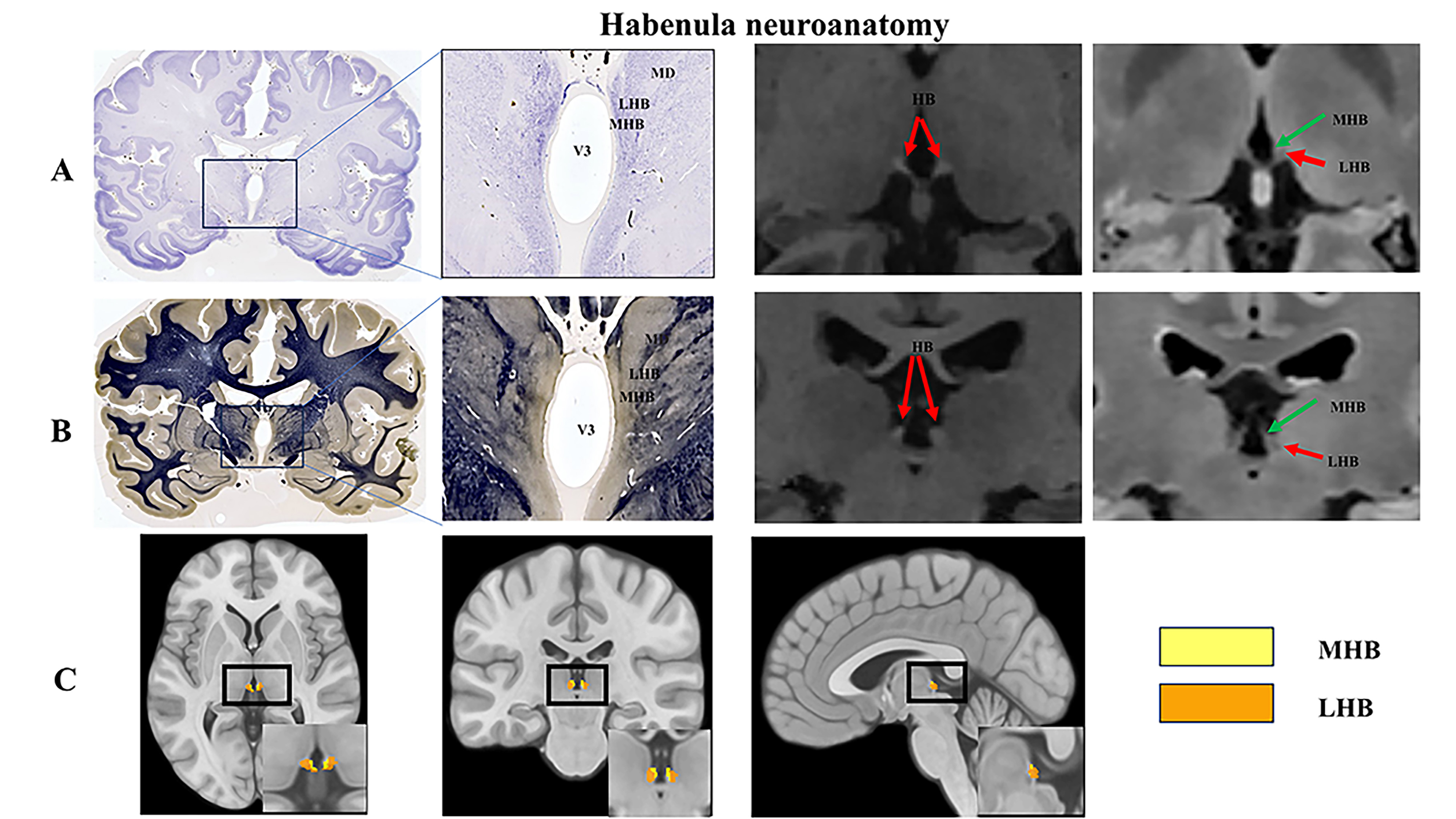
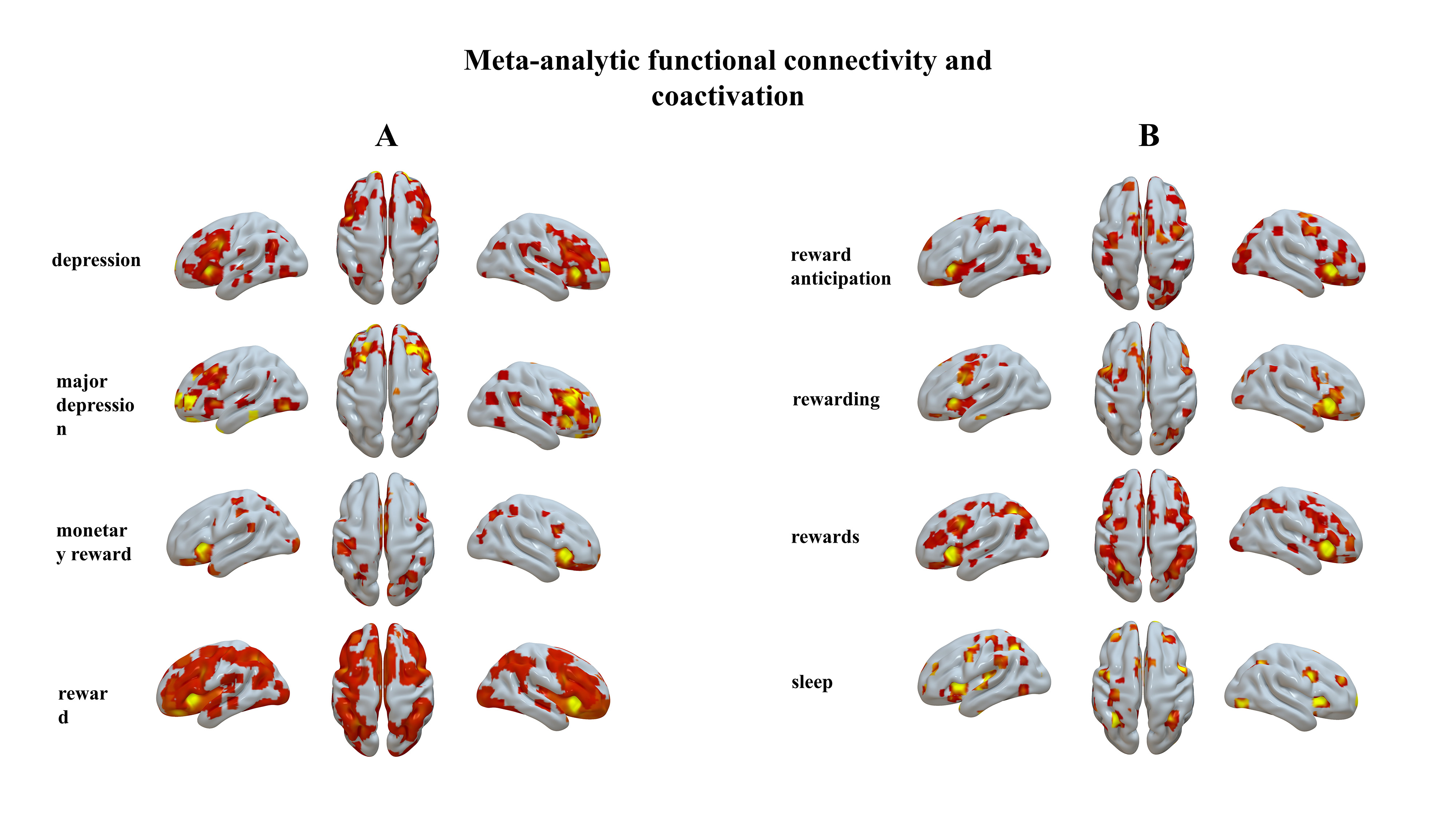
In a group of 300 healthy adults aged between 18 and 85 years. Self-reported sleep quality was used to assess individual sleep quality, including sleep duration, habitual sleep efficiency, sleep disturbances, sleep medication, and daytime dysfunction. MRI data were obtained using a state-of-the-art whole-body 5.0 T MRI scanner (uMR Jupiter, UIH, China) in Zhongnan Hospital of Wuhan University. In order to take advantage of 5T MRI resolution, we segmented the bilateral habenula on the basis of individual 1.5 mm3 functional imaging and 0.7 mm3 structural imaging resolution, referencing multiple sequence contrast information, and used them as seed regions to calculate whole-brain resting-state functional connectivity. Preprocessing of the resting-state fMRI data employed a standard pipeline (SI-Methods) embedded in the Data Processing & Analysis of Brain Imaging (DPABI, http://rfmri.org/DPABI) To gain a deeper understanding of the coupling network systems and functional roles associated with the identified GM atrophy and asymmetry clusters, we utilized the NeuroSynth database (https://www.neurosynth.org) Also, we parcellated lateral and medial portions of the Hb and calculated structural and functional connectivity on the basis of these parcellations using diffusion model and Lesion network mapping.Results
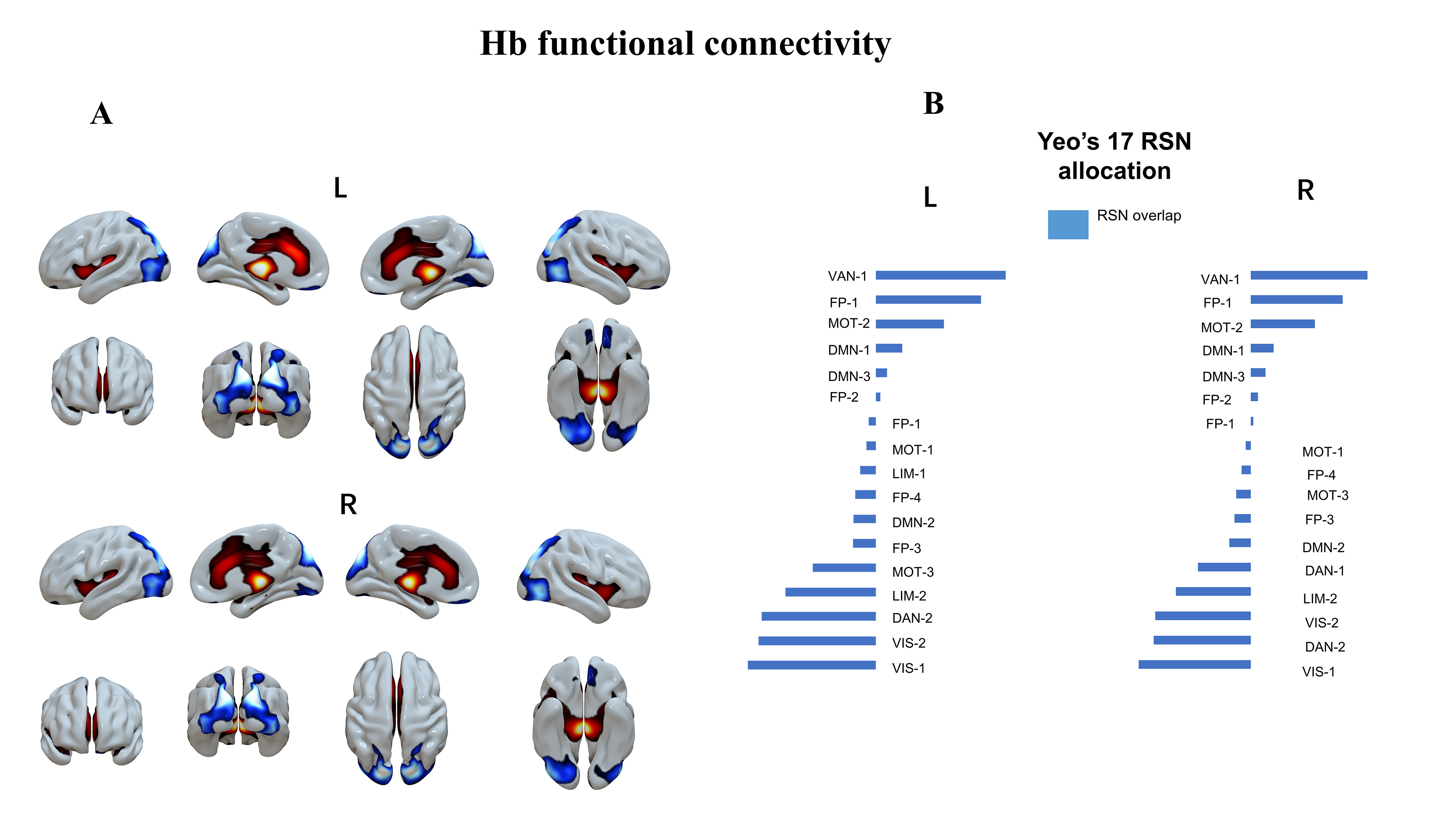
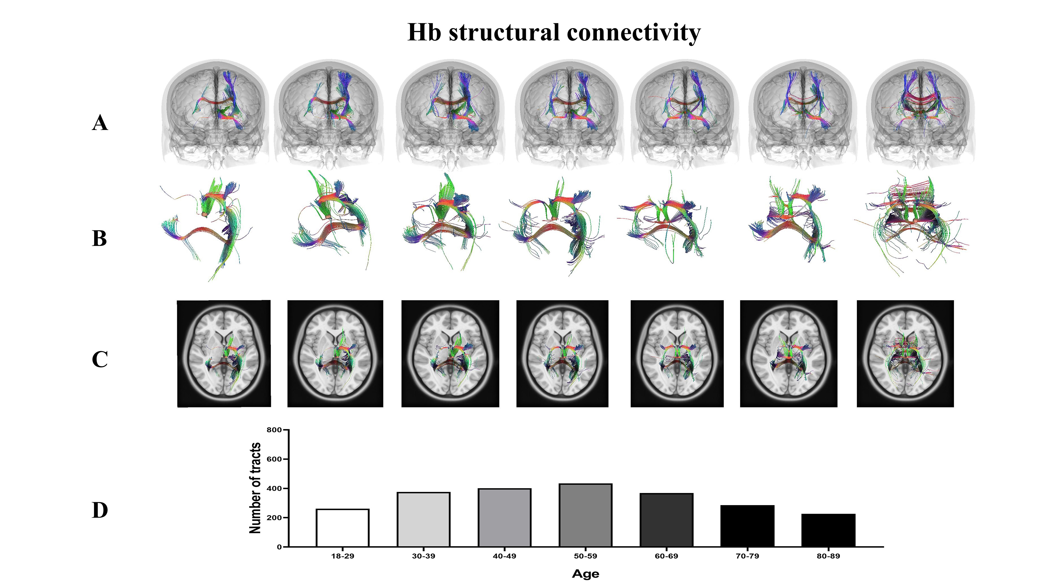
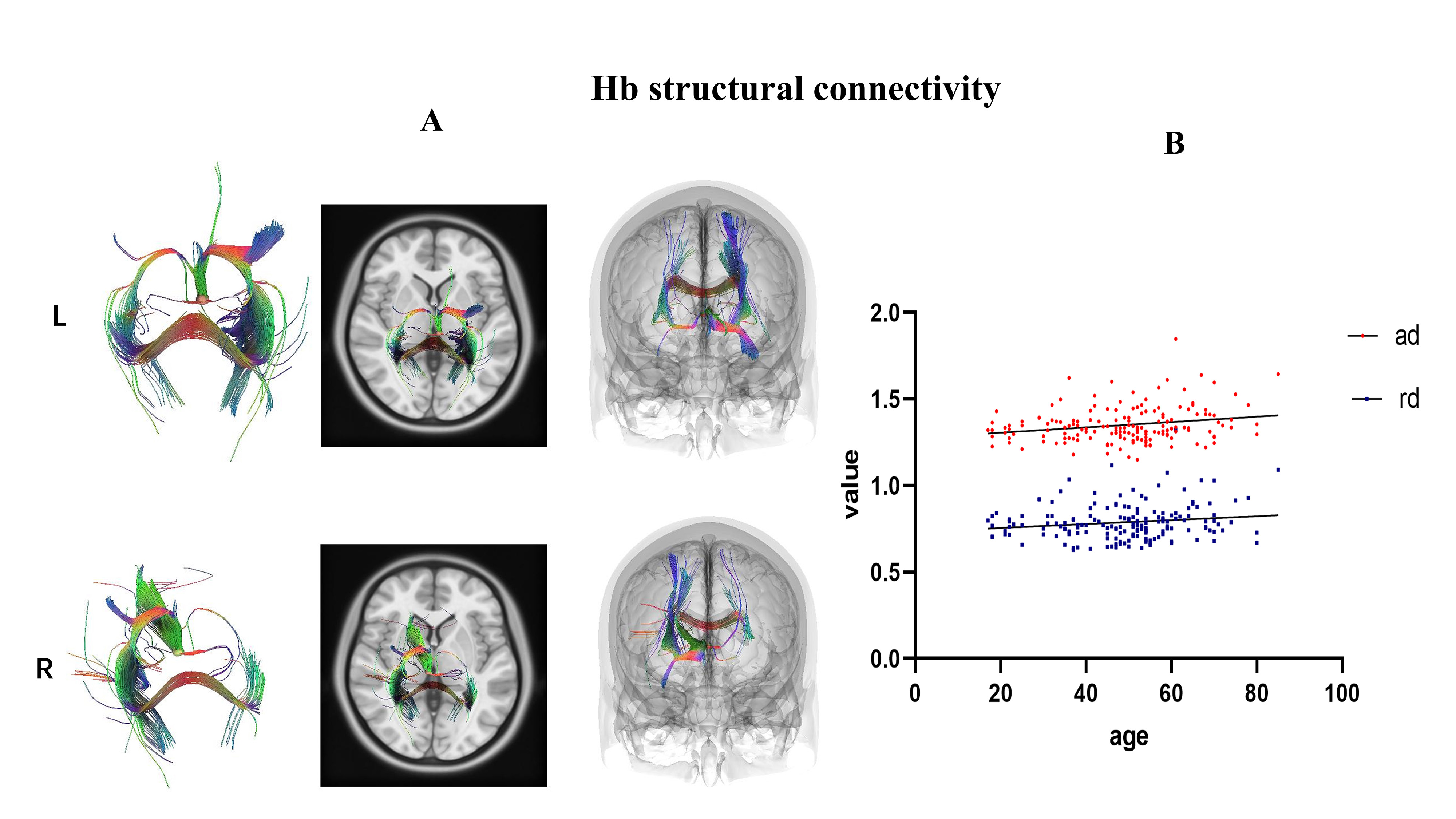
Due to the insufficient resolution and signal-to-noise ratio, low field strength magnetic resonance imaging has certain limitations in distinguishing ultrafine brain structure.(Strotmann et al., 2014) Thus, 5T ultrahigh-resolution MRI was used to analysis the structural and functional. High-resolution 3D T1-weighted fast spoiled gradient echo (T1-gre-fsp-iso) anatomic sequence(0.4 mm*0.6 mm*0.8mm)made an outstanding performance in Hb structure imaging. In addition, to verify that our results were reliable, we also validated them using the standard MNI stereospecific coordinates (-2.8, -24.2, 2.3) and (4.8, -24.1, 2.2) recommended by previous studies. We parcellated lateral and medial portions of the Hb and calculated Hb structural and functional connectivity on the basis of these parcellations. Firstly, we have identified that the lateral and medial portions of the Hb are structurally connected to the insula and ventral prefrontal cortices; Then, we examined whole brain FC pattern with the habenula. The lateral and medial portions of the Hb are functionally anticorrelated to the visual and salience networks. This pattern identified positive connectivity with salience, limbic and part of somatomotor networks, and negative connectivity with bilateral visual cortices; Also, in the association analysis with individual sleep amounts, we found that average sleep amount rather than last night’s sleep time predicted the anticorrelation between the visual and salience networks. Further lifespan trajectory analysis revealed that these anticorrelations are age-dependent.Conclusion
In conclusion, advanced imaging of Hb may provide significant benefits to our understanding of sleep disorder. Using functional and structural imaging of the Hb, we revealed the critical role of the Hb in the regulation of cognitive and emotional processes. Significant correlations were also found between average sleep and the visual and salience networks, which may provide an explanation for the loss in attention and memory caused by sleep disorder.Acknowledgements
We thank imaging technician of Department of Radiology, Zhongnan Hospital of Wuhan University Wuhan for MRI scanning and subject recruitmentReferences
References:
Aizawa, H., Cui, W., Tanaka, K., Okamoto, H., 2013. Hyperactivation of the habenula as a link between depression and sleep disturbance. Frontiers in Human Neuroscience 7, 826. Boulos, L.J., Darcq, E., Kieffer, B.L., 2017. Translating the Habenula-From Rodents to Humans. BIOLOGICAL PSYCHIATRY 81, 296-305. Killgore, W.D.S., 2013. Self-Reported Sleep Correlates with Prefrontal-Amygdala Functional Connectivity and Emotional Functioning. SLEEP 36, 1597-1608. Li, C., Fronczek Poncelet, J., Lange, D., Hennecke, E., Kroll, T., Matusch, A., Aeschbach, D., Bauer, A., Elmenhorst, E.M., Elmenhorst, D., 2020. Impact of acute sleep deprivation on dynamic functional connectivity states. HUMAN BRAIN MAPPING 41, 994-1005. Namboodiri, V.M., Rodriguez-Romaguera, J., Stuber, G.D., 2016. The habenula. CURRENT BIOLOGY 26, R873-R877. Strotmann, B., Heidemann, R.M., Anwander, A., Weiss, M., Trampel, R., Villringer, A., Turner, R., 2014. High-resolution MRI and diffusion-weighted imaging of the human habenula at 7 tesla. JOURNAL OF MAGNETIC RESONANCE IMAGING 39, 1018-1026. Verweij, I.M., Romeijn, N., Smit, D.J., Piantoni, G., Van Someren, E.J., van der Werf, Y.D., 2014. Sleep deprivation leads to a loss of functional connectivity in frontal brain regions. BMC NEUROSCIENCE 15, 88.
Figures