2041
Cardiorespiratory fitness is associated with cerebral myelin content in aging1Laboratory of Clinical Investigation, National Institute on Aging, Baltimore, MD, United States
Synopsis
Keywords: White Matter, Relaxometry, Myelin
Motivation: While higher cardiorespiratory fitness (CRF) is recognized as vital for brain health, its specific connection with white matter integrity, especially cerebral myelination, remains unclear.
Goal(s): This study aims to investigate the association between CRF and myelin in cognitively unimpaired adults spanning a wide age range.
Approach: We employed our advanced multicomponent MR relaxometry method to measure myelin water fraction, a direct proxy of myelin content, while CRF was assessed using the maximum rate of oxygen consumption, peak VO2.
Results: Our results indicate that higher peak VO2 is associated with greater myelin content across several white matter structures, particularly among older adults.
Impact: This work lays the foundation for future investigations to further elucidate the mechanisms underlying the relationship between cardiorespiratory fitness and cerebral myelination, as well as its potential as an interventional target in addressing age-related neurodegeneration, including in Alzheimer’s disease.
Introduction
Cardiorespiratory fitness (CRF) is a modifiable lifestyle factor that reflects the capacity of an individual’s circulatory and respiratory systems to deliver oxygen to skeletal muscles during sustained moderate to vigorous physical activity1. Growing evidence suggests that higher CRF is associated with preserved and enhanced neural structure and function2, improved cognitive performance3, and delayed neurodegenerative disease progression, including Alzheimer’s disease4. However, previous literature on the relationship between CRF and white matter integrity, particularly cerebral myelination, is limited. In this study, we evaluated the relationship between peak oxygen consumption (VO2), the gold standard measure of CRF, and myelin water fraction (MWF), a direct and specific MRI metric of myelin integrity. The overarching goal of this study is to develop further insights into how CRF promotes brain health and mitigates white matter degeneration in normative aging.Methods
Data AcquisitionEach participant underwent our BMC-mcDESPOT protocol for whole brain MWF mapping. The acquisition details of this protocol can be found in our previous studies5-6. Cardiorespiratory fitness was determined from a graded maximal treadmill test. Oxygen consumption was calculated every 30 seconds, and the highest value was termed peak VO2, expressed in milliliters per kilogram body weight per minute. The complete fitness protocol can be found in previous Baltimore Longitudinal Study of Aging (BLSA) literature7.
Data Processing and Statistical Analysis
For each participant, a whole-brain MWF map was generated using the BMC-mcDESPOT analysis from the SPGR, bSSFP, and DAM images8,9. The averaged SPGR image over FAs was nonlinearly registered to the MNI space using FSL software10. The derived deformation matrix was then applied to the corresponding MWF map. Fourteen white matter (WM) regions of interest (ROIs) were defined from the MNI structural atlas. For each ROI, the effect of peak VO2 on MWF was investigated using a multiple linear regression model. The dependent variable was the mean value of MWF in each ROI. Age, peak VO2, sex, systolic blood pressure (SBP), and a continuous age*VO2 interaction term were included as independent variables. All continuous variables were z-scored.
Results & Discussion
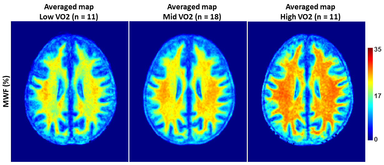
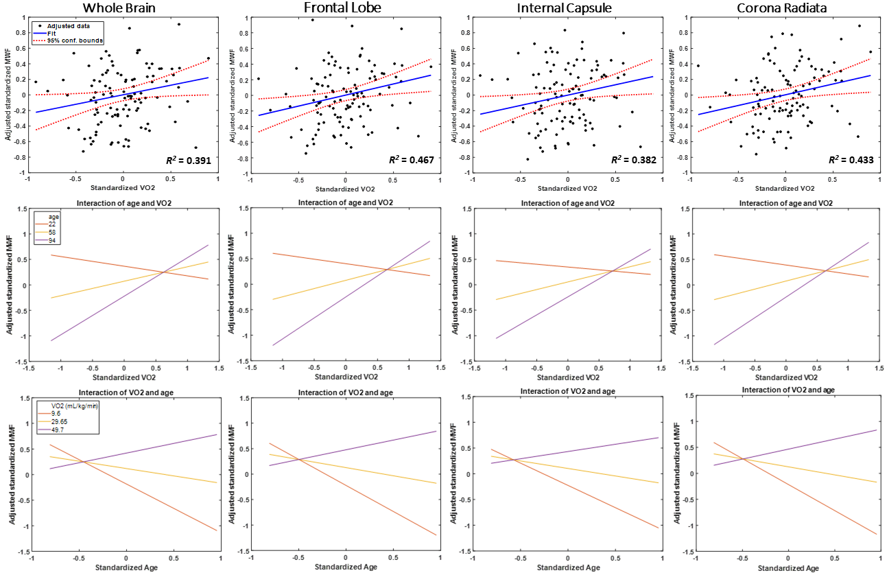
After excluding subjects with cognitive impairments, severe motion artifacts, or missing VO2 data, the final study cohort consisted of 110 cognitively unimpaired participants, ranging in age from 22 to 94 years (55.1 ± 20.4 years). Sixty-five (59%) were men and fifty-five (41%) were women. Mean ± standard deviations of SBP and peak VO2 were 116.1 ± 14.0 mmHg and 28.2 ± 8.10 mL/kg/min, respectively.We found positive correlations between peak VO2 and MWF (Figs. 1, 2), indicating that higher peak VO2 levels are associated with greater cerebral myelin content. The associations between peak VO2 and MWF were positive and statistically significant (p < 0.05) or close-to-significant (p < 0.1) in most white matter brain regions investigated, even after adjustment for covariates. The correlations were strongest in several critical white matter regions, particularly the frontal lobes (ꞵ = 0.28, p = 0.11), internal capsule (ꞵ = 0.27, p = 0.03), and corona radiata (ꞵ = 0.28, p = 0.02). These results suggest that higher CRF may provide the most substantial benefits to myelination in these brain regions compared to other cerebral structures. Indeed, previous structural and functional MRI studies have consistently shown positive relationships between CRF and neural integrity of frontal brain regions2, while the internal capsule and corona radiata have been reported as particularly sensitive to cerebral blood flow, which may be modulated and enhanced through aerobic fitness11-15.
Additionally, the interaction between age and peak VO2 was strong and significant in almost all white matter brain regions investigated, including the frontal lobes (ꞵ = 0.57, p = 0.001), internal capsule (ꞵ = 0.46, p = 0.01), and corona radiata (ꞵ = 0.56, p = 0.002) (Fig. 2). The interaction between age and peak VO2 exhibited the steepest positive slope in the old age group, suggesting that the effect of peak VO2 on MWF increases with age. Further, the interaction between age and peak VO2 exhibited a positive slope in the high VO2 group and negative slopes in the middle and low VO2 groups, indicating that high VO2 levels are associated with greater myelination.
Conclusion
In this exploratory study examining the association between cardiorespiratory fitness and myelin integrity, we show that higher peak VO2 is associated with greater myelination in a cohort of cognitively healthy adults. Our study provides new insights on the potential protective role of CRF in preserving white matter integrity and myelination in normative aging and offers a potential therapeutic target against concomitant cognitive and physical declines associated with white matter demyelination16,17.Acknowledgements
This work was supported by the Intramural Research Program of the National Institute on Aging of the National Institutes of Health.References
1. Hayes SM, Salat DH, Forman DE, Sperling RA, Verfaellie M. Cardiorespiratory fitness is associated with white matter integrity in aging. Annals of Clinical and Translational Neurology. 2015;2(6):688-698.
2. Hayes SM, Hayes JP, Cadden M, Verfaellie M. A review of cardiorespiratory fitness-related neuroplasticity in the aging brain. Front Aging Neurosci. 2013;5:31.
3. Smith PJ, Blumenthal JA, Hoffman BM, et al. Aerobic exercise and neurocognitive performance: a meta-analytic review of randomized controlled trials. Psychosom Med. 2010;72:239-252.
4. Perea RD, Vidoni ED, Morris JK, Graves RS, Burnes JM, Honea RA. Cardiorespiratory fitness and white matter integrity in Alzheimer’s disease. Brain Imaging and Behavior. 2016;10:660-668.
5. Bouhrara M, Rejimon AC, Cortina LE, Khattar N, Bergeron CM, Ferrucci L, et al. Adult brain aging investigated using BMC-mcDESPOT-based myelin water fraction imaging. Neurobiol Aging. 2020;85:131-9.
6. Bouhrara M, Spencer RG. Rapid simultaneous high-resolution mapping of myelin water fraction and relaxation times in human brain using BMC-mcDESPOT. Neuroimage. 2017;147:800-11.
7. Talbot LA, Morell CH, Metter J, Fleg JL. Comparison of cardiorespiratory fitness versus leisure-time physical activity as predictors of coronary events in men aged ≤ 65 years and> 65 years. Am J Cardiol. 2002;89-1187-1192.
8. Bouhrara M, Spencer RG. Improved determination of the myelin water fraction in human brain using magnetic resonance imaging through Bayesian analysis of mcDESPOT. Neuroimage. 2016;127:456-71.
9. Deoni SC, Rutt BK, Peters TM. Rapid combined T1 and T2 mapping using gradient recalled acquisition in the steady state. Magn Reson Med. 2003;49(3):515-26.
10. Jenkinson M, Beckmann CF, Behrens TE, Woolrich MW, Smith SM. FSL. Neuroimage. 2012;62(2):782-90.
11. Laporte JP, Faulkner ME, Gong Z, Palchamy E, Akhonda MABS, Bouhrara M. Investigation of the association between central arterial stiffness and aggregate g-ratio in cognitively unimpaired adults. Front Neurol. 2023;14:1170457.
12. Laporte JP, Faulkner ME, Gong Z, Akhonda MABS, Ferrucci L, Egan JM, Bouhrara M. Hypertensive adults exhibit lower myelin content: a multicomponent relaxometry and diffusion magnetic resonance imaging study. Hypertension. 2023;80:1728-1738.
13. Kiely M, Triebswetter C, Gong Z, Laporte JP, Faulkner ME, Akhonda MABS, Alsameen MH, Spencer RG, Bouhrara M. Evidence of an association between cerebral blood flow and microstructural integrity in normative aging using a holistic MRI approach. J Magn Reason Imaging. 2023;58-284-293.
14. Bouhrara M, Triebswetter C, Kiely M, Bilgel M, Dolui S, Erus G, Meirelles O, et al. Association of cerebral blood flow with longitudinal changes in cerebral microstructural integrity in the coronary artery risk development in young adults (CARDIA) study. JAMA Network Open. 2022;5(9):e2231189.
15. Bouhrara M, Alisch JSR, Khattar N, Kim RW, Rejimon AC, Cortina LE, Qian W, et al. Association of cerebral blood flow with myelin content in cognitively unimpaired adults. BMJ Neurology Open. 2020;2(1):e000053.
16. Gong Z, Bilgel M, Kiely M, Triebswetter C, Ferrucci L, Resnick SM, Spencer RG, Bouhrara M. Lower myelin content is associated with more rapid cognitive decline among cognitively unimpaired individuals. Alzheimers Dement. 2023;19(7):3098-3107.
17. Faulkner ME, Laporte JP, Gong Z, Akhonda MABS, Triebswetter C, Kiely M, Palchamy E, Spencer RG, Bouhrara M. Lower myelin content is associated with lower gait speed in cognitively unimpaired adults. J Gerontol A Biol Sci Med Sci. 2023;7(8):1339-1347.
Figures