2031
GABA in the anterior cingulate cortex mediates the association of white matter hyperintensities with executive function1Department of Radiology,Union Hospital,Tongji Medical College,Huazhong University of Science and Technology, Wuhan, China, 2Clinical & Technical Support, Philips Healthcare, Beijing 100600, China, Beijing, China
Synopsis
Keywords: White Matter, White Matter, Gamma-aminobutyric acid
Motivation: The alteration of cortical GABA levels in WMH patients and whether it mediates the association of WMH volume with executive function remain unclear
Goal(s): We investigated the cortical GABA levels, and whether cortical GABA mediates the association between WMH and executive function in the WMH people.
Approach: We used the independent samples test,general linear model,partial correlation analyses and mediation analysis.
Results: Patients with moderate to severe WMH showed lower GABA+/Cr in the anterior cingulate cortex (ACC) and worse executive function than mild WMH patients. And the GABA level in ACC mediates the association between white matter hyperintensities and executive function in WMH patients.
Impact: The GABA+/Cr level in the the anterior cingulate cortex had a critical role. And it mediates the association between white matter hyperintensities and executive function in the WMH patients.
Introduction
White matter hyperintensities (WMH), one of the most important radiological manifestations of cerebral small vessel disease 1,2, was related to cognitive function, especially executive function 3. Gamma-aminobutyric acid (GABA), an amino acid neurotransmitter in the central nervous system, plays a role in inhibiting neuronal excitability and reducing energy consumption 4. Numerous studies not only demonstrate a close interrelation between gray and white matter but also indicate that cortical alterations serve as mediating factors for the cognitive decline observed in patients with WMH. The purpose of this study was to investigate the cortical GABA level, including the anterior cingulate cortex (ACC) and posterior cingulate cortex (PCC), and whether cortical GABA mediates the association between WMH and executive function in WMH people.Methods
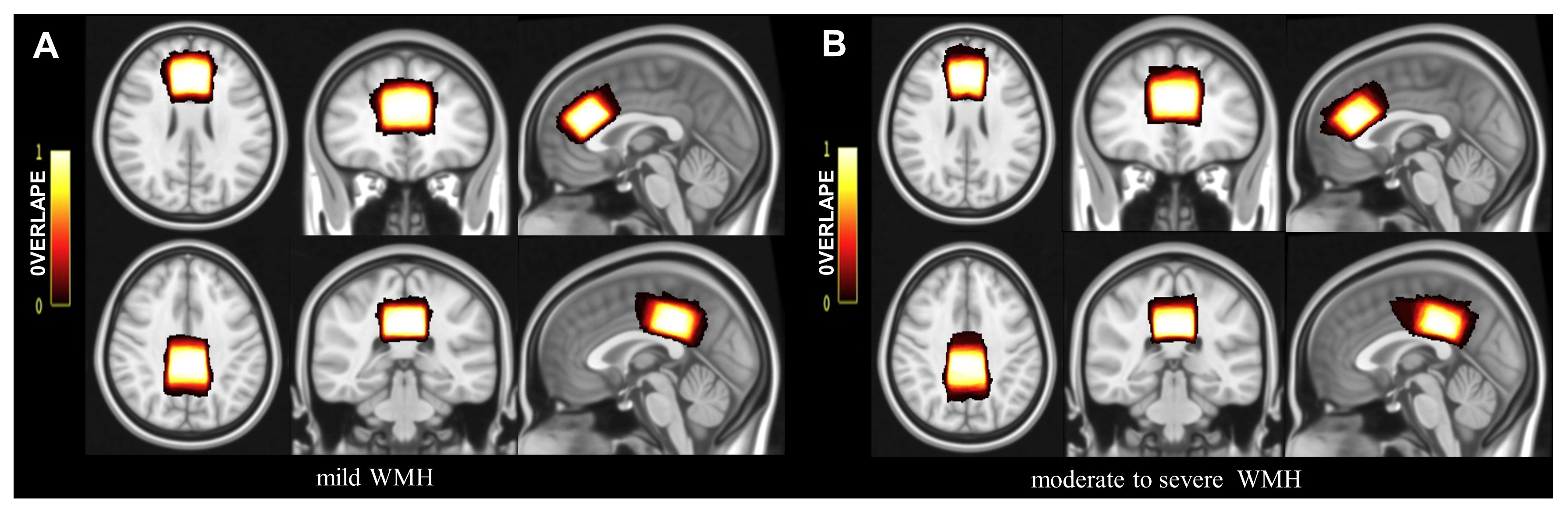
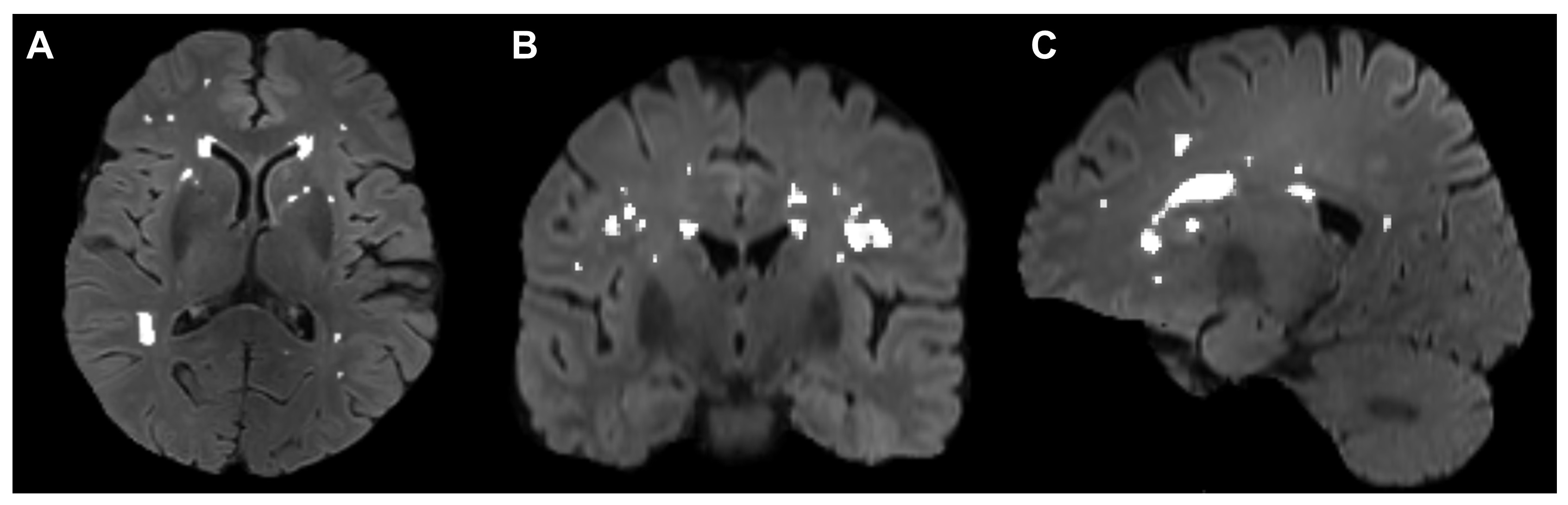
In this study, a total of 87 patients with WMH, including 46 mild WMH patients and 41 patients with moderate to severe WMH, were finally included. Compare the levels of gamma-aminobutyric acid plus relative to creatine (GABA+/Cr) and glutamate-glutamine relative to creatine (Glx/Cr) in the ACC and PCC (Figure 1A,1B) measured using the Meshcher-Garwood point resolved spectroscopy (MEGA-PRESS) sequence, WMH volume (Figure 2) and executive function between groups. Partial correlation analyses were performed to detect correlations of metabolites, WMH volume, and executive function, with age, gender, and education in years as covariates. Mediation analysis was performed to explore whether the GABA levels in the ACC and PCC mediate the association between WMH volume and executive function in the WMH patients.Results
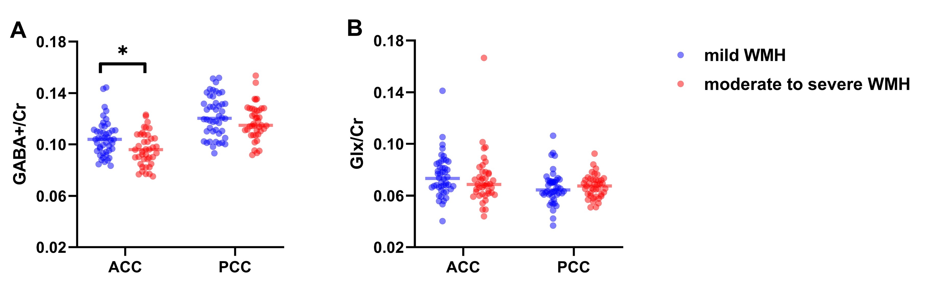
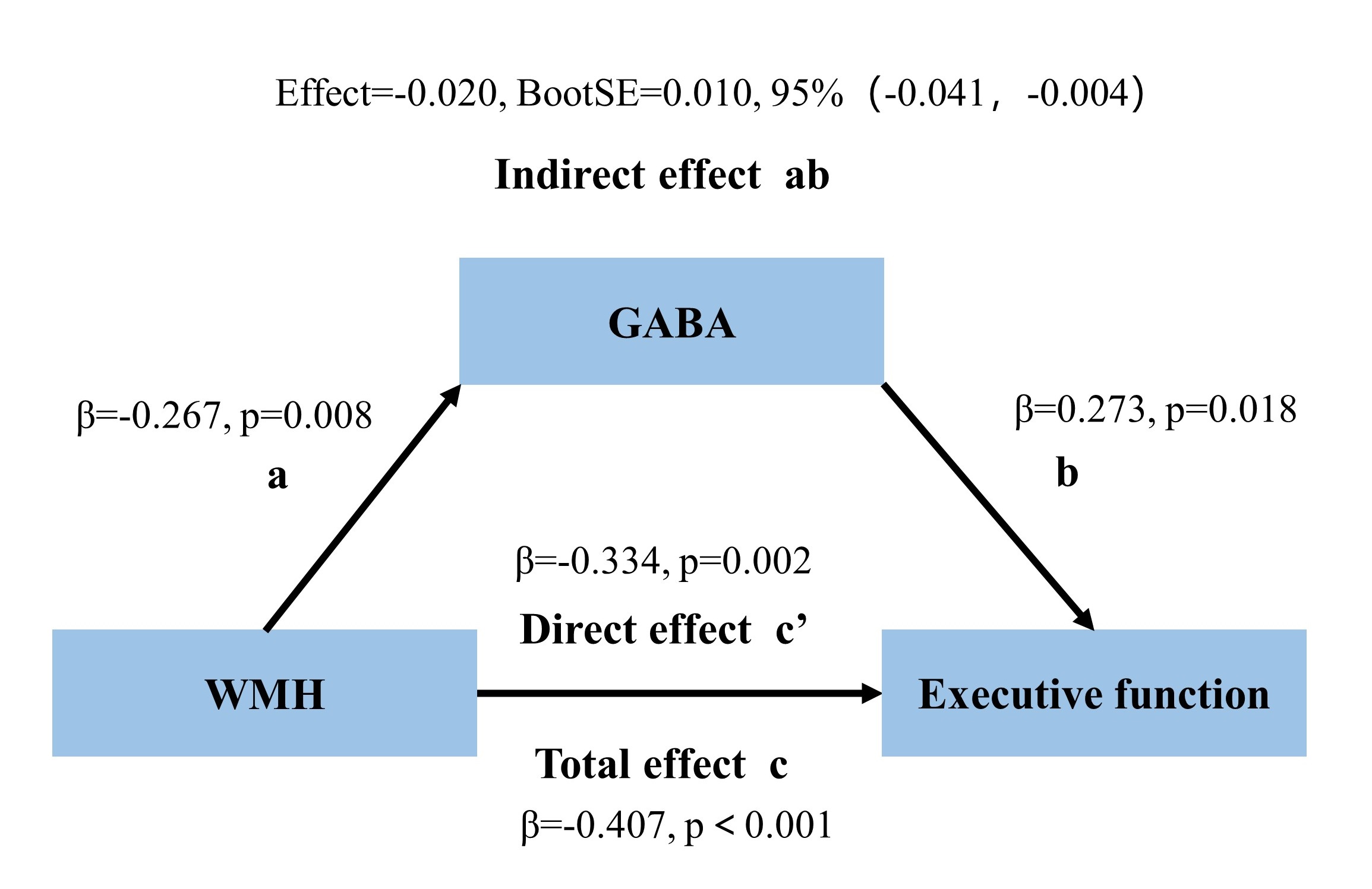
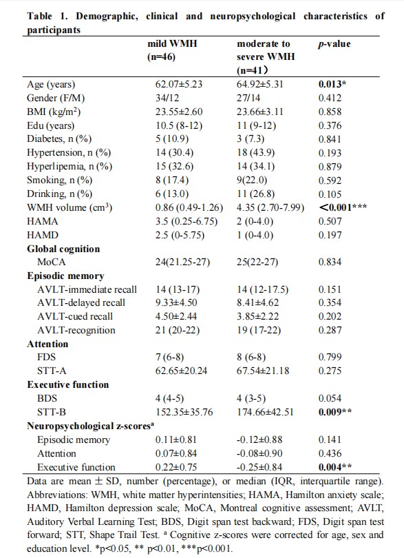
The moderate to severe WMH patients were older than mild WMH patients (Table 1). Patients with moderate to severe WMH showed lower GABA+/Cr in the ACC (p=0.034, Figure 3) and worse executive function (p=0.004, Table 1) than mild WMH patients, with age, gender, and education in years as covariates. There were no significant differences in memory and attention between groups. In all WMH patients, correlations among WMH burden, GABA levels in the ACC, and executive function were also been found (all p values<0.05). And GABA levels in the ACC mediated the association between WMH and executive function in patients with WMH (Figure 4).Discussion
There was a significantly decreased GABA+/Cr level in the ACC in the moderate to severe WMH patients. Previous studies demonstrated decreased levels of GABA or Glx in the ACC or PCC within individuals with disorders, such as multiple sclerosis, schizophrenia, and mild cognitive impairment 5-7. Our study found that moderate to severe WMH patients had terrible executive function. Therefore, the decreased GABA level in the ACC observed in this study showed that moderate to severe WMH was also one of the CNS disorders. Moreover, in physiological mechanisms 4, the decrease in GABA+/Cr observed in our study was probably because of neuronal loss, decreased GABA/Glu synthesis, or altered astrocyte cycle of GABA, Glu, and Gln. Our study demonstrated that the moderate to severe WMH patients had worse executive function compared to the mild WMH group, which was similar to the previous study 8. We also supported that a negative correlation between WMH volume and executive function in all WMH patients. White matter comprises structural connections that link various regions of gray matter throughout the brain. This correlation may be attributed to that as the volume of the WMH increases, it will lead to a greater degree of damage to the pathways responsible for information transmission. We revealed a robust inverse relationship between GABA levels in ACC and WMH volume, as well as a positive correlation with executive function in all WMH patients. Moreover, we first demonstrated that the GABA levels in the ACC mediated the relationship between WMH and executive function, although many studies showed disrupted brain networks and brain atrophy mediated the relationship between WMH and cognitive impairment8,9. This mediating result may reflect that CSVD leads to disruption of the integrity of white matter fiber bundles, which showed up as WMH in FLAIR imaging images. Due to secondary degeneration, the damaged fiber bundles also affected the axonal cytoskeleton 10 and cortex, where the cortex was characterized by high concentrations of neuronal cell bodies, such as GABA neurons, astrocytes, etc. Subsequently, alterations in GABA levels driven changes in executive function.Conclusion
The GABA+/Cr levels in the ACC had a critical role in the WMH patients. Moreover, the mediational analysis further indicated that the GABA levels in the ACC could serve as a protective factor or potential target for preventing the occurrence and progression of executive function decline in WMH people.Acknowledgements
We thank all individuals who participated in this study as well as the investigators who contributed to data collection.References
1. Gorelick PB, Scuteri A, Black SE, et al. Vascular contributions to cognitive impairment and dementia: a statement for healthcare professionals from the american heart association/american stroke association. Stroke. 2011;42(9):2672-2713.
2. Cipollini V, Troili F, Giubilei F. Emerging Biomarkers in Vascular Cognitive Impairment and Dementia: From Pathophysiological Pathways to Clinical Application. Int J Mol Sci. 2019;20(11).
3. Camerino I, Sierpowska J, Reid A, et al. White matter hyperintensities at critical crossroads for executive function and verbal abilities in small vessel disease. Hum Brain Mapp. 2021;42(4):993-1002.
4. Sood A, Preeti K, Fernandes V, Khatri DK, Singh SB. Glia: A major player in glutamate-GABA dysregulation-mediated neurodegeneration. J Neurosci Res. 2021;99(12):3148-3189.
5. Oeltzschner G, Wijtenburg SA, Mikkelsen M, et al. Neurometabolites and associations with cognitive deficits in mild cognitive impairment: a magnetic resonance spectroscopy study at 7 Tesla. Neurobiol Aging. 2019;73:211-218.
6. Reid MA. Glutamate and Gamma-Aminobutyric Acid Abnormalities in Antipsychotic-Naive Patients With Schizophrenia: Evidence From Empirical and Meta-analytic Studies Using Magnetic Resonance Spectroscopy. Biol Psychiatry. 2021;89(3):e1-e3.
7. Kantorova E, Hnilicova P, Bogner W, et al. Neurocognitive performance in relapsing-remitting multiple sclerosis patients is associated with metabolic abnormalities of the thalamus but not the hippocampus- GABA-edited 1H MRS study. Neurol Res. 2022;44(1):57-64.
8. Zhu W, Huang H, Yang S, et al. Cortical and Subcortical Grey Matter Abnormalities in White Matter Hyperintensities and Subsequent Cognitive Impairment. Neurosci Bull. 2021;37(6):789-803.
9. Wang Y, Yang Y, Wang T, Nie S, Yin H, Liu J. Correlation between White Matter Hyperintensities Related Gray Matter Volume and Cognition in Cerebral Small Vessel Disease. J Stroke Cerebrovasc Dis. 2020;29(12):105275.
10. Jones DK, Lythgoe D, Horsfield MA, Simmons A, Williams SC, Markus HS. Characterization of white matter damage in ischemic leukoaraiosis with diffusion tensor MRI. Stroke. 1999;30(2):393-397.
Figures