2029
In Vivo Reproducibility of tNOE of Human Brain Using Tx/Rx Coil at 3T1Center for Metabolic Imaging in Precision Medicine (CAMIPM), Department of Radiology, University of Pennsylvania, Philadelphia, PA, United States
Synopsis
Keywords: White Matter, Magnetization transfer, NOE
Motivation: Nuclear Overhauser Effect (NOE) is based on dipolar cross relaxation mechanism that enables the indirect detection of aliphatic protons via the water proton signal. This work focuses on determining the reproducibility of the transient-Nuclear-Overhauser-Effect (tNOE) of the healthy human brain at 3 Tesla.
Goal(s): To establish reproducibility of tNOE in the brain regions of healthy subjects at 3T.
Approach: We scanned three healthy subjects multiple times to determine inter-day reproducibility of tNOE on 3T.
Results: The inter-subject coefficient of variations (CoV) of tNOE from GM and WM were 5.12% and 3.97%, respectively. The intra-subject tNOE CoV range for GM and WM was 0.24%-4.0%.
Impact: This work will facilitate the use of tNOE at 3T to investigate macromolecular (lipid and proteins) derangements in different diseases with significantly reduced scan time and improved specificity compared to steady state NOE.
Introduction
Lipids are a significant component of the human brain, constituting around 50% of its dry weight, with most of these lipids found in the form of myelin1. These lipids are key components of cellular membranes including synapses, myelin sheath and can regulate a range of biological processes. Consequently, imbalances and disturbances in lipid levels have been associated with numerous neurological disorders, including Alzheimer's disease, Parkinson's disease, and many others2. A reliable, resilient, and replicable imaging biomarker is necessary to monitor changes in lipid metabolism during the development of neurological disorders and in instances of central nervous system injuries. Steady-state nuclear Overhauser (ssNOE) is increasingly becoming a popular approach to measure in vivo lipid3, which relies on the NOE emerging from dipolar coupling between aliphatic moieties (methyl and methylene protons) of macromolecules (proteins and lipids) and bulk water4 . This NOE contribution manifests as a broad peak spanning 2 to 5 ppm up field of water, thought to occur via relayed exchange mechanisms. Recently, we demonstrated an alternative approach to measuring NOE contribution from macromolecule in brain using transient NOE (tNOE) at 7T, with underlying mechanism being the cross-relaxation between aliphatic moieties and bulk water5. In this study, we investigated the reproducibility of tNOE at 3T, showing superb white matter (WM)–gray matter (GM) contrast.Methods
All human studies were conducted under an approved University of Pennsylvania Institutional Review Board protocol. Three healthy volunteers (3 males) participated in the study. All imaging was performed on a 3T whole body scanner (MAGNETOM Prisma, Siemens Healthcare, Erlangen, Germany) with a 32-channel transmit/receive proton birdcage head coil (MR Tools, Germany). Two intra-day scans were performed on each subject for assessment of reproducibility. To accurately obtain consistent slice locations between subjects, we used an in-house developed co-registration program, ImScribe6. The tNOE imaging involved a magnetization-preparation module consisting of a frequency-selective inversion pulse (hyperbolic secant adiabatic pulse with TBWP= 12.8), followed by a single-shot tfl readout with centric phase-encoding order and tfl-TR = 3.5 ms, tfl-TE = 1.47 ms, receiver bandwidth (rBW) = 550 Hz/pixel, with SHOT-TR of 6s. Imaging parameters for tNOE experiment in human study were as follows: resolution = 1×1×5 mm3, matrix size = 128 × 128, thickness = 5 mm, FOV phase = 100%, averages = 4, rBW = 550 Hz/pixel, TR = 6 s, tfl TE = 1.62 ms, tfl TR = 3.5 ms, and tfl flip angle = 4°. Data processing was performed using in-house MATLAB (version R2019a) scripts where tNOE was calculated as: $$$ tNOE(\%) = 100* \frac{S_{0}- S_{-3.5} }{S_{0} } $$$, where where S0 and S(-3.5) are the signal intensities without inversion and with inversion pulse applied at -3.5ppm ppm, respectively. Brain subregions were obtained via automated segmentation of an acquired MPRAGE image, with the following steps: 1) bias field correction using ANTS7 ;2) brain masking using HD-BET8; 3) segmentation for subcortical gray matter regions using FSL FIRST9. The intra and inter-subject coefficient of variation (CoV) for each volunteer were calculated as: $$$ CoV (\%) = \frac{\sigma}{\mu}$$$ , where σ is the standard deviation of the mean contrast from each scan and µ is the mean of the pixels across the slab.Results
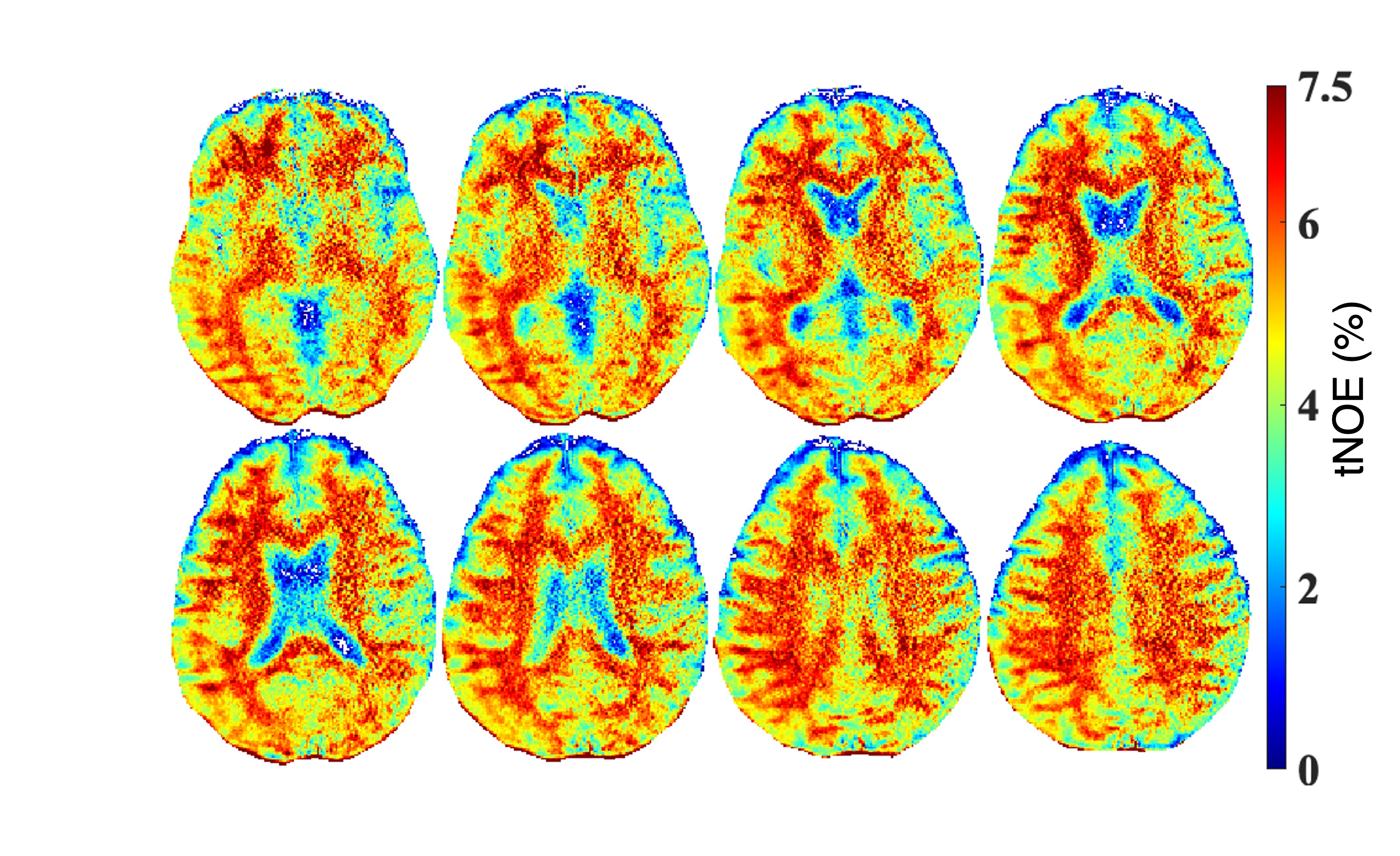
We found dominant contribution of the tNOE signal comes from the WM regions (Figure 1). We also found that the mean tNOE contributions from GM and WM values were 4.75% and 5.16%, respectively (Table 1). The inter-subject CoV of tNOE from GM and WM were 5.12% and 3.97%, respectively. The intra-subject tNOE CoV range for GM and WM were 0.24% - 4.0%.Discussion & Conclusion
Our research results indicate that the tNOE signal predominantly originates from WM regions, in line with earlier investigations3,5. Additionally, we have shown that the tNOE signal exhibits remarkable reproducibility, with CoVs of less than 5% in both GM and WM regions. In addition, compared to the ssNOE, the tNOE comes with substantially attenuated MT effects thereby improved dynamic range for the lipid component, significantly reduced scan time and hence enhanced patient tolerability. These findings suggest that this technique holds great promise for detecting lipid degeneration in various disease types, such as multiple sclerosis (MS), Alzheimer's disease, and potentially others. This could have significant implications for the diagnosis and monitoring of these conditions, offering valuable insights into the progression and characterization of lipid-related changes in these diseases.Acknowledgements
This work was supported by the National Institute of Biomedical Imaging and Bioengineering of the National Institutes of Health under award Number P41EB029460 and by the National Institute of Aging of the National Institutes of Health under award numbers R01AG071725 and R01AG063869.References
1. Hamilton JA, Hillard CJ, Spector AA, Watkins PA. Brain uptake and utilization of fatty acids, lipids and lipoproteins: application to neurological disorders. J Mol Neurosci. 2007; 33: 2-11.
2. Williams TI, Lynn BC, Markesbery WR, Lovell MA. Increased levels of 4-hydroxynonenal and acrolein, neurotoxic markers of lipid peroxidation, in the brain in mild cognitive impairment and early Alzheimer's disease. Neurobiol Aging. 2006; 27: 1094-1099.
3. Jones CK, Huang A, Xu J, et al. Nuclear Overhauser enhancement (NOE) imaging in the human brain at 7T. Neuroimage. 2013; 77: 114-124.
4. Overhauser, Albert W. (1953). "Polarization of Nuclei in Metals". Physical Review. 92 (2): 411–5.
5. Kumar, D, Benyard, B, Soni, ND, et al. Feasibility of transient nuclear Overhauser effect imaging in brain at 7 T. Magn Reson Med. 2023; 89: 1357-1367.
6. Wolf, D. H., Satterthwaite, T. D., Loughead, et al. Amygdala abnormalities in first-degree relatives of individuals with schizophrenia unmasked by benzodiazepine challenge. Psychopharmacology. 2011, 218(3), 503–512.
7. Avants BB, Yushkevich P, Pluta J, et al. The optimal template effect in hippocampus studies of diseased populations. Neuroimage. 2010 Feb 1;49(3):2457-66.
8. Isensee F, Schell M, Tursunova I, et al. Automated brain extraction of multi-sequence MRI using artificial neural networks.1901.11341, 2019.
9. Patenaude, B., Smith, S.M., Kennedy, D., and Jenkinson M. A Bayesian Model of Shape and Appearance for Subcortical Brain NeuroImage, 56(3):907-922, 2011.
Figures