2024
Optimizing pseudo-continuous ASL at 7 Tesla using dynamic low order-shim update1Siemens Medical Solutions USA Inc., Malvern, PA, United States, 2A.A. Martinos Center of Biomedical Imaging, Department of Radiology, Massachusetts General Hospital, Charlestown, MA, United States, 3Harvard Medical School, Boston, MA, United States, 4Siemens S.A., Madrid, Spain, 5Siemens Healthcare GmbH, Erlangen, Germany, 6Department of Neurology, University of Pennsylvania, Philadelphia, PA, United States, 7Department of Radiology, University of Pennsylvania, Philadelphia, PA, United States
Synopsis
Keywords: Perfusion, Arterial spin labelling, shims
Motivation: The labeling efficiency of pseudo-continuous ASL (pCASL) is reduced in the presence of large B0 off-resonance, which makes pCASL challenging at ultra-high fields due to susceptibility-induced off-resonance.
Goal(s): We aim to significantly reduce the labeling efficiency dependence on B0 off-resonance of pCASL to achieve highest allowed labeling efficiency at a given labeling flip angle.
Approach: 0th- (frequency) and 1st-order (x,y,z) shim components were used dynamically in the duration of labeling to mitigate B0 off-resonance without affecting imaging readout.
Results: Improved labeling efficiency was achieved, which enabled higher-resolution perfusion imaging using pCASL. Selective labeling of blood was also demonstrated possible using our approach.
Impact: Our approach provides a simple method to substantially remove the labeling-efficiency dependence on B0 off-resonance for pCASL at 7T. This enables high-quality ASL-based perfusion imaging at ultra-high fields for study of brain function, physiology, and pathology.
Purpose
Pseudo-continuous arterial spin labeling (pCASL) is a unique MR technique as its two major components – blood labeling and imaging readout – occur at distinct spatial locations1. Since both labeling and imaging degrade in the presence of B0-field inhomogeneity, pCASL presents significant challenges at 7-Tesla, where susceptibility-induced off-resonance effects are more prominent. Prioritizing B0 for imaging using the scanner system shim (usually up to 3rd order) often leaves the field at the labeling-plane suboptimal, resulting in compromised labeling efficiency and poor perfusion measurements. Prior studies have explored B0 off-resonance effects on pCASL and have provided methods to address them2-6, including at 7T7,8. These approaches offset the phase drift of blood water spins caused by off-resonance during labeling using either additional phase shift factors in the labeling RF pulses or an additional gradient moment. In this work, we introduce an alternate approach that aims to explicitly address B0 off-resonance by providing improved B0 shimming for labeling without affecting imaging.Methods
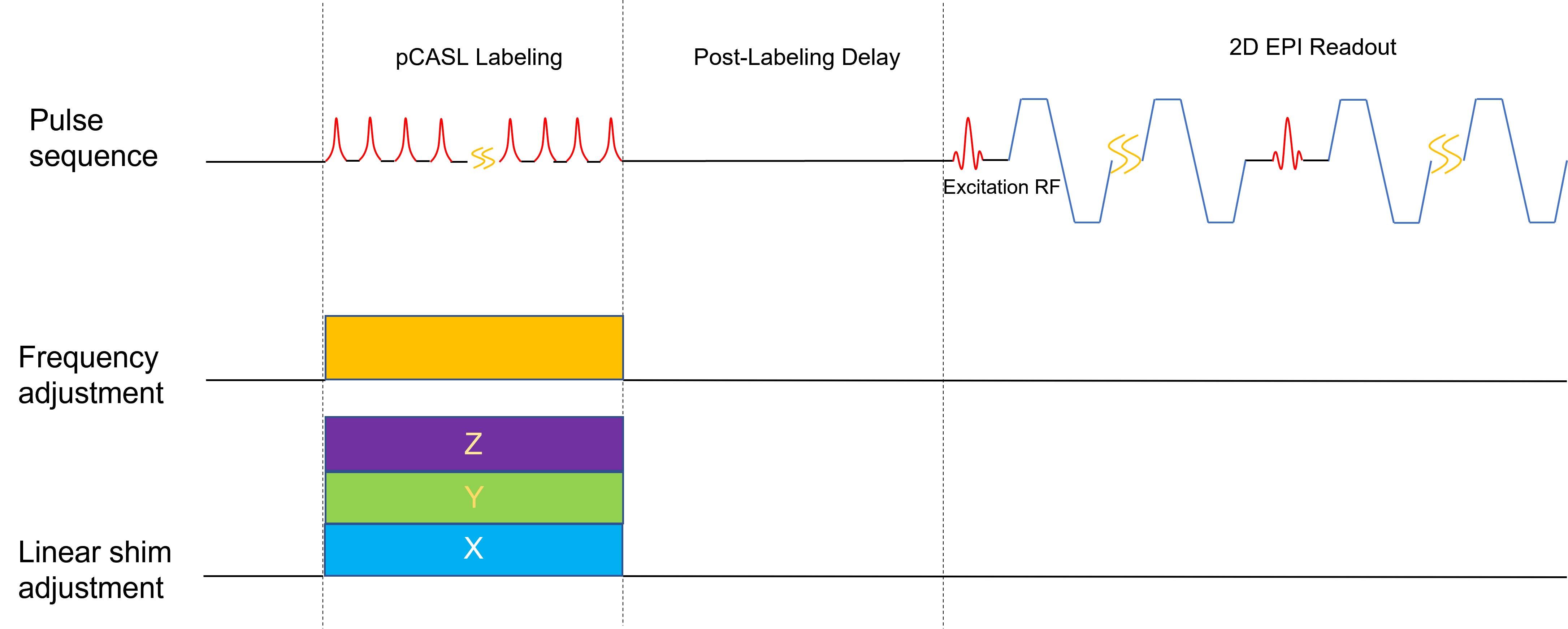
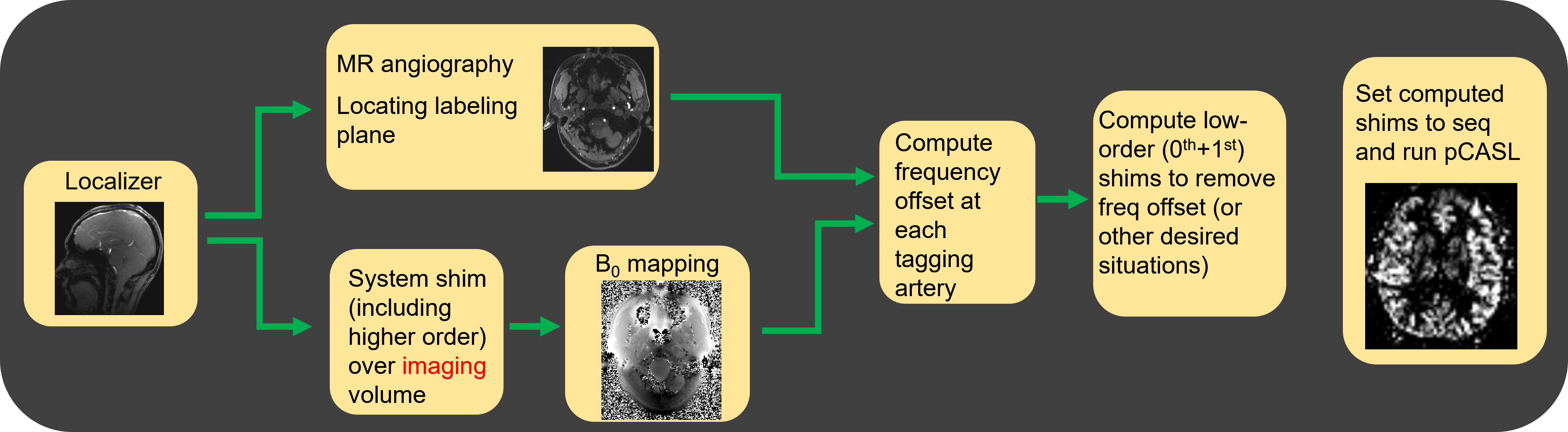
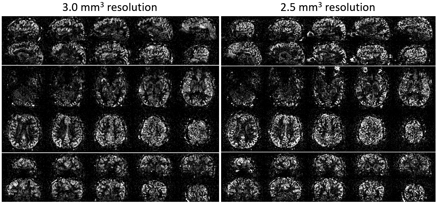
Theory: In a modern MR scanner, the low-order shim components, consisting of the frequency (0th order) and the 1st order linear terms (x,y,z) can be switched and settled in real-time (i.e., <<1 ms), suitable for shimming a specific volume of the subject in real-time during a scan. Specific for pCASL, they can be switched on during only the labeling module (Fig. 1) to shim the labeling plane.Experiments: Participants (n=3) were scanned in a total of 7 times on a 7T scanner (MAGNETOM Terra, Siemens Healthcare, Erlangen, Germany) using an 8-channel transmit, 32-channel receive coil (8Tx32Rx) (Nova Medical, MA, USA) as part of an IRB-approved study. Informed written consent was obtain from each participant. Only the bird-cage mode of the transmit system was used. Scanner system shim (up to 3rd order) was first used to optimize B0 of the imaging volume. B0 maps covering the labeling plane were then obtained and were used to determine B0 off-resonance at the arteries identified with MR angiography. First, the effects of dynamic frequency adjustment were tested by varying the system off-resonance from -400 Hz to 400 Hz at a 100 Hz interval. Next, linear terms (mainly x-shim) were added to achieve (1) optimal labeling efficiency over all arteries and (2) spatially selective labeling. Finally, higher-resolution (3.0 mm3, 2.5 mm3) images were acquired using the optimal shimming conditions from #1. The workflow using the shim is shown in Fig. 2.
ASL sequence: Switching of the low-order shim was implemented in a prototype pCASL sequence with 2D gradient-echo EPI readout9. Balanced pCASL was used with labeling duration 1.2 s, post-labeling delay (PLD)=1.7 s, RF duration=500 µs, gradient ramp time=60 µs, gap=380 µs, maximum/average gradient=8.0/1.0 mT/m, effective flip angle=13° (estimated based on B1+ maps). No background suppression was used. Readout uses 3× in-plane acceleration, matrix size=54×54, spatial resolution=3.5 mm3, 32 slices, TR/TE=4850/8.4 ms, bandwidth=2375 Hz/pixel, 12 label-control pairs, acquisition time=2.2 min. For 3.0 mm3-resolution scan, matrix=64×64, TR/TE=4920/9.4 ms, PLD 1.56 s, 32 pairs in 5.5 min; For 2.5 mm3-resolution scan, matrix=76×76, TR/TE=5050/11 ms, PLD 1.4 s, 31 pairs in 5.5 min.
Results
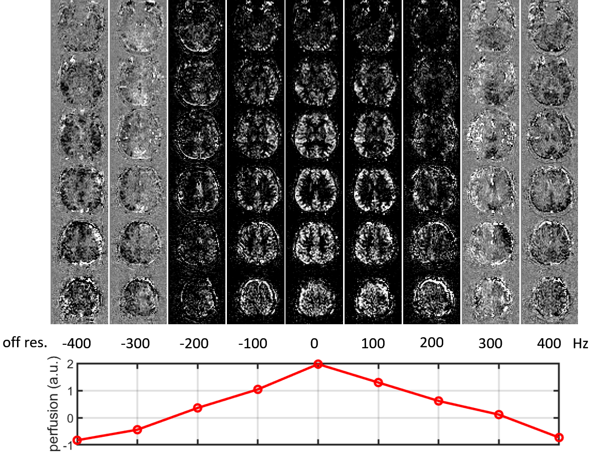
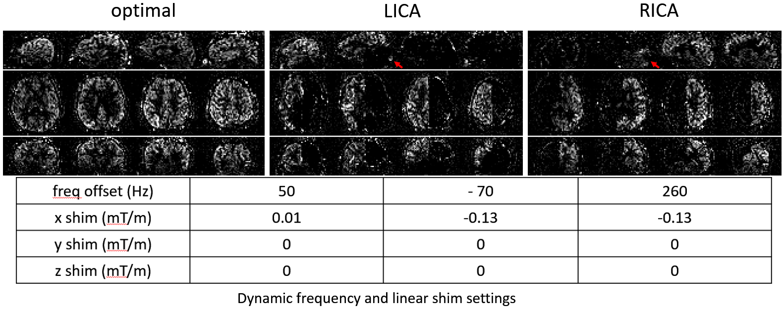
The effects of frequency adjustment are shown in Fig. 3. In this example, the optimal frequency offset likely falls between 0 and 100 Hz. The “null-labeling” condition is achieved near 300 Hz off-resonance. The effects of linear shims are shown in Fig. 4. In this example, the fields at the two internal carotid arteries (ICAs) were 64 and 37 Hz off-resonance; given they were 4.5 cm apart along the x-axis, a 0.01 mT/m x shim together with a 50 Hz off-resonance were necessary to bring the fields in both vessels near 0 Hz off-resonance ("optimal"). On the other hand, a 0.13 mT/m x shim would separate the fields by about 250 Hz, providing opportunities for partially-selective labeling ("LICA", "RICA"). Using the optimal shim condition, higher-resolution perfusion scans were acquired and shown in Fig. 5.Discussion
In this work, low-order scanner shim was used to optimize and control pCASL labeling efficiency at 7T. This approach does not rely on any custom hardware, although additional shimming components would provide greater field control10. Labeling efficiency may be further improved under SAR constrictions by using special transmit coils4 or advanced RF pulse design11,12. Compared to the phase-shift methods3,7,8, our approach does not require the modification of the labeling module of the pulse sequence.Conclusion
We demonstrated that the four fast switching, low-order system shim components can be used in pCASL to reduce off-resonance-induced labeling efficiency loss, which reduces pCASL dependence on the field and improves the reliability of pCASL at 7T.Acknowledgements
NIH K01AG070318References
1. Dai W, Garcia D, de Bazelaire C, Alsop DC. Continuous flow-driven inversion for arterial spin labeling using pulsed radio frequency and gradient fields. Magn Reson Med 2008;60:1488–14972.
2. Jahanian H, Noll DC, Hernandez-Garcia L. B0 field inhomogeneity considerations in pseudo-continuous arterial spin labeling (pCASL): effects on tagging efficiency and correction strategy. NMR Biomed 2011;24:1202–1209.3.
3. Shin DD, Liu TT, Wong EC, Shankaranarayanan A, Jung Y. Pseudo-continuous arterial spin labeling with optimized tagging efficiency: optimized PCASL (OptPCASL) Method. Magn Reson Med. 2012;68:1135-1144.4.
4. Stafford RB, Woo MK, Oh SH, et al. An Actively Decoupled Dual Transceiver Coil System for Continuous ASL at 7 T. Imaging Systems and Technology 2016;26:106-1155.
5. Zhao L, Vidorreta M, Soman S, Detre JA, Alsop DC. Improving the Robustness of Pseudo-Continuous Arterial Spin Labeling to Off-Resonance and Pulsatile Flow Velocity, Magn Reson Med 2017;78:1342-13516.
6. Berry ESK, Jezzard P, Okell TW. Off-resonance correction for pseudo-continuous arterial spin labeling using the optimized encoding scheme. NeuroImage 2019;199:304-312.7.
7. Luh WM, Talagala SL, Li TQ, Bandettini PA, Pseudo-Continuous Arterial Spin Labeling at 7 T for Human Brain: Estimation and Correction for Off-Resonance Effects Using a Prescan. Magn Reson Med 2013;69:402-4108.
8. Saib G, Koretsky AP, Talagala SL. Optimization of pseudo-continuous arterial spin labeling using off-resonance compensation strategies at 7T. Magn Reson Med 2022;87:1720-17309.
9. Vidorreta M, Wang Z, Rodriguez I, Pastor MA, Detre JA, Fernandez-Seara MA, Comparison of 2D and 3D single-shot ASL perfusion fMRI sequences. NeuroImage 2013;66:662-67110.
10. Craven-Brightman et al. Regulating labeling efficiency in arterial spin labeling using a multi-coil B0 shim array: Application to territory mapping. ISMRM 2021:046911.
11. Tong Y, Jezzard P, Okell TW, Clarke WT. Improving PCASL at ultra-high field using a VERSE-guided parallel transmission strategy. Magn Reson Med. 2020:84:777-78612.
12. Wang K, Ma SJ, Shao X, et al., Optimization of pseudo-continuous arterial spin labeling at 7T with parallel transmission B1 shimming. Magn Reson Med. 2022;87:249-262
Figures