2021
Arterial Transit Time and Cerebral Blood Flow Estimation in Multi-delay Pseudo-continuous Arterial Spin Labeling1College of Biomedical Engineering & Instrument Science, Zhejiang University, Hangzhou, China, 2GE Healthcare, Shanghai, China
Synopsis
Keywords: Arterial Spin Labelling, Arterial spin labelling
Motivation: The inherent low SNR of ASL limits the accuracy of cerebral blood flow (CBF) and arterial transit time (ATT) quantification. Although a postlabeling delay weighted delay method has been proposed, its benefits are not clear and cannot be used in general delay protocols.
Goal(s): To compare multi-delay protocols and estimators.
Approach: A general weighted delay estimator was proposed. Its performance was evaluated using Monte Carlo simulations and in-vivo experiments, compared to the direct model fitting.
Results: The multi-PLD/LD protocols provided a superior estimation compared to the other protocols. The L2-norm fitting and the GWD estimator may provide improved CBF and ATT estimation, respectively.
Impact: The multi-PLD/LD protocols provided superior accuracy and precision which may provide a feasible option for quantifying the dynamic characteristics of perfusion. The L2-norm fitting and the general weighted delay method may provide improved CBF and ATT estimation, respectively.
Introduction
Arterial spin labeling (ASL) MRI is limited by its inherently low signal-to-noise ratio (SNR). Although cerebral blood flow (CBF) and arterial transit time (ATT) can be estimated with multiple delays protocols, an effective estimator is highly demanded. A postlabeling delay (PLD)-weighted delay estimator1 has been proposed to provide stable ATT estimation compared to the model fitting with L2-norm. Borogovac et al.2 suggested that multi-labeling duration (LD) protocols with a fixed PLD may have a higher SNR. Zhao et al.3 also proposed protocols with variable PLDs and LDs. However, the advantages of the weighted delay estimator are not clear and a general weighted delay method is demanded. In this work, the performance of the weighted delay method and the L2-norm fitting was evaluated in numerical simulations and in-vivo scans.Methods
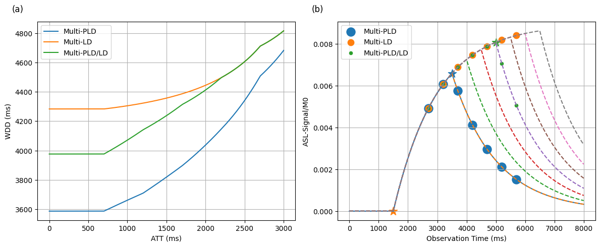
A general weighted delay (GWD) method was proposed to achieve a monotonic function, similar to the Dai’s1 work. When the monotonic function was not affected by the CBF, ATT can be estimated using a lookup table approach.$$GWD=[\sum_{i=1}^{7}(LD_i+PLD_i)\Delta M(AT T,LD_i,PLD_i)]/[\sum_{i=1}^{7}\Delta M(AT T,LD_i,PLD_i)]$$
Where $$$\Delta M(AT T,LD_i,PLD_i)$$$ was the pCASL signal with ATT at $$$LD_i$$$ and $$$PLD_i$$$. When LD was fixed in the multi-PLD protocol, the GWD estimator degraded to Dai’s model with a constant scaling factor of LD. The ATT can be calibrated from the GWD curve, Figure 1a. The CBF was calculated based on the ATT and it was averaged across multiple delays. As a benchmark, CBF and ATT were estimated directly by fitting the model with L2-norm using the differential evolution from the SciPy.optimize package.
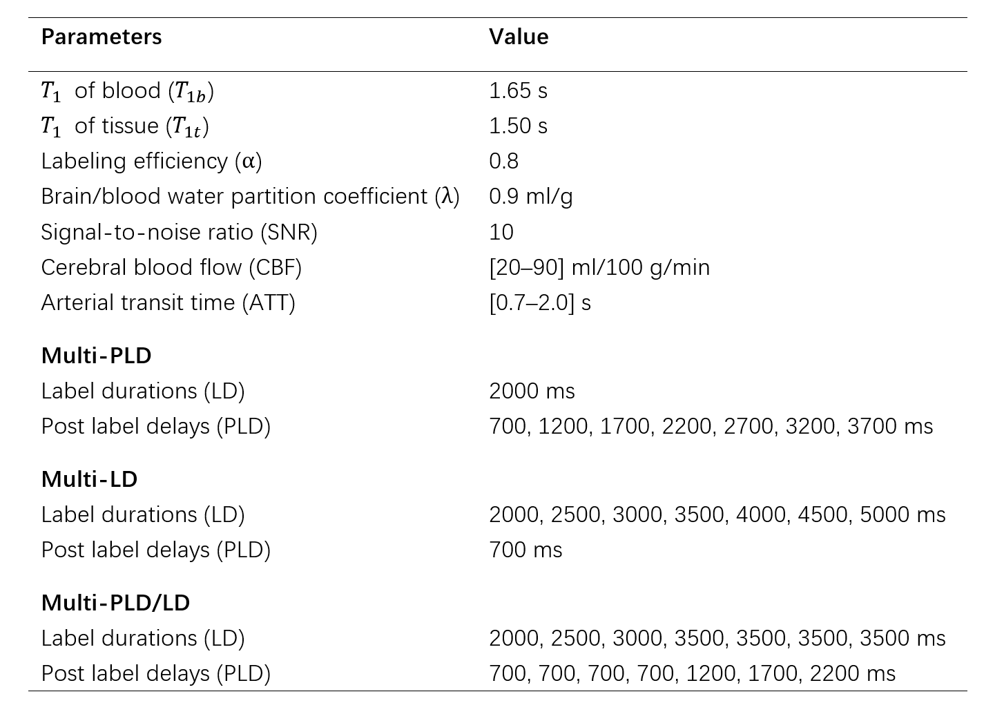
The pCASL signal model derived from the general kinetic model of Buxton et al.4 was used to generate the ASL signal with the parameters in Table 1. Three pCASL protocols were simulated using Monte Carlo including Multi-PLD, Multi-LD, and Multi-PLD/LD. All three protocols had the same PLD+LD duration in each scan. Two noise models were investigated, including Gaussian noise with a SNR of 10 and Rician noise with a SNR of 5. Each signal was simulated 1000 times.
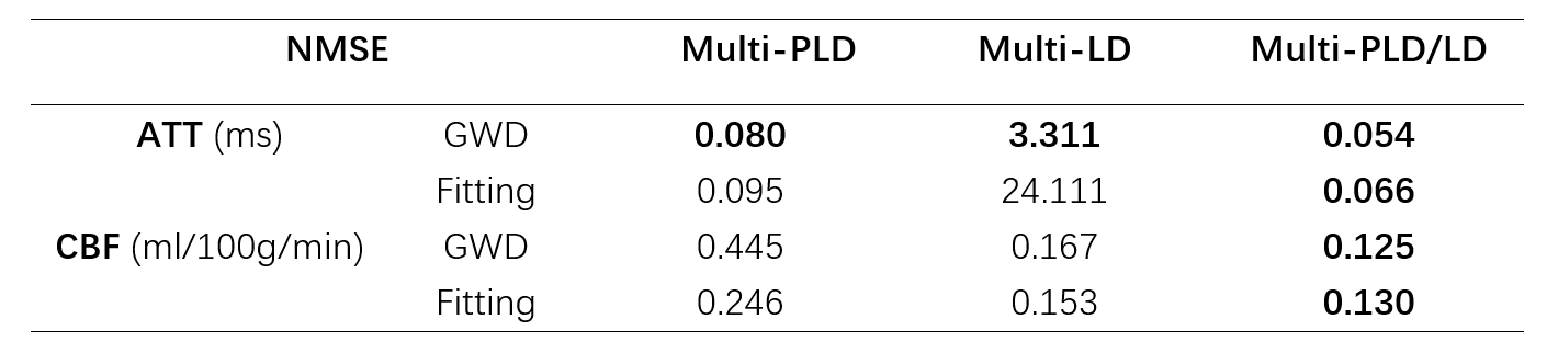
One healthy volunteer (male, 20 years old) was scanned on a 3.0T GE SIGNA Architect scanner. The pCASL images were acquired using a 3D fast spin echo stack of spiral with three arms, 34 slices with 4 mm thickness, and 8 averages. The LDs and PLDs were the same as Table 1. Normalized root mean square error (NRMSE) of the estimation was calculated based on the data with 4 averages and the multi-PLD/LD fitting maps of 8 averages were selected as the ground truth with a high SNR. The mean error was calculated in the gray matter mask threshold by the CBF at 30ml/100g/min.
Results
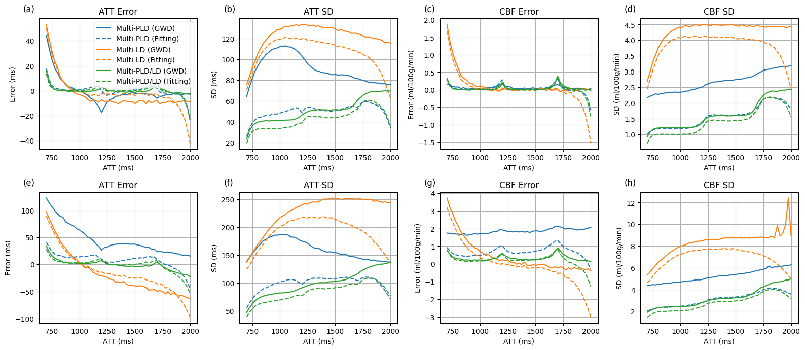
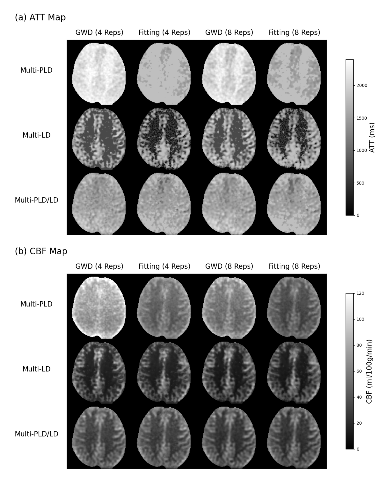
The simulation results are demonstrated in Figure 2. The multi-PLD/LD protocol provided accurate estimation with the smallest bias and variance, compared to the other protocols. In addition, the L2-norm fitting reduced the standard deviation (SD) of ATT and CBF in all protocols. The GWD method worked similarly to the L2-norm fitting, but with less bias at the large ATTs. ATT estimation errors were observed at ATT = PLD or LD+PLD, which were the turning points of the model. The estimated ATT was biased with the multi-LD protocol at the low CBF. With Rician noise and a low SNR, the estimations became biased, and variances were increased in most protocols and estimators.The ATT and CBF maps of the volunteer scan are shown in Figure 3, and the NRMSE is shown in Table 2. The multi-PLD/LD protocol provided the best ATT and CBF estimation compared to the other protocols, for both the proposed GWD method and the L2-norm fitting. The GWD estimator provided reduced ATT estimation error in all the protocols. The L2-norm fitting showed a notable improvement in the CBF estimation with the multi-PLD protocol. Underestimated ATT was found in the white matter region with the multi-LD protocol which was consistent with the simulation results.
Discussion and Conclusion
In this work, the multi-PLD/LD protocol provided a superior estimation compared to the other protocols. The L2-norm fitting is a maximum likely estimator in the Gaussian noise. Therefore, it provided reduced variance in theory and no worse estimation compared to the GWD method in the CBF estimation in the scan. However, the model fitting can be time-consuming and sensitive to the initial values, which inhibits its usage in practice. The flat section of the GWD curve limits ATT estimation which may lead to the wrong ATT estimation of WM with multi-LD protocols. The protocols used in this work can be optimized further using the Cramer-Rao Lower Bound approach.Acknowledgements
This work is supported by the National Key R&D Program of China (2022ZD0118004), the Alzheimer's Association (AARF-18-566347), Zhejiang Provincial Natural Science Foundation of China (LGJ22H180004, 202006140, and 2022C03057), and the MOE Frontier Science Center for Brain Science & Brain-Machine Integration, Zhejiang University.References
1. Dai W, Robson P M, Shankaranarayanan A, et al. Reduced resolution transit delay prescan for quantitative continuous arterial spin labeling perfusion imaging[J]. Magnetic resonance in medicine, 2012, 67(5): 1252-1265.
2. Borogovac A, Habeck C, Small S A, et al. Mapping brain function using a 30-day interval between baseline and activation: a novel arterial spin labeling fMRI approach[J]. Journal of Cerebral Blood Flow & Metabolism, 2010, 30(10): 1721-1733.
3. Zhao L, Taso M, Dai W, et al. Non-invasive measurement of choroid plexus apparent blood flow with arterial spin labeling[J]. Fluids and Barriers of the CNS, 2020, 17: 1-11.
4. Buxton R B, Frank L R, Wong E C, et al. A general kinetic model for quantitative perfusion imaging with arterial spin labeling[J]. Magnetic resonance in medicine, 1998, 40(3): 383-396.
Figures