2020
Improving the Robustness of Pseudo-Continuous Arterial Spin Labeling in the Intraoperative MRI Setting.1Radiology, Clínica Universidad de Navarra, Pamplona, Spain, 2Siemens Healthcare, Madrid, Spain, 3Neurosurgery, Clínica Universidad de Navarra, Pamplona, Spain, 4Anesthesia, Perioperative Medicine and Critical Care, Clínica Universidad de Navarra, Pamplona, Spain
Synopsis
Keywords: Arterial Spin Labelling, Arterial spin labelling
Motivation: Improvement of the intraoperative pseudo-continuous arterial spin labeling images for resection control in brain tumor surgery at 3T.
Goal(s): Evaluate the effect of shortening RF pulse duration and gap on the labeling efficiency of PCASL and to test it in the intraoperative setting.
Approach: A study in 10 healthy volunteers was done approximating the off-resonance effects at the labeling plane. 2 PCASL sequences were tested in 2 patients.
Results: A PCASL sequence robust to off-resonance effects is obtained by shortening RF duration and gap.
Impact: Shortening the RF duration and gap improves the PCASL labeling efficiency at high B0 off-resonance values at the labeling plane.
Introduction
Previous work has assessed the clinical utility of intraoperative pseudo-continuous arterial spin labeling (PCASL) for resection control in brain tumor surgery 1,2. Although both studies showed the potential of the technique, PCASL image quality was poor in some patients, which could be associated with ineffective labeling due to off-resonance effects 1, that are exacerbated in the intraoperative setting.For high off-resonance at the labeling plane, shortening RF pulse duration and gap should improve labeling efficiency because the phase error accumulated between RF pulses is directly proportional to ([RF [duration +gap] * off-set) 3. A similar strategy has been successfully employed at 7T 4.
Therefore, the aim of this study was to evaluate the effect of shortening RF pulse duration and gap on the labeling efficiency of PCASL and to test this strategy in the intraoperative setting.
Methods
Numerical SimulationsSimulations were performed based on the Bloch equations to compute the labeling efficiency. The RF duration varied from 300 µs to 700 µs in intervals of 100 µs keeping B1 mean and duty cycle (50%) constant. A range of average gradients (Gave) 0.1-2 mT/m and ratios between the selective gradient during the RF pulse (Gmax) and average gradient, 2-14, were evaluated. Off-resonance frequencies were varied from 0 to 420 Hz.
Weighted average labeling efficiency was calculated for carotid artery blood velocity profile used in Zhao et al 3.
Healthy volunteers
Subjects: 10 healthy volunteers (5 female; 24 ± 4 years [mean ± standard deviation]).
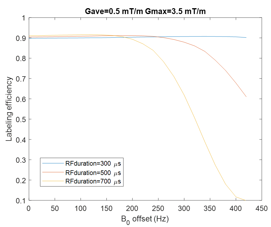
Gave of 0.5 mT/m and Gmax of 3.5 mT/m (ratio = 7) were selected because they have been demonstrated to be robust to off-resonance in previous work 3.
Off-resonance effects approximation was done by adding an extraphase (Φ) offset between RF pulses: Φ=foff T, where foff is off-resonance frequency and T is RF spacing 3.
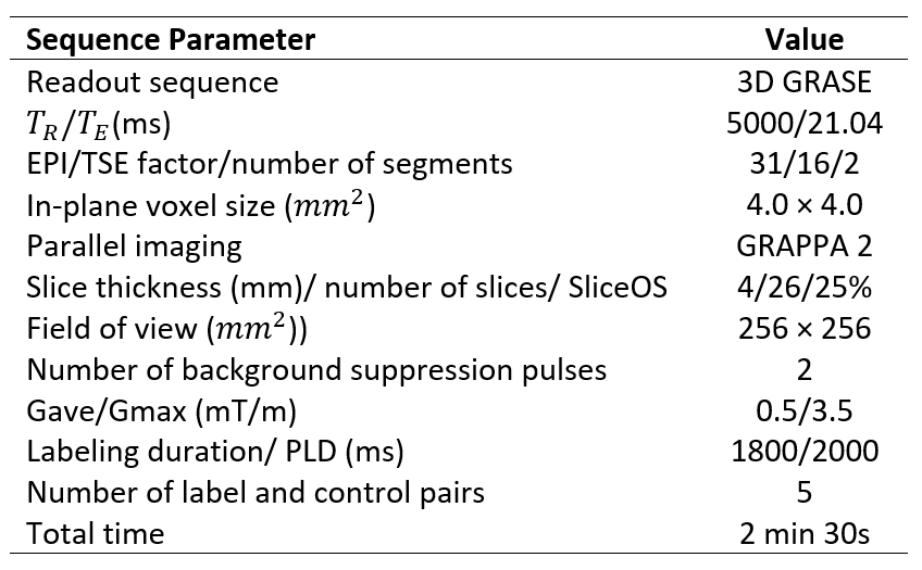
Scanning protocol: Was performed in a 3T MRI scanner (Skyra, Siemens Healthineers) using a 32-channel head coil. The protocol included 3D T1-weighted MPRAGE anatomical image and 15 PCASL sequences with RF durations: 300, 500 and 700 µs and varying off-resonance frequency between 0-400 Hz with 100 Hz step size. PCASL sequence parameters are in Table 1.
Patients
Subjects: 2 patients (2 Female, 74 and 44 years), subject to intraoperative MRI, during tumor surgery as part of the clinical protocol.
Scanning protocol: Was performed in a 3T MRI scanner (Skyra, Siemens Healthineers) using 2 4-channel flexible coils. 2 PCASL sequences were added to the intraoperative protocol, with RF duration of 300 and 500 µs with the same parameters of Table 1 except, in this case, 12 ASL pairs were acquired.
Data preprocessing and analysis
ASL images were realigned to the control image that was acquired before the T1-weighetd image. Perfusion-weighted images were obtained. For patients, realignment was not necessary.
Binary gray matter masks were attained by segmenting the anatomical image and applying an intensity filter with a threshold of 0.7.
Two way ANOVA was performed to test the effects of RF duration, off-resonance frequency, and their interaction on perfusion signal. Subsequently, one way ANOVAs were performed for data with off resonance frequencies of 200,300 and 400 Hz to assess the difference in the perfusion signal across RF durations followed by Post Hoc comparisons corrected by Bonferroni. A p-value less than 0.05 was considered significant.
Results
Numerical simulationsWith RF duration of 300 µs, labeling efficiency remains practically constant across off- sets (Figure 1).
Healthy volunteers
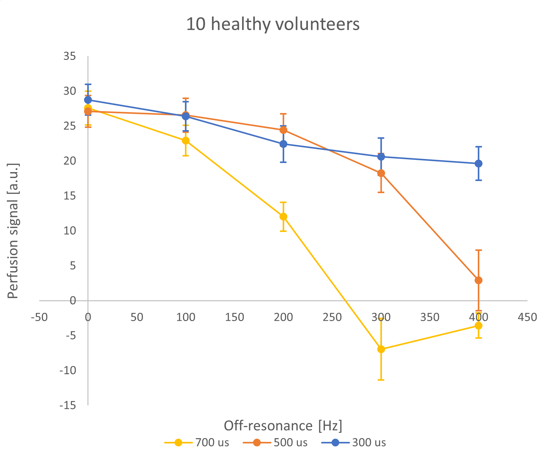
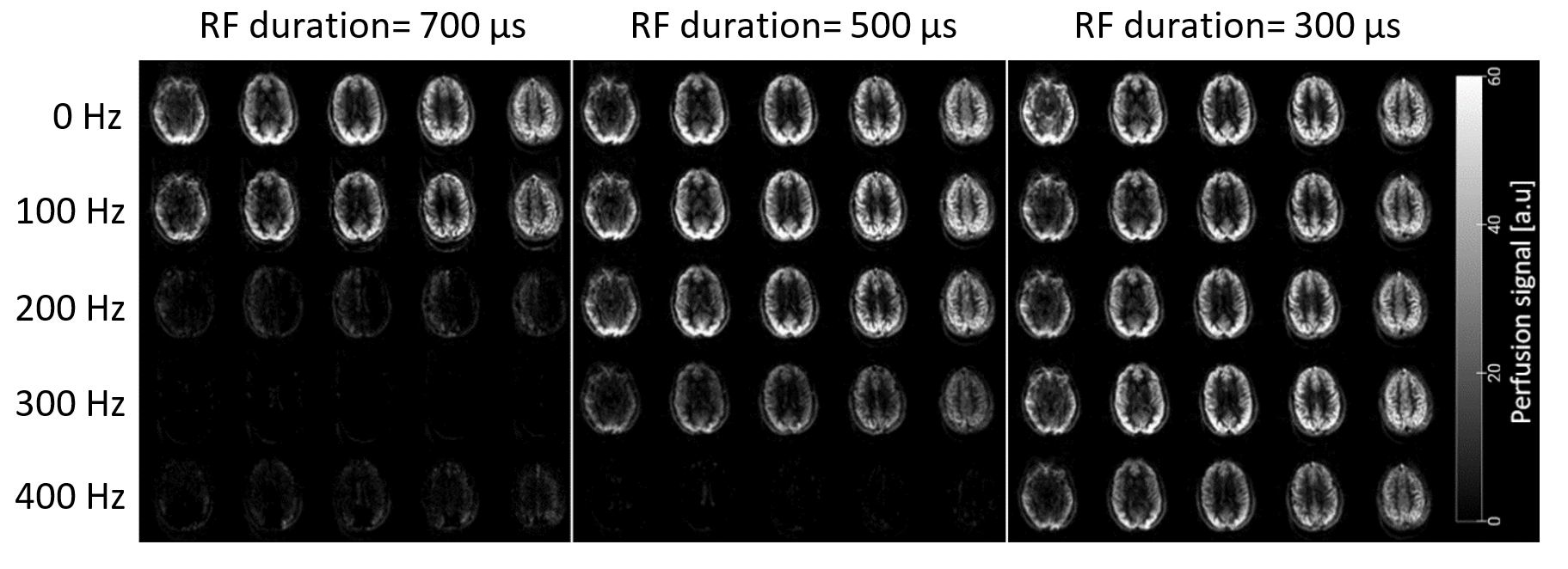
In Figure 2 group mean perfusion values in the gray matter, are displayed. Figure 3 shows perfusion maps for a representative subject obtained with the different RF durations and B0 off-sets. A similar pattern was found for the 10 subjects.
Patients
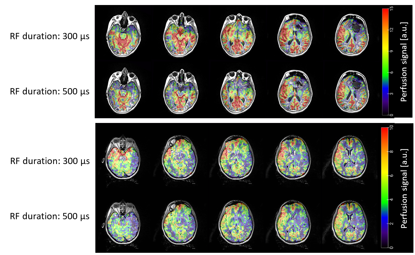
Perfusion increases are observed in Figure 4, for the RF duration of 300 us, for both patients.
Statistical analysis
ANOVA results showed that the effects of the RF duration, off resonance and the interaction between them were significant. Also showed significant differences for the 3 RF durations at 200, 300 and 400 Hz.
For 400 Hz, the RF duration of 300 µs was significantly different from 500 and 700 µs. Therefore, the sequence with shortest RF duration obtained higher perfusion values.
Discussion
Shortening the RF pulse duration and gap increased the labeling efficiency making the sequence more robust to off-resonance effects, as demonstrated in healthy volunteers and patients.Other issues must be considered to optimize the sequence for the intraoperative setting, such as the reduction in blood velocity observed in patients, due to anesthesia 5.
Conclusion
A PCASL sequence robust to off-resonance effects is obtained by shortening RF duration and gap.Acknowledgements
Carmen Sánchez-Albardíaz receives Ph.D. grant support from Siemens Healthcare Spain. Spanish Ministry of Science, Innovation and Universities (grant PI18/00084).References
1. Calvo-Imirizaldu, M. et al. Clinical utility of intraoperative arterial spin labeling for resection control in brain tumor surgery at 3 T. NMR Biomed 36, (2023).
2. Lindner, T. et al. Intraoperative resection control using arterial spin labeling — Proof of concept, reproducibility of data and initial results. Neuroimage Clin 15, 136–142 (2017).
3. Zhao, L., Vidorreta, M., Soman, S., Detre, J. A. & Alsop, D. C. Improving the robustness of pseudo-continuous arterial spin labeling to off-resonance and pulsatile flow velocity. Magn Reson Med 78, 1342–1351 (2017).
4. Wang, K. et al. Optimization of pseudo-continuous arterial spin labeling at 7T with parallel transmission B1 shimming. Magn Reson Med 87, 249–262 (2022).
5. Conti, A. et al. Cerebral haemodynamic changes during propofol-remifentanil or sevoflurane anaesthesia: Transcranial Doppler study under bispectral index monitoring. Br J Anaesth 97, 333–339 (2006).
Figures