2013
Whole-Brain SNR-Efficient Pseudo-Continuous Arterial Spin Labeling at 7T1Wellcome Centre for Integrated Neuroimaging, FMRIB, Nuffield Department of Clinical Neuroscience, University of Oxford, Oxford, United Kingdom, 2Institute of Science and Technology for Brain-inspired Intelligence, Fudan University, Shanghai, China
Synopsis
Keywords: Arterial Spin Labelling, Perfusion
Motivation: The high SAR burden of PCASL at 7T typically requires long TRs and short label durations, reducing SNR efficiency and perfusion measurement accuracy.
Goal(s): Our goal was to maximize the SNR efficiency of PCASL at 7T.
Approach: We optimized the PCASL B1+/gradient parameters and background suppression pulses to maximize SNR efficiency (i.e., balancing labeling efficiency and SAR), and utilized VERSE and dynamic B0 shimming to reduce SAR.
Results: Compared to optimizing the PCASL parameters for maximum labeling efficiency, our max-SNR efficiency optimized parameters increased SNR efficiency by 38% and reduced TRs by 41% in vivo.
Impact: The improved SNR efficiency of our optimized 7T PCASL parameters increases the advantages of ultra-high field perfusion measurements, enabling robust and high-SNR perfusion measurement in clinically viable scan times.
Introduction
The higher SNR and longer T1 at 7T has the potential to increase the SNR of pseudo-continuous ASL (PCASL)1 perfusion measurements by 2-4x compared to 3T.2 This could enable higher spatial resolution imaging3 and robust measurement of white matter perfusion.4 However, PCASL is a high-SAR labeling method, so short label durations (LDs) and deadtime to extend the TR are used to mitigate the increase in SAR at 7T.5–9 This decreases the SNR and number of averages that can be acquired within a given scan duration, reducing the benefit compared to typical 3T protocols.Previous studies8,9 optimized the PCASL parameters and background suppression (BGS) pulses to maximize PCASL labeling efficiency (LE) and BGS inversion efficiency (IE) over a large range of off-resonance (±200 Hz), but this came with large increases in SAR which required short LDs and prolonged TRs.
Here, we instead optimize the PCASL parameters and BGS pulses to maximize the SNR efficiency (SNReff), such that we maximize the SNR for a given scan time, where $$$SNR_{eff}=\frac{SNR}{\sqrt{TR}}\propto\frac{SNR}{\sqrt{RF_{power}}}$$$, $$$SNR\propto LE_{PCASL}\cdot IE_{BGS}^2$$$, and $$$RF_{power}=\int_0^{TR}|B_1^+(t)|^2\cdot dt\propto \int_0^{TR}SAR\cdot dt$$$. We optimize the PCASL parameters over a much smaller range of off-resonance (±50 Hz) to reduce the need for high B1+ and instead use dynamic B0-shimming10 of the labeling plane to reduce off-resonance. Together with VERSE and optimized BGS pulses,11 we achieve superior SNR within a 4-minute scan with shorter TRs.
Methods
The PCASL LE was calculated for a range of PCASL parameters using Bloch simulations: B1+mean=0.1-2µT, Gmax=3-15mT/m, Gmean=0.1-2mT/m, RF spacing=0.5-1.16ms, 50% RF duty-cycle. Simulations assumed pulsatile laminar flow,12 B0±50Hz, T1/T2=2.1s/0.06s. Minimum-SAR-VERSE was used.11 Parameter combinations that exceeded hardware limitations or perturbed static spins >1.5cm from the labeling plane were excluded. Two sets of PCASL parameters were identified: one which maximized SNReff and one which maximized LE (Figure 1).Two non-selective BGS pulses were considered (Figure 1): a standard hyperbolic-secant (HS-BGS) pulse; and a 25% SAR-reduced VERSE HS pulse with the phase-waveform optimized using optimal-control (OC-BGS).11 In each case, the pulses (HS parameters/phase-waveform) were optimized to maximize IE across B1+±50% and B0±500Hz using Bloch simulations (T1/T2=2.1s/0.06s).
In vivo scans were performed in three volunteers on a Siemens 7T Magnetom Plus with a 8Tx/32Rx head-coil at 1st-level SAR constraints. Saturation-inversion-recovery experiments were used to measure the IE of the BGS pulses. Four-minute white-paper13 PCASL data (LD=1.8s, PLD=1.8s, GE-EPI readout, 24 slices, 3.4x3.4x5mm3) were acquired to compare the SNR-efficiencies of the PCASL parameter sets and BGS pulses, where the TR was minimized for each volunteer and scan. Dynamic B0-shimming of the labeling plane reduced resonance offsets to <50 Hz (results presented elsewhere). Using 3DREAM,14 the reference voltage was set to accommodate the mean B1+ within the middle EPI slice and the nominal PCASL flip angle was increased to achieve, on average, the desired flip angle within the feeding arteries at the labeling plane.
Results
The optimized PCASL parameters and BGS pulses are described in Figure 1. Although the maxSNReff parameters have a lower simulated LE than the maxLE case, the PCASL RFpower was reduced by a factor of 5.5, resulting in a 20% higher simulated SNReff. The use of VERSE reduced PCASL RFpower by 23-25%.The in vivo inversion efficiency of the BGS pulses is shown in Figure 2. The OC-BGS pulse had a 25% lower RFpower and only slightly reduced inversion efficiency, in agreement with simulations (Figure 1). However, the OC-BGS pulse was less robust to very low B1+, as expected from simulations (not shown).
The in vivo SNReff results are shown in Figures 3 and 4. To satisfy the scanner's 10s-SAR limit, the PCASL flip angle for the maxLE scan had to be reduced by a factor of 1.61±0.06 (mean±SD). The maxSNReff+HS-BGS scan achieved both 41% shorter TRs and 38% higher SNReff. With the use of the OC-BGS pulse, the TRs were further shortened, but at the cost of some SNReff due to the lower IE compared to the HS-BGS pulse.
Example whole-brain quantified CBF maps are shown in Figure 5.
Discussion
Optimizing for SNReff (i.e., balancing LE and SAR), instead of simply aiming to maximizing LE, led to much higher SNR and improved temporal resolution. By using dynamic B0-shimming, we were able to optimize the PCASL parameters over a smaller range of off-resonances (±50Hz rather than ±200 Hz in8,9) reducing the need for high B1+mean and reducing SAR.We further reduced the TR by using a reduced-SAR OC BGS pulse. However, this came with a reduction in SNReff in vivo, in contrast to simulations (not shown), suggesting the IE was lower than expected and requiring further investigation.
Acknowledgements
This study was supported by a Sir Henry Dale Fellowship jointly funded by the Wellcome Trust and the Royal Society (Grant Number 220204/Z/20/Z). The Wellcome Centre for Integrative Neuroimaging is supported by core funding from the Wellcome Trust (203139/Z/16/Z).References
1. Dai W, Garcia D, De Bazelaire C, Alsop DC. Continuous flow-driven inversion for arterial spin labeling using pulsed radio frequency and gradient fields. Magnetic Resonance in Medicine. 2008;60(6):1488-1497. doi:10.1002/mrm.217902. Teeuwisse WM, Webb AG, Van Osch MJP. Arterial spin labeling at ultra-high field: All that glitters is not gold. International Journal of Imaging Systems and Technology. 2010;20(1):62-70. doi:10.1002/ima.20218
3. Shao X, Guo F, Shou Q, et al. Laminar perfusion imaging with zoomed arterial spin labeling at 7 Tesla. NeuroImage. 2021;245:118724. doi:10.1016/j.neuroimage.2021.118724
4. Gardener AG, Jezzard P. Investigating white matter perfusion using optimal sampling strategy arterial spin labeling at 7 Tesla. Magnetic Resonance in Medicine. 2015;73(6):2243-2248. doi:10.1002/mrm.25333
5. Saïb G, Koretsky AP, Talagala SL. Optimization of pseudo‐continuous arterial spin labeling using off‐resonance compensation strategies at 7T. Magn Reson Med. November 2021:mrm.29070. doi:10.1002/mrm.29070
6. Meixner CR, Eisen CK, Schmitter S, et al. Hybrid ‐shimming and gradient adaptions for improved pseudo‐continuous arterial spin labeling at 7 Tesla. Magn Reson Med. August 2021:mrm.28982. doi:10.1002/mrm.28982
7. Boland M, Stirnberg R, Pracht ED, Stöcker T. Robust and SAR-efficient whole-brain pseudo-continuous ASL at 7T. In: Proceedings of the 29th Annual Meeting of the ISMRM, Virtual. ; 2019:4963.
8. Wang K, Ma SJ, Shao X, et al. Optimization of pseudo-continuous arterial spin labeling at 7T with parallel transmission B1 shimming. Magnetic Resonance in Medicine. 2022;87(1):249-262. doi:10.1002/mrm.28988
9. Zhao C, Shao X, Shou Q, et al. Whole-Cerebrum distortion-free three-dimensional pseudo-continuous arterial spin labeling at 7T. NeuroImage. 2023;277:120251. doi:10.1016/j.neuroimage.2023.120251
10. Blamire AM, Rothman DL, Nixon T. Dynamic shim updating: A new approach towards optimized whole brain shimming. Magnetic Resonance in Medicine. 1996;36(1):159-165. doi:10.1002/mrm.1910360125
11. Woods JG, Chiew M, Okell TW. Minimizing SAR for SNR-efficient pseudo-continuous Arterial Spin Labeling at 7T. In: Proceedings of the 31st Annual Meeting of the ISMRM, Toronto, Canada. ; 2023:0371.
12. Zhao L, Vidorreta M, Soman S, Detre JA, Alsop DC. Improving the robustness of pseudo-continuous arterial spin labeling to off-resonance and pulsatile flow velocity. Magnetic Resonance in Medicine. 2017;78(4):1342-1351. doi:10.1002/mrm.26513
13. Alsop DC, Detre JA, Golay X, et al. Recommended implementation of arterial spin-labeled perfusion MRI for clinical applications: A consensus of the ISMRM perfusion study group and the European consortium for ASL in dementia. Magnetic Resonance in Medicine. 2015;73(1):102-116. doi:10.1002/mrm.25197
14. Ehses P, Brenner D, Stirnberg R, Pracht ED, Stöcker T. Whole‐brain B1‐mapping using three‐dimensional DREAM. Magnetic Resonance in Medicine. 2019;(November 2018):mrm.27773. doi:10.1002/mrm.27773
15. Jenkinson M, Beckmann CF, Behrens TEJ, Woolrich MW, Smith SM. FSL. NeuroImage. 2012;62(2):782-790. doi:10.1016/j.neuroimage.2011.09.015
Figures
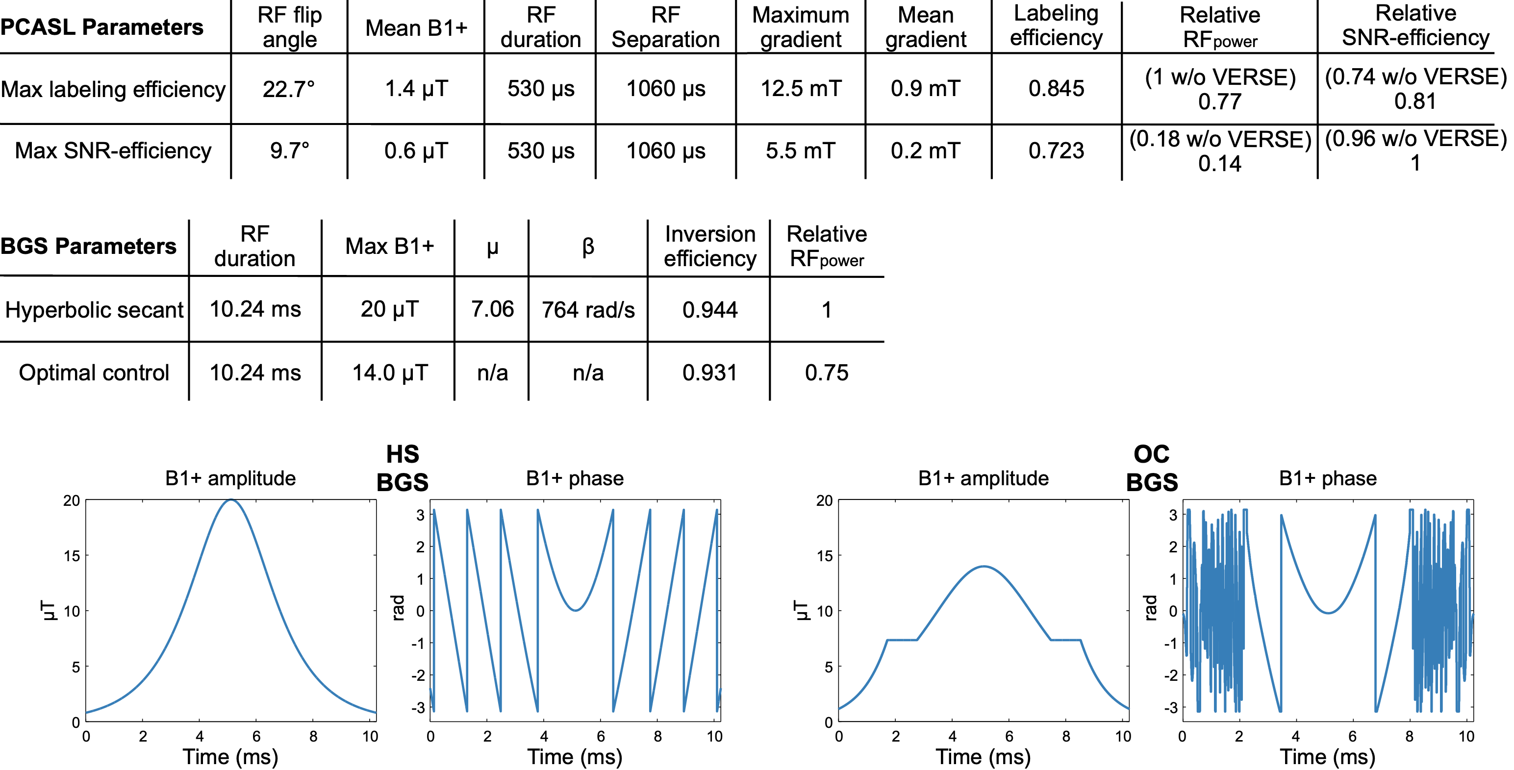
Figure 1: Top: parameters for the PCASL pulse train when optimized for SNReff (i.e. accounting for SAR) or maximum LE (i.e. ignoring SAR). The labeling and SNR efficiencies were calculated using Bloch simulations. The SNR efficiency accounts for the total sequence SAR. The maximum PCASL gradient parameter is before application of VERSE. Bottom: the parameters for the BGS-HS inversion pulse and the lower SAR OC-BGS inversion pulse. The inversion efficiencies were calculated using Bloch simulations. The B1+ amplitude and phase waveforms are shown for both BGS pulses.
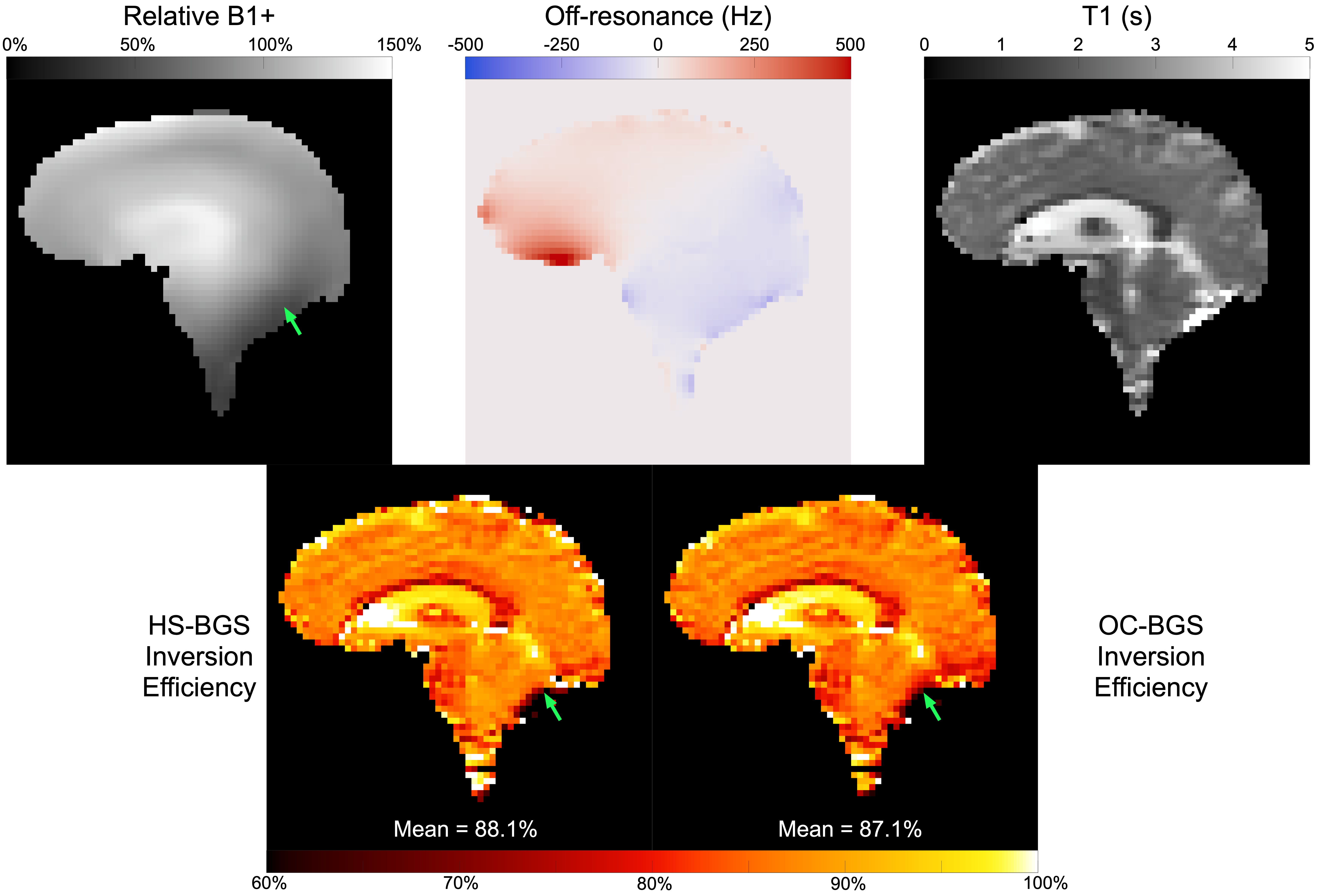
Figure 2: Representative results from the in vivo BGS inversion efficiency experiment. The OC-BGS pulse resulted in slightly lower IE than the HS-BGS pulse, especially in regions of low B1+ (green arrows), but overall maintained reasonable IE. The EPI data were undistorted using a B0-fieldmap and FSL's FUGUE tool.15 The IE and T1 values were jointly estimated by non-linear least squares fitting of the multi-TI saturation-inversion recovery EPI data. The mean IE within the brain mask is displayed at the bottom of each figure. M0 was measured with a long-TR EPI readout.
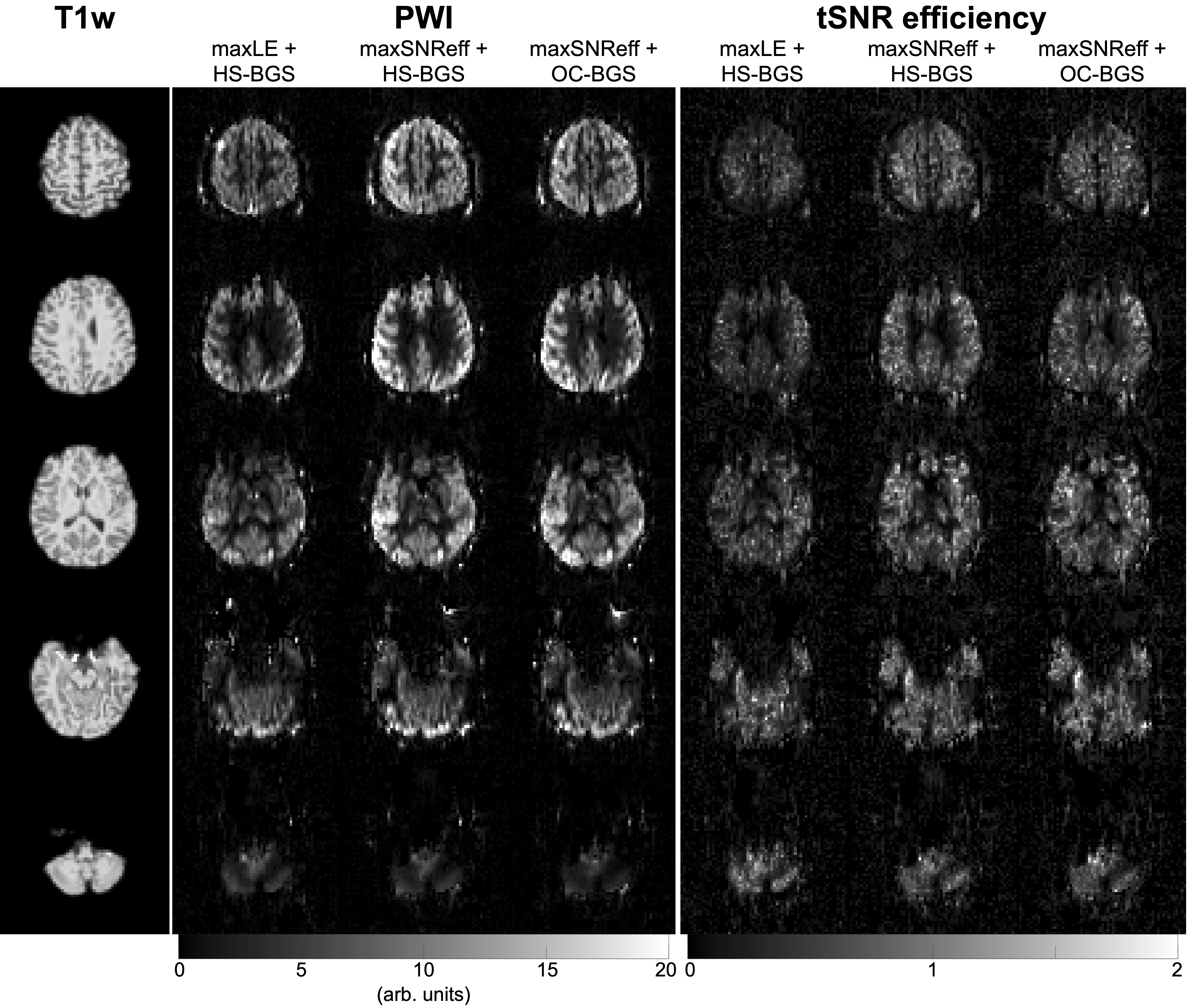
Figure 3: Representative in vivo T1-weighted structural images, perfusion-weighted images (PWI), and SNR efficiency maps (calculated as the temporal SNR (tSNR) divided by the square-root of the protocol TR) for each of the three protocols in one subject. Both maxSNReff scans have higher tSNR efficiency than the maxLE scan, demonstrating the SNR advantage of this approach.
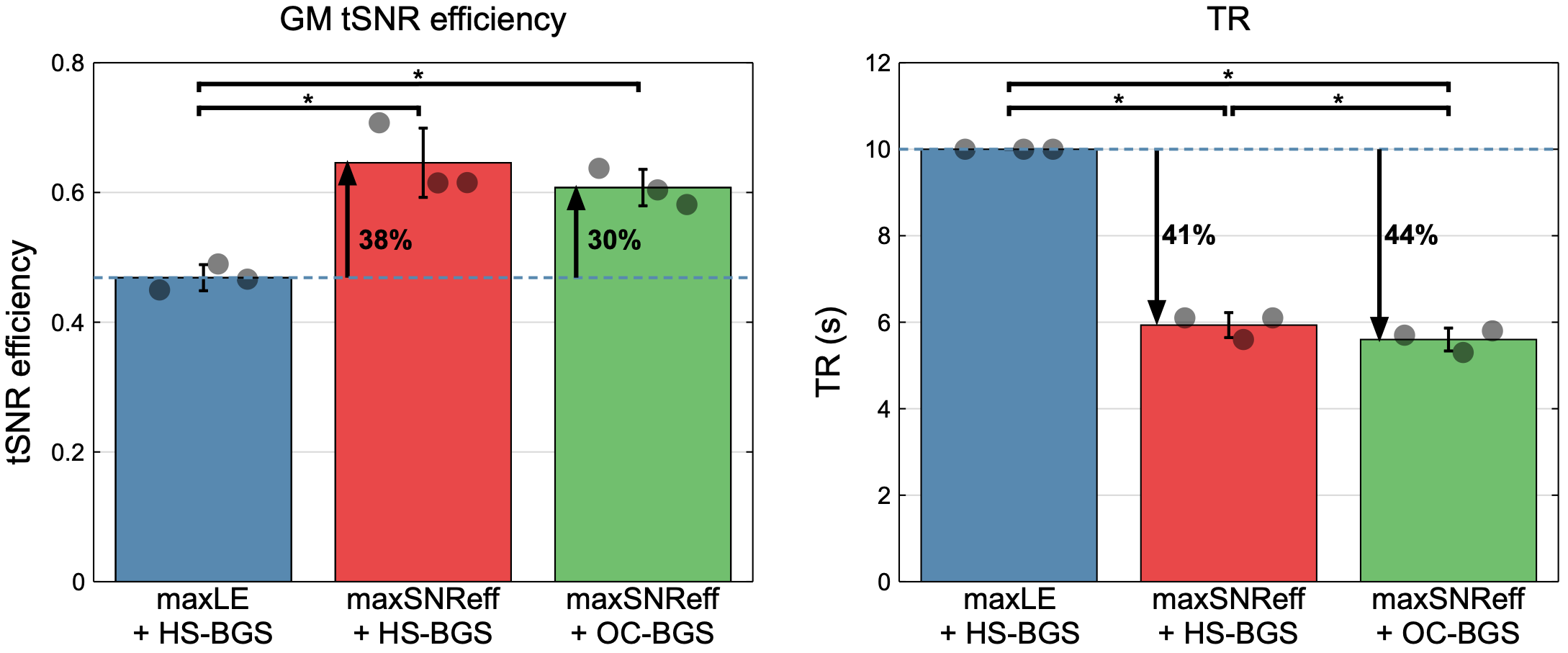
Figure 4: In vivo mean gray matter (GM) temporal SNR efficiency and the minimum achieved TR for the three volunteers. For the maxLE scan, the TR always had to be set to a minimum of 10 s and the PCASL flip angle reduced by a factor of 1.61±0.06 (mean±SD across subjects) to satisfy the 10 s SAR limit, otherwise it would not have been possible to run the scan. When using the maxSNReff parameters with either BGS pulse, the GM tSNR efficiency was significantly higher than for the maxLE PCASL scan.
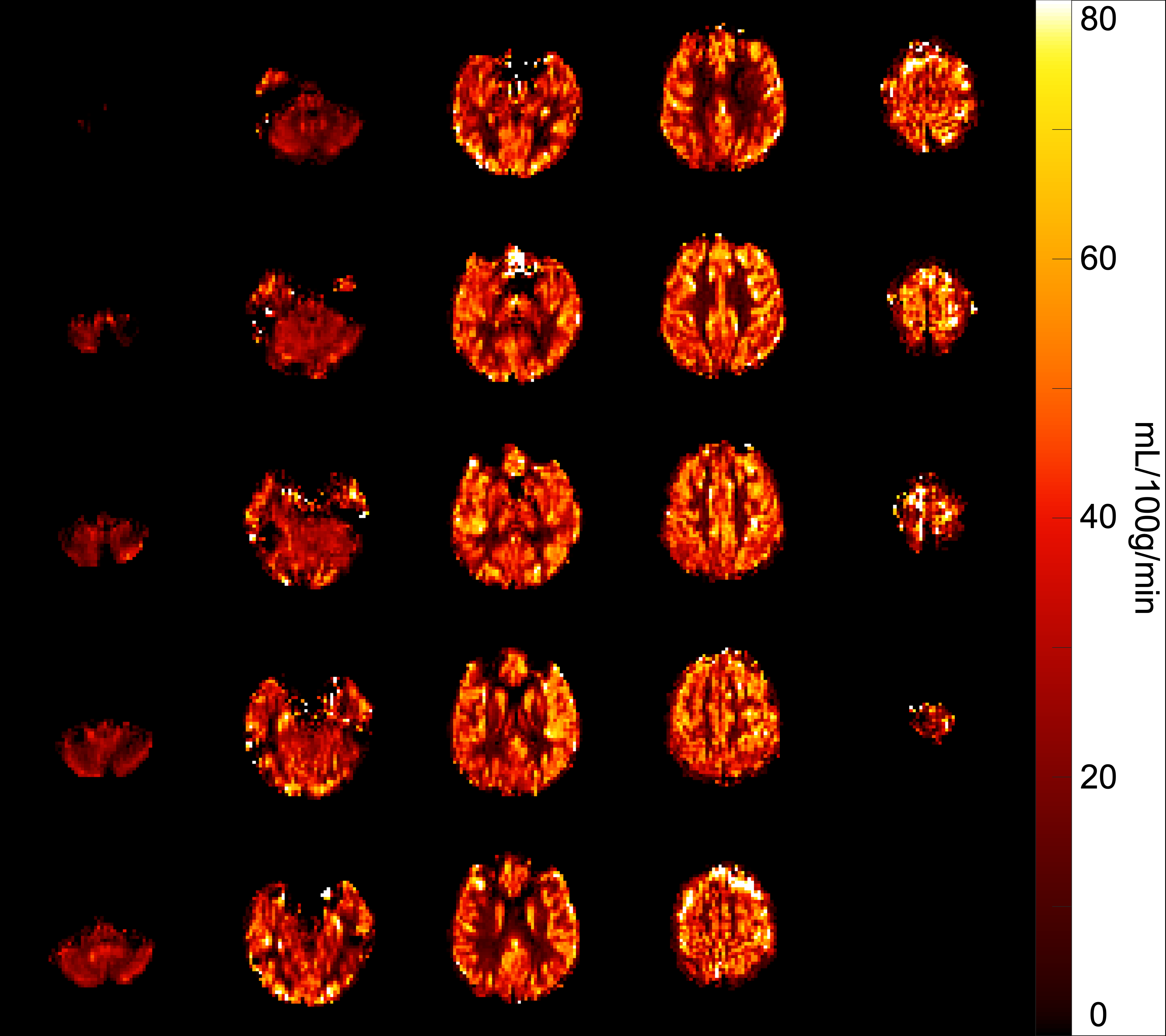
Figure 5: Example in vivo whole brain quantified CBF maps for the maxSNReff + HS-BGS protocol. The gray matter CBF median ± IQR is 39 ± 25 mL/100g/min which is in line with the expected value. This suggests that the PCASL labeling efficiency and HS-BGS inversion efficiency are close to the expected values from simulations.