2009
Calibrated fMRI with a background-suppressed PCASL and multi-echo BOLD dual-acquisition sequence1Basque center on Cognition Brain and Language (BCBL), Donostia-San Sebastián, Spain, 2Siemens Healthineers, Madrid, Spain, 3Clínica Universidad de Navarra, Pamplona, Spain, 4Instituto de Investigación Sanitaria de Navarra (IdiSNA), Pamplona, Spain
Synopsis
Keywords: Arterial Spin Labelling, Arterial spin labelling, calibrated fMRI, BOLD, multi-echo, sequence, brain
Motivation: Calibrated fMRI quantitatively estimates cerebral metabolic rate of oxygen (CMRO2) by simultaneously measuring cerebral blood flow (CBF) with arterial spin labeling (ASL) and BOLD. Pseudo-continuous ASL (PCASL) with background suppression (BS) and 3D readout is recommended for CBF while multi-echo (ME) 2D BOLD has gained popularity. Nowadays, no calibrated fMRI sequence integrates both.
Goal(s): Our goal is to combine them in a single sequence.
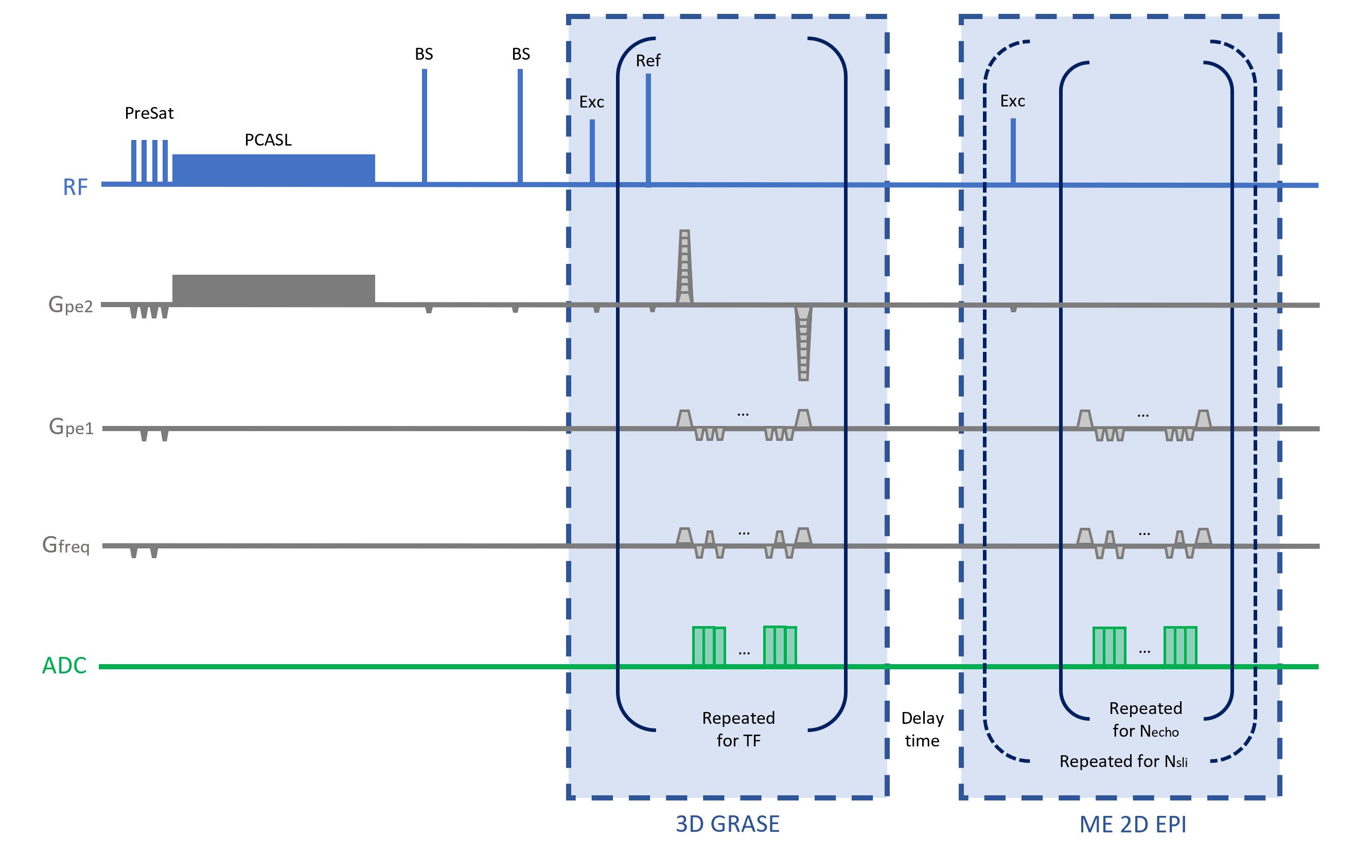
Approach: We introduce a novel dual-acquisition calibrated fMRI sequence integrating BS-PCASL-3D GRASE for CBF and 2D multi-echo EPI BOLD.
Results: Resting-state data in a healthy volunteer showed CBF values concordant with literature and improved BOLD tSNR and connectivity maps.
Impact: We propose a novel sequence for calibrated fMRI that optimizes both CBF and BOLD measurements with a dual acquisition scheme, thus overcoming the limitations of previous ASL-BOLD calibrated fMRI sequences and showing promise for improved accuracy and reliability.
Introduction
Calibrated fMRI is a quantitative alternative to BOLD fMRI, enabling the estimation of cerebral metabolic rate of oxygen (CMRO2) by simultaneously measuring cerebral blood flow (CBF), using arterial spin labeling (ASL) and BOLD data, along with gas challenges [1,2]. Thus, these measurements can provide a more comprehensive view of brain activity and metabolism [3]. To obtain these measurements, ASL sequences with dual-echo (DE) or multi-echo (ME) readouts are frequently employed [4,5]. However, a dual-acquisition (DA) approach provides an opportunity to optimize both CBF and BOLD components. Implementing a background-suppressed (BS) pseudo-continuous ASL (PCASL) with a 3D readout, as recommended in the ASL consensus paper [6], along with a standard 2D EPI BOLD module has shown to significantly enhance perfusion and BOLD data quality for calibrated fMRI with a hypercapnic challenge [7]. The goal of this work is to improve this sequence by incorporating a multi-echo (ME) module for BOLD acquisition, thereby enhancing BOLD sensitivity and enabling TE-dependent denoising [8,9].Methods
We implemented a novel DA sequence (Figure 1) consisting on a BS pCASL single-shot 3D GRASE for CBF acquisition and 2D ME EPI for BOLD acquisition. Whole-brain data acquisitions were performed on a healthy volunteer during eyes-open resting state in a 3T Siemens PrismaFit scanner using a 64-channel receiver head coil with TR = 7 s, voxel resolution = 4x4x5.5 mm3 , matrix size = 64x64, 16 slices and BW = 3004 Hz/Px. For pCASL, 72 label-control pairs were acquired with TE=31 ms, FA(exc/ref)=90º/160º, TF=12, slice oversampling=27.5%, slice Partial Fourier factor= 5/8, labeling duration=1.8 s, postlabeling delay=1.8 s. For BOLD data, three TEs were collected at 17, 45 and 73 ms. A delay time of 1.3 s was set between both parts to allow for recovery of the longitudinal magnetization. Raw data was reconstructed offline using a MATLAB in-house code. Preprocessing (skull-stripping and volume registration) and computation of the CBF map (single-compartment model [10]) were performed using AFNI, and multi-echo T2*-weighted combination, usually known as optimally combined (OC) [8], and denoising with ME-Independent Component Analysis were done using TEDANA [9] and RICA [11]. Temporal SNR (tSNR) maps, defined as the voxelwise mean divided by its standard deviation, and seed-based functional connectivity maps (AFNI instacorr using Pearson correlation, high-pass filter 0.01 Hz plus detrending with linear and quadratic trends, and spatial smoothing with FWHM gaussian kernel of 4 mm radius) were computed from BOLD data.Results and discussion
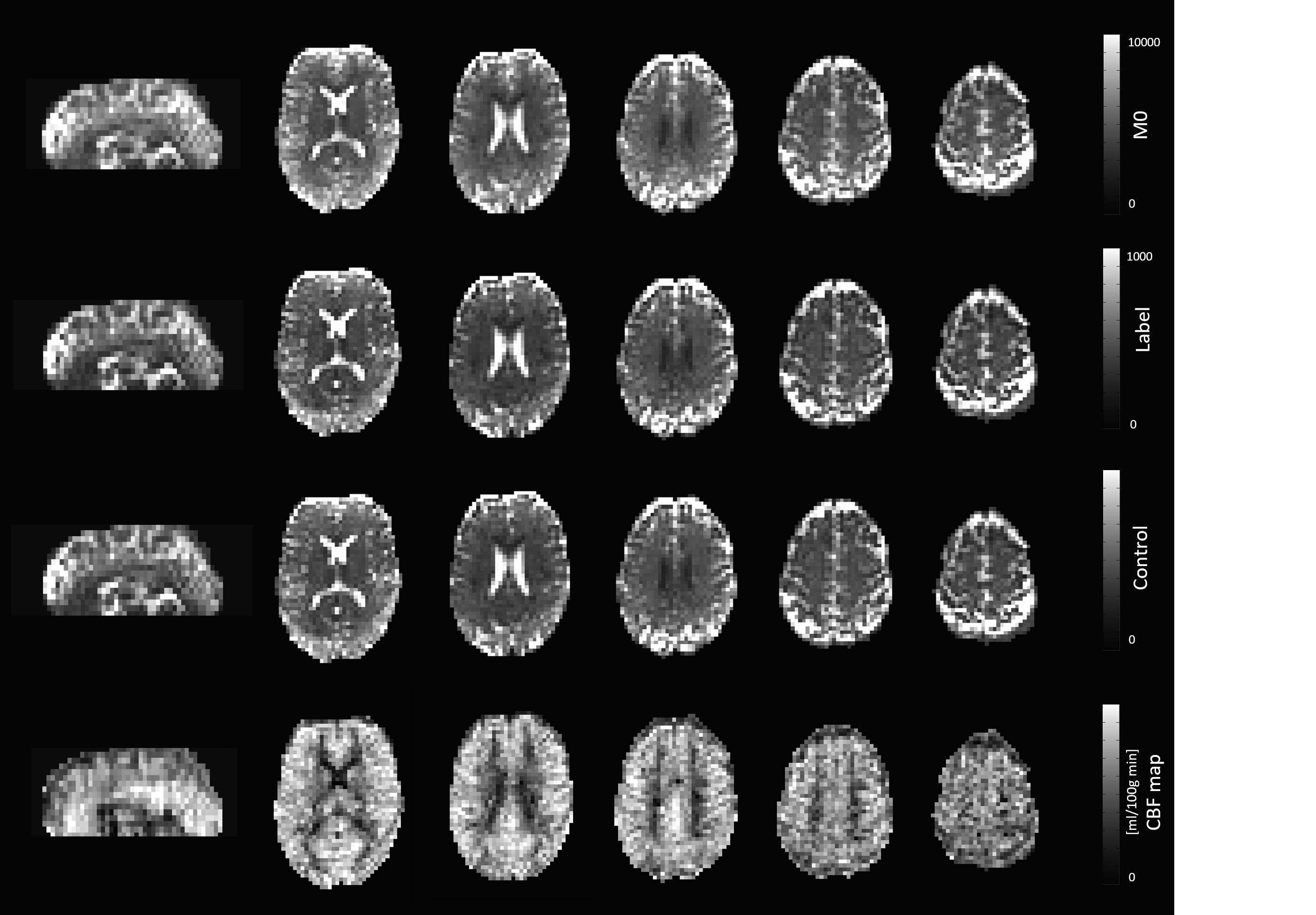
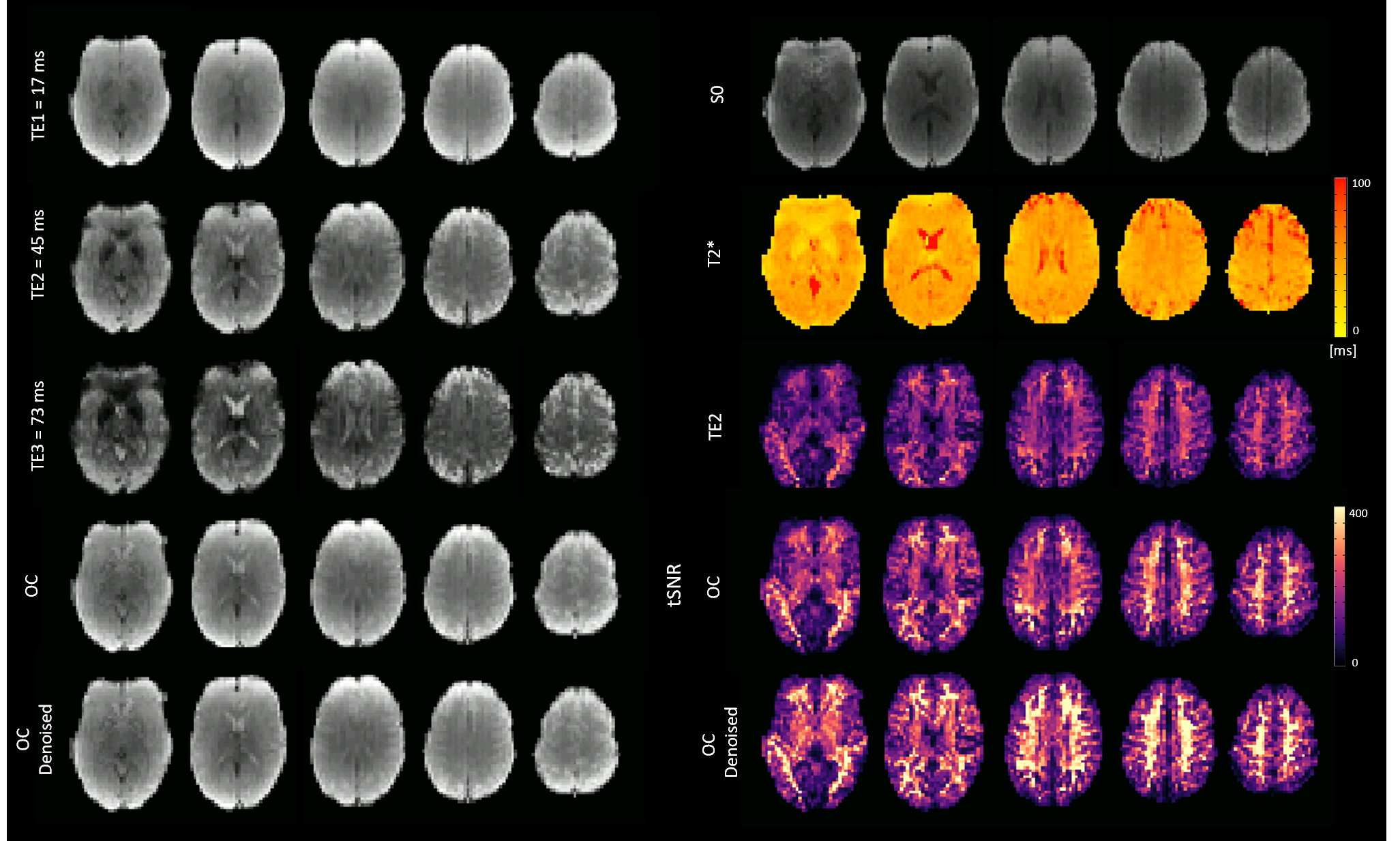
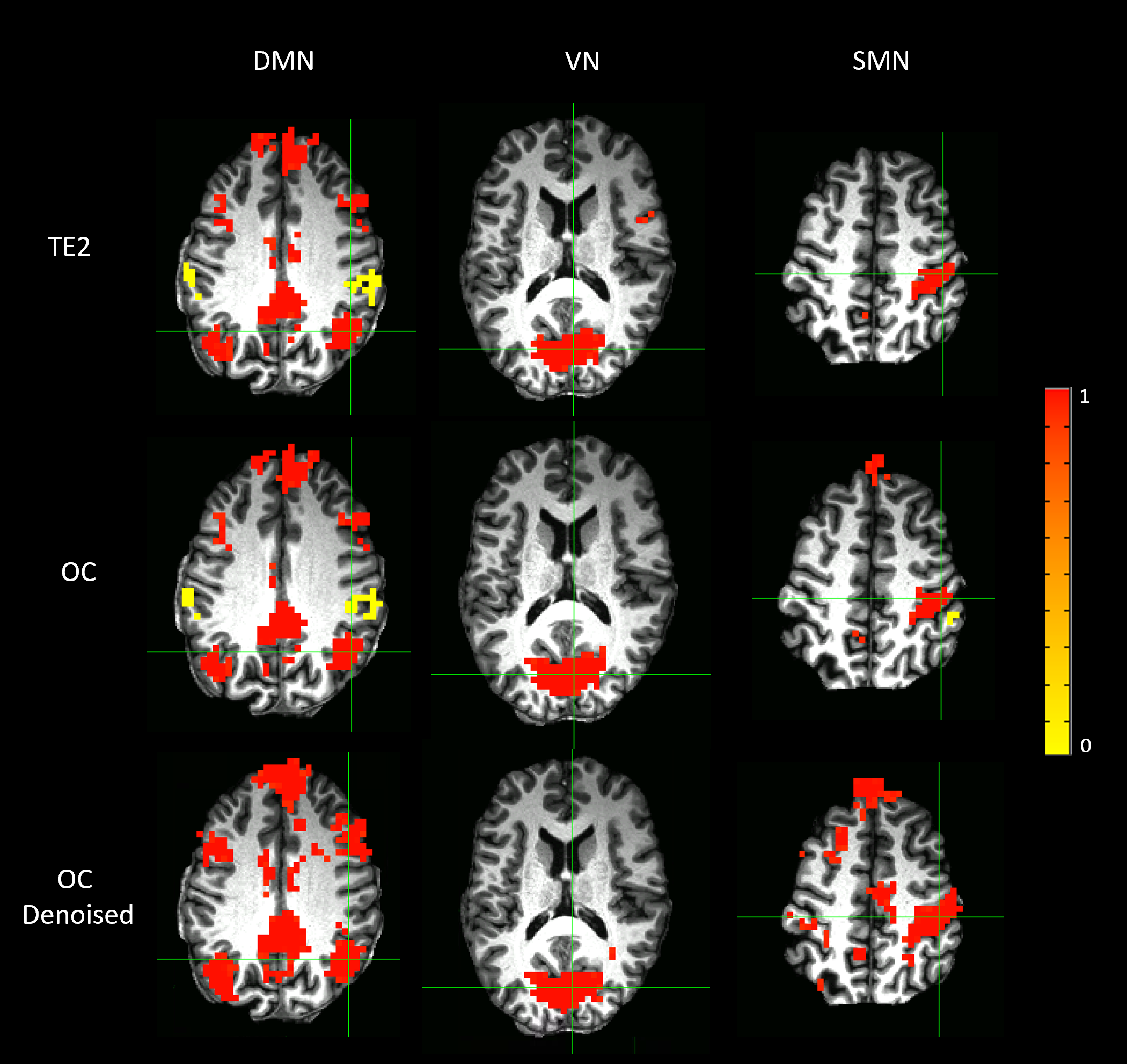
Figure 2 shows the M0, and representative label and control images alongside the corresponding CBF maps. Average CBF values in whole-brain, gray and white matter were 44.75 ± 16.97, 51.20 ± 15.31 and 33.38 ± 12.33 ml/100g min, respectively. The CBF maps exhibit the anticipated quality and values concordant with literature [7, 12]. Figure 3 displays three example slices of each individual echo, the estimated S0 and T2* maps, the T2*-weighted combination (OC), and after ME-ICA denoising (OC denoised). The tSNR maps in Fig. 3 illustrate increased values for OC compared to using the second echo (TE2), as a proxy of a conventional single-echo acquisition, which are further heightened for OC denoised. The mean tSNR values in whole-brain, gray and white matter are respectively 110.11±64.18, 99.70±54.67 and 146.08±69.65 for TE2; 146.09±84.78, 131.02±69.49 and 202.83±92.33 for OC, and 175.43±105.18, 154.43±83.79 and 247.67±117.31 for OC denoised. Finally, the seed-based functional connectivity maps shown in Figure 4 show the typical patterns observed for the default mode network (DMN), primary visual network (VN) and sensorimotor network (SMN). It is visible that connectivity strength and network size are increased with OC and ME-ICA denoising, compared with a single-echo acquisition (e.g., notice the connectivity between bilateral primary motor and supplementary motor regions in the map of the SMN for OC denoised).In conclusion, these initial results illustrate the potential of the proposed sequence for significantly enhancing the quality of calibrated fMRI with simultaneous ASL and BOLD data acquisition. Anticipated work will evaluate the proposed sequence in task-based and resting-state calibrated fMRI studies, including model calibration with a gas challenge inducing a state of hypercapnia, and benchmark it against conventional dual-echo ASL and BOLD sequences.Acknowledgements
This study was supported by the Spanish Ministry of Economy and Competitiveness (Ramon y Cajal Fellowship, RYC-2017–21845), theSpanish State Research Agency (BCBL “Severo Ochoa” excellence accreditation CEX2020-001010/AEI/10.13039/501100011033) and the Basque Government (BERC 2022–2025).
This research has been made possible through a Formación de Personal Investigador (FPI) contract for the completition of doctoral theses, granted by the Spanish Ministry of Economyand Competitiveness (PRE2019-090025), affiliated with the project (SEV-2015-0490-19-3).
References
[1] Davis, T.L., et al. (1998) Proc Natl Acad Sci U S A, 95(4), 1834-1839. https://doi.org/10.1073/pnas.95.4.1834
[2] Hoge, R.D., et al. (2012) Neuroimage, 62: 930-937. https://doi.org/10.1016/j.neuroimage.2012.02.022
[3] Fukunaga, M.., et al. (2008) J. Cereb. Blood Flow Metab., 28 (7),1377-1387. 10.1038/jcbfm.2008.25
[4] Cohen, A.D., et al. (2017) PLoS One,12(3):e0169253. https://doi.org/10.1371/journal.pone.0169253
[5] Leontiev, O. & Buxton, R.B. (2007) NeuroImage,35(1): 175-184. https://doi.org/10.1016/j.neuroimage.2006.10.044
[6] Alsop, D.C., et al. (2015) Magn. Reson. Med., 73(1), 102–116. https://doi.org/10.1002/mrm.25197
[7] Fernández-Seara, M., et al. (2016) Neuroimage, 142:474-482. https://doi.org/10.1016/j.neuroimage.2016.08.007
[8] Posse S., et al. (1999) Magn. Reson. Med., 42:87-97. https://doi.org/10.1002/(SICI)1522-2594(199907)42:1<87::AIDMRM13>3.0.CO;2-O
[9] DuPre, E., et al. (2021) JOSS, 6(66):3669. https://doi.org/10.21105/joss.03669
[10] Wang, J., et al. (2005) Radiology, 235(1), 218-228. https://doi.org/10.1148/radiol.2351031663
[11] Uruñuela, E. (2021). Rica (Version v1.0.17) [Computer software]. https://doi.org/10.5281/zenodo.5788350
[12] Vidorreta M., et al. (2013) Neuroimage, 66:662-671. https://doi.org/10.1016/j.neuroimage.2012.10.087
Figures