2007
Age-Related Hemodynamic Variations in Children: A Study Using Single- and Multi-delay ASL1Department of Pathophysiology, the Second Faculty of Medicine, Charles University, Prague, Czech Republic, 2University Hospital Motol, Prague, Czech Republic, 3Institute of Radiopharmaceutical Cancer Research, Helmholtz-Zentrum Dresden-Rossendorf, Dresden, Germany, 4Department of Neuroradiology, Hospital Beatriz Ângelo, Lisbon, Portugal, 5Department of Radiology, Stanford University, Stanford, CA, United States, 6Department of Neurosurgery, Stanford University, Stanford, CA, United States
Synopsis
Keywords: Arterial Spin Labelling, Perfusion, Arterial Spin Labelling, Normal Development
Motivation: There is lacking understanding of hemodynamic changes in children and their impact on MRI perfusion quantification.
Goal(s): We aim to investigate age-related hemodynamic changes in developing brain.
Approach: Perfusion parameters from single- and multi-delay Arterial Spin Labeling (ASL) MRI of MR-negative children are analyzed.
Results: We found significant age-dependent differences between perfusion quantification with single- and multi-delay approaches. ATT followed a non-linear distribution in age and was heterogeneous across vascular territories Our findings support the use of multi-delay ASL for improved perfusion assessment in children and provide better understanding of hemodynamic changes in developing brain.
Impact: This is the first study to investigate age-related arterial transit-time changes in children and their impact on perfusion quantification with perfusion MRI. Our findings prompt improved understanding of age-related perfusion changes and standardization of hemodynamic parameters in the pediatric cohort.
INTRODUCTION
Increasing clinical availability and application of perfusion MRI in pediatric brain imaging emphasizes the need for better understanding of perfusion changes in healthy development. Arterial Spin Labeling (ASL) is of special interest in pediatric cohorts due to its non-invasiveness, which allows easier clinical implementation, perfusion imaging of preterms and neonates, and repeatability in longitudinal studies. The recent position paper by the ISMRM Perfusion Study Group1 argued that pediatric population requires protocol adoption from the adult standards. Furthermore, a higher, age-related, heterogeneity of cerebral blood blow (CBF) and arterial transit time (ATT) in the developing brain needs to be considered for optimal scanning and interpretation2. Here, we investigate the age-related changes of CBF and ATT and the performance of single- versus multi-delay ASL in MR-negative children.METHODS
Thirty-one pediatric subjects (0.7-17.9 years, mean 9.2±5.3, 19 male) admitted to Lucile Packard Children’s Hospital between July 2021 and March 2022 were retrospectively analyzed. Inclusion criteria were: age<18 years, clinically confirmed MR-negative status, single- and/or multi-delay ASL. MR-negative status was defined as: no structural or functional impairment, confirmed by a pediatric neuroradiologist (ET). 3D T1-weighted and ASL scans were acquired on 3T GE Premier MRI system. 3D background-suppressed single-delay (labeling duration (LD) 1450 ms, post-labeling delay (PLD) 1525 or 2025 ms) and multi-delay (3 PLD - 300, 2000, 3700 ms, effective LD 500 ms; or/and 7 PLD - 700, 1250, 1800, 2350, 2900, 3450, 4000 ms, effective LD 550 ms) pseudo-continuous ASL was was available for 10, 19, 31, and 18 subjects respectively3,4. ASL images were processed with Explore ASL 1.11.05, including healthy tissue segmentation, motion correction, and CBF and ATT quantification using FSL-BASIL toolbox (University of Oxford, UK) with partial volume correction. ASL was quality-checked by two researchers (JP, YP). Data with severe motion/labeling artifacts were excluded. Mean CBF values in GM and WM, and mean ATT in GM and individual vascular territories (ACA, MCA, and PCA) were extracted. The relative difference between ASL measurements (ΔGM-CBF) was calculated as a ratio of the difference between multi-delay GM-CBF3PLD and single-delay GM-CBF2025ms to the GM-CBF2025ms. Spearman correlation test and linear regression models were used to investigate the association between relative ΔGM-CBF and ATT and age. Furthermore, per-voxel coefficient of variation (CoV) was calculated for GM-ATT and GM-CBF in multi-delay subdatasets as a ratio of the standard deviation to the mean across subjects. Finally, paired Student’s t-test and effect size estimated as Cohen’s d were used to analyze the ATT difference between multi-delay sequences and the GM- and WM-CBF difference between single- and multi-delay sequences.RESULTS
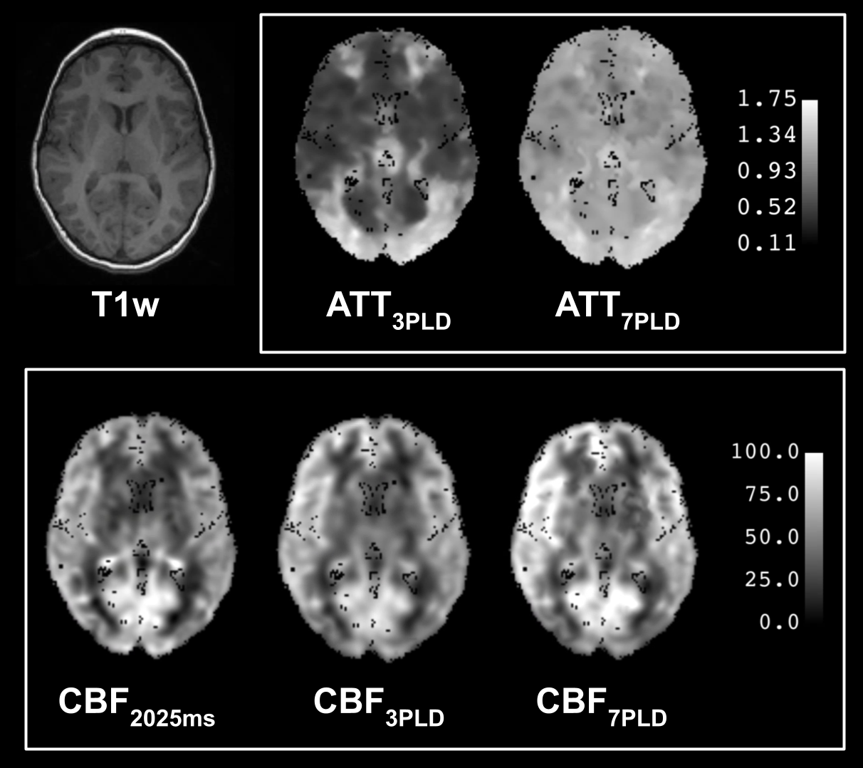
CBF distribution across age is in Figure 1A. In GM, CBF7PLD was higher than CBF3PLD (d=1.01, p=1.9e-5), while there was no significant difference between CBF3PLD and CBFPLD2025 (d=0.20, p=0.128). In WM, CBF7PLD was also consistently higher than CBF3PLD (d=0.58, p=0.003), but CBF3PLD was also higher than CBFPLD2025 (d=0.24, p=0.011). Correlation analysis results are in Figure 1B. ΔGM-CBF was negatively associated with age (beta=-20.02, p=0.004), but there was no significant association with GM-ATT (beta=-0.78, p=0.09) (Figure 1C).GM-ATT7PLD had significantly higher values compared with GM-ATT3PLD (d=2.77, p=1.98e-13) (Figure 2A). GM-ATT7PLD was on average 65.6% higher GM-ATT3PLD. Mean ATT3PLD distribution in ACA and PCA vascular territories was not significantly different (d=0.03, p=0.6) (Figure 2B), while ATT7PLD in ACA territory was consistently shorter than in PCA (d=0.81, p=8.13e-07) (Figure 2C). Visual illustration of ATT CoV spatial distribution highlights consistently increased ATT heterogeneity in caudate nucleus and putamen (Figure 3), while there was no observed region-specific heterogeneity for CBF (Figure 4). Figure 5 depicts a representative case, which highlights the improved CBF and ATT quantification by 7-PLD ASL.
DISCUSSION
The observed relationship between hemodynamics and age may prompt insights into pediatric neural development, such as the rapid proliferation of neural connections and synaptic pruning. Differences in CBF and ATT quantification between the multi-delay sequences underscore the advantages of improved hemodynamic modeling and will apply particularly in cerebrovascular pathologies in children.This study has certain limitations: 1) hemodynamic parameters were analyzed for the entire cohort instead of per age groups to account for the non-linear association between age and perfusion7; 2) confounding effects of sedation8 and sex9 were not addressed; 3) our single-center, single-vendor study does not addresssite variability.
CONCLUSION
ATT follows a non-linear distribution in age and is heterogeneous across vascular territories. The negative correlation between ΔGM-CBF and age, and a positive correlation between mean ATT and age emphasize the importance of using multi-delay ASL in children to mitigate transit artifacts. This preliminary study underscores the importance of the multi-delay approach for accurate perfusion quantification in children.Acknowledgements
YP has been awarded the ISMRM Exchange Program grant. This work is supported by American Heart Association Grant #23SCEFIA1141920, project nr. LX22NPO5107 (MEYS): Financed by EU – Next Generation EU and by grant nr. NU23-08-00460 from Czech Health Research Council.References
1 Lindner, T., Bolar, D. S., Achten, E., Barkhof, F., Bastos‐Leite, A. J., Detre, J. A., ... & ISMRM Perfusion Study Group. (2023). Current state and guidance on arterial spin labeling perfusion MRI in clinical neuroimaging. Magnetic Resonance in Medicine, 89(5), 2024-2047.
2 Zhao, M. Y., Tong, E., Duarte Armindo, R., Woodward, A., Yeom, K. W., Moseley, M. E., & Zaharchuk, G. (2023). Measuring Quantitative Cerebral Blood Flow in Healthy Children: A Systematic Review of Neuroimaging Techniques. Journal of Magnetic Resonance Imaging.
3 Zhao, M. Y., Tong, E., Duarte Armindo, R., Fettahoglu, A., Choi, J., Bagley, J., ... & Zaharchuk, G. (2023). Short‐and Long‐Term MRI Assessed Hemodynamic Changes in Pediatric Moyamoya Patients After Revascularization. Journal of Magnetic Resonance Imaging.
4 Zhao, M. Y., Fan, A. P., Chen, D. Y. T., Ishii, Y., Khalighi, M. M., Moseley, M., ... & Zaharchuk, G. (2022). Using arterial spin labeling to measure cerebrovascular reactivity in Moyamoya disease: Insights from simultaneous PET/MRI. Journal of Cerebral Blood Flow & Metabolism, 42(8), 1493-1506.
5 Mutsaerts, H. J., Petr, J., Groot, P., Vandemaele, P., Ingala, S., Robertson, A. D., ... & Barkhof, F. (2020). ExploreASL: an image processing pipeline for multi-center ASL perfusion MRI studies. Neuroimage, 219, 117031.
6 Zhao, M. Y., Yeom, K., Tong, E., Duarte, R. M., Moseley, M., Zaharchuk, G. “(ISMRM 2022) Comparing Single and Multiple Delays methods for ASL Perfusion Quantification on Pediatric Neuroimaging.” Accessed: Nov. 06, 2023. [Online]. Available: https://archive.ismrm.org/2022/1223.html
7 Carsin-Vu, A., Corouge, I., Commowick, O., Bouzillé, G., Barillot, C., Ferré, J. C., & Proisy, M. (2018). Measurement of pediatric regional cerebral blood flow from 6 months to 15 years of age in a clinical population. European journal of radiology, 101, 38-44.
8 Makki, M. I., O'Gorman, R. L., Buhler, P., Baledent, O., Kellenberger, C. J., Sabandal, C., ... & Schmitz, A. (2019). Total cerebrovascular blood flow and whole brain perfusion in children sedated using propofol with or without ketamine at induction: An investigation with 2D‐Cine PC and ASL. Journal of Magnetic Resonance Imaging, 50(5), 1433-1440.
9 Liu, Y., Zhu, X., Feinberg, D., Guenther, M., Gregori, J., Weiner, M. W., & Schuff, N. (2012). Arterial spin labeling MRI study of age and gender effects on brain perfusion hemodynamics. Magnetic resonance in medicine, 68(3), 912-922.
Figures
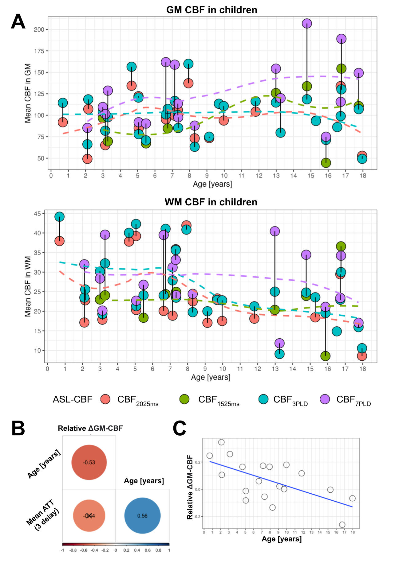
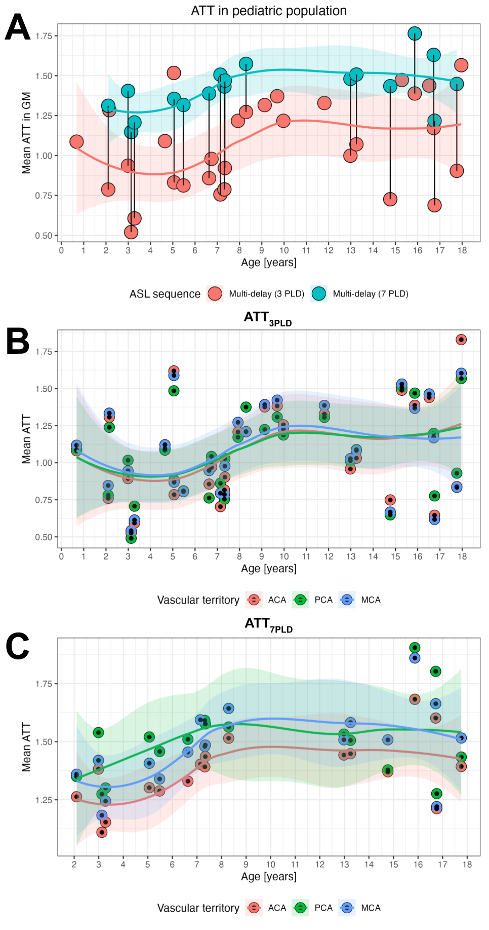
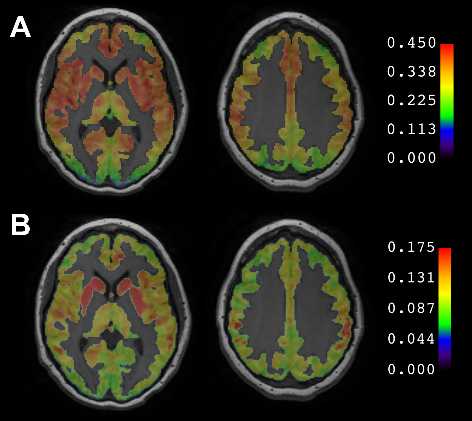
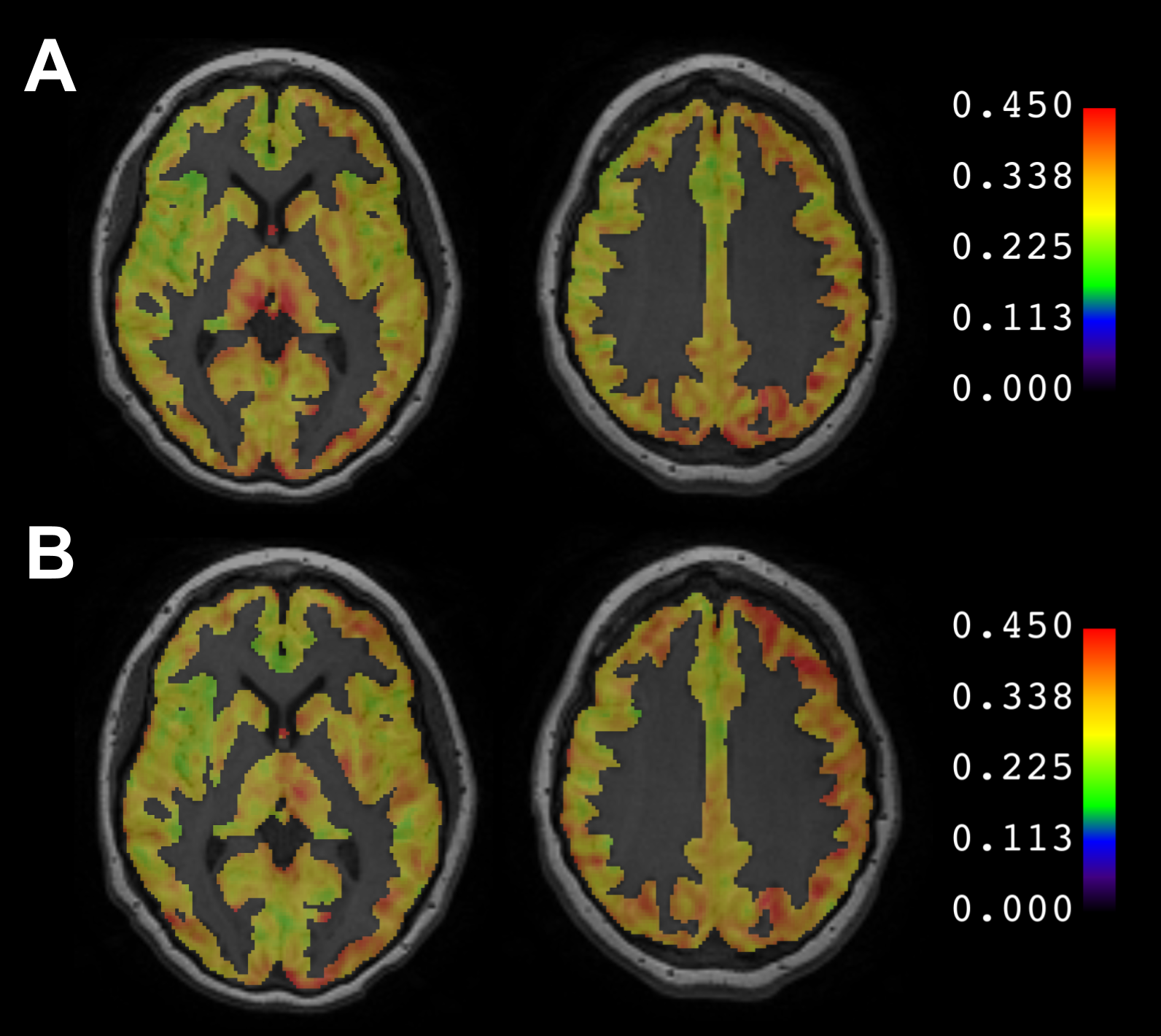
Figure 4. A. Spatial distribution of GM-CBF3PLD CoV (n=31). B. Spatial distribution of GM-CBF7PLD (n=18). CoV highlights the variability of perfusion within subjects and across age.