2005
Off-resonance effects on infant pCASL1College of Biomedical Engineering & Instrument Science, Zhejiang University, Hangzhou, China, 2Department of Radiology, Children's Hospital, Zhejiang University School of Medicine, National Clinical Research Center for Child Health, Hangzhou, China, 3Department of Computer Science, State University of New York at Binghamton, Binghamton, NY, United States, 4Radiology, Beth Israel Deaconess Medical Center and Harvard Medical School, Boston, MA, United States
Synopsis
Keywords: Arterial Spin Labelling, Arterial spin labelling, infant; labeling efficiency; off-resonance
Motivation: The image quality of ASL in infants is variable.
Goal(s): To investigate the labeling efficiency in infant pCASL.
Approach: Labeling efficiency was simulated using the Bloch equation. Prospective scans were performed on 11 subjects, with pCASL at 60mm and 90mm below the AC-PC line, MRA, and B0 map.
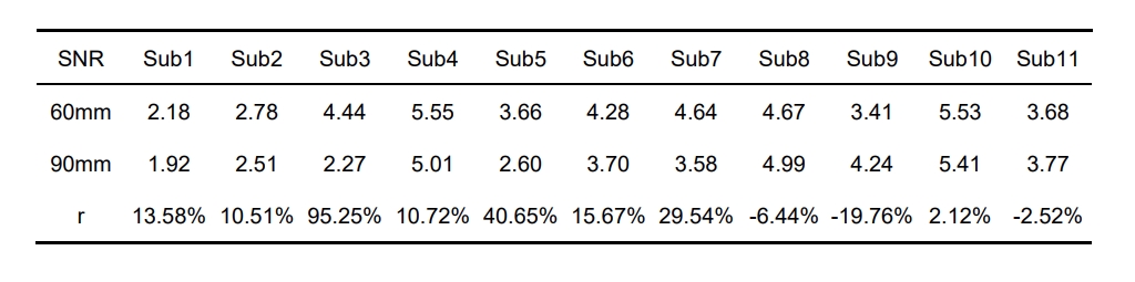
Results: Blood flow velocity has negligible effects on the labeling efficiency at the on-resonance frequency. The combined effects of off-resonance and the high blood flow velocity are the primary cause of the variable image quality. pCASL images at 60mm showed improved SNR in 8 /11 infants, compared to that of 90mm.
Impact: This study examined the factors contributing to variable ASL images in infant and provided a preliminary solution by selecting the labeling plane.
Introduction
Without radiation or exogenous tracer injection, Arterial spin labeling (ASL) presented an ideal approach for assessing cerebral blood flow in infants. However, the image quality of infant ASL is variable. Especially for pseudo continuous ASL (pCASL), in which the labeling location and gradients were originally optimized for adults. Possible reasons are being investigated, such as brain size, blood flow velocity, and pulsatility in infants1. This study investigated the flow velocity and off-resonance effects on the pCASL labeling efficiency and tested on 11 subjects.Methods
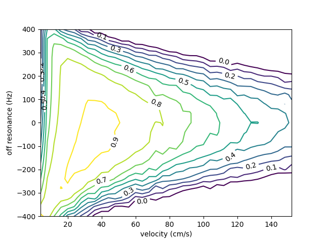
Bloch equations were used to simulate the pCASL labeling efficiency as a function of blood velocity, pulsatile flow, and off-resonance frequency. Infant pulsatile flow was modeled by proportionally scaling the adult flow profile2. The blood flow velocity in the internal carotid artery for infants was assumed to be 1.5 times that of adults3,4.Simulation 1: the labeling efficiency was investigated at off-resonance frequencies from -400Hz to 400Hz and across the flow velocities from 0.1 to 150 cm/s.
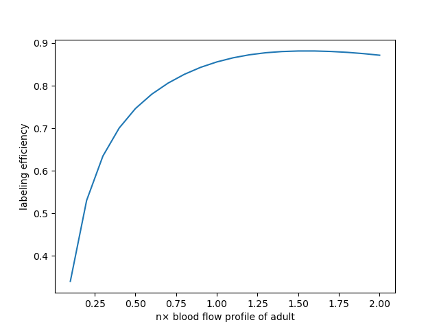
Simulation 2: to investigate the effects of blood flow velocity and pulsatile, labeling efficiency was simulated at pulsatile flow velocities ranging from 0.1x to 2.0x times that of adults (76cm/s). Overall labeling efficiency was calculated by taking into account the effect of laminar flow and the blood volume contributions at different velocities5.
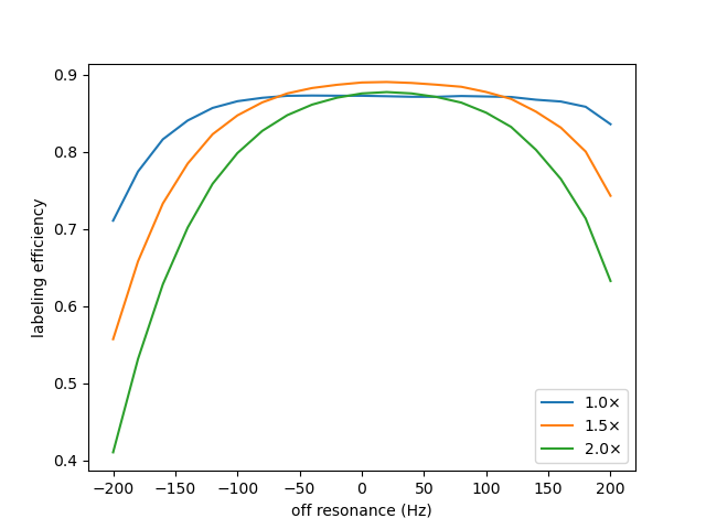
Simulation 3: overall labeling efficiency was calculated by averaging the labeling efficiency at the off-resonance frequencies from -200Hz to 200Hz and velocities of 1.0x, 1.5x, and 2.0x times that of adults.
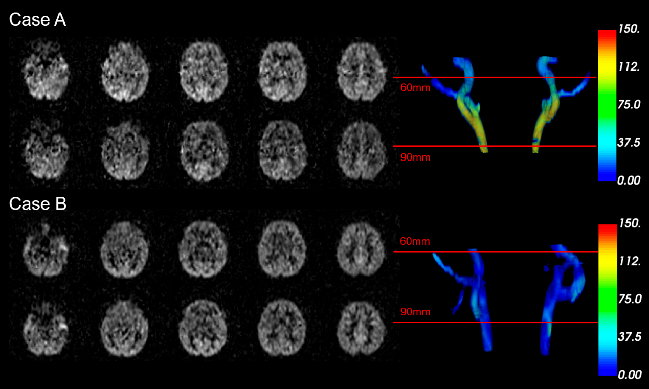
11 toddlers (2 female, 7.3±6.1 months) were scanned on a Philips Achieva 3.0T scanner. pCASL was acquired at two different labeling planes, the 60mm (about the C2/C3 segment and above the bifurcation) and the 90mm (default) below the AC-PC line. Other parameters were TR 3927ms, TE 11ms; bandwidth 3885Hz/px, FOV 200mm×200mm, resolution 3.75mm×3.75mm, and 10 slices with 6mm thickness. B0 map imaging was acquired on 4 of the 11 subjects using fast field echo, with TR 650ms, TE 7.0ms, bandwidth 434Hz/px, FOV 180mm×180mm, resolution 2.0mm×2.0mm, and 24slices with 3.5mm thickness. MRA was acquired with fast field echo with TR 15ms, TE 3.5ms, bandwidth 289Hz/px, FOV 160mm×160mm, resolution 1.0mm×1.0mm, and 30slices with 3mm thickness.
The grey matter region was segmented using the Otsu thresholding method. Signal-to-noise ratios (SNR) were calculated as the ratio of the mean signal in the grey matter to the standard deviation in the background region. The SNR improvement rate was calculated as r=(SNR60-SNR90)/SNR90. The carotid arteries were segmented from the MRA images using the 3D slicer. The B0 map was then registered to the MRA image using SPM12, and the off-resonance frequency was extracted and rendered using an in-house Python script.
Results
Greatly reduced labeling efficiency was observed at off-resonance frequencies. A more severe loss of labeling efficiency was observed at higher velocities, as indicated by a narrower region of high labeling efficiency, Fig. 1. Simulations indiciated that blood flow velocity and pulsatility had negligible effects on the labeling efficiency at on-resonance frequency, Fig. 2. The labeling efficiency decreased less than 8% across the range from 0.7x to 2.0x the adult blood flow profile. Furthermore, it was found that larger losses in labeling efficiency occurred at higher off-resonance frequencies and higher blood flow velocities, Fig. 3.Figure 4 shows ASL and the overlay of B0 map onto the MRA of two selected subjects. In subject A (16 months old), off-resonance frequencies were -144.63Hz and -55.19Hz at the labeling plane of 90mm and 60mm, respectively. Higher image quality was observed at the 60mm labeling plane. In subject B (13 months old), the off-resonance frequency was similar at both labeling planes, which resulted in similar image quality. Improved SNR was observed in 8 out of 11 infants (Table 1).
Discussion
Although flow velocities are associated with labeling efficiency, the contribution of peak velocity to labeling efficiency is low due to the pulsatile flow throughout the cardiac cycle and the laminar distribution across the vessel. Reduced labeling efficiency was observed at off-resonance frequency and was more severe at higher flow velocities. This may be the primary cause of reduced ASL image quality in infants. Additionally, the off-resonance effects varied among subjects, resulting in variable ASL image quality. By changing the labeling plane, the present work may help avoid the off-resonance frequency location associated with brain anatomy, Fig 4 and Table 1.Conclusion
High off-resonance frequency in the labeling plane primarily caused instability in infant ASL. Selecting a labeling plane based on anatomic features may result in a lower off-resonance frequency, which can effectively improve the ASL image.Acknowledgements
This work is supported by the National Key R&D Program of China (2022ZD0118004), the Alzheimer's Association (AARF-18-566347), Zhejiang Provincial Natural Science Foundation of China (LGJ22H180004, 202006140, and 2022C03057), and the MOE Frontier Science Center for Brain Science & Brain-Machine Integration, Zhejiang University.References
[1] Hu, Z., Jiang, D., Shepard, J., et al. Macrovasculature-suppressed ASL MRI in neonates: quantification of cerebral blood flow and arterial transit time. In Proceedings of the International Society for Magnetic Resonance in Medicine (ISMRM). 2023; Abstract 0047.
[2] Holdsworth D W, Norley C J D, Frayne R, et al. Characterization of common carotid artery blood-flow waveforms in normal human subjects. Physiological measurement. 1999; 20(3): 219.
[3] Yazici B, Erdogmus B, Tugay A. Cerebral blood flow measurements of the extracranial carotid and vertebral arteries with Doppler ultrasonography in healthy adults. Diagnostic and interventional radiology. 2005; 11(4): 195.
[4] Bode H, Wais U. Age dependence of flow velocities in basal cerebral arteries. Archives of disease in childhood. 1988; 63(6): 606-611.
[5] Zhao L, Vidorreta M, Soman S, et al. Improving the robustness of pseudo‐continuous arterial spin labeling to off‐resonance and pulsatile flow velocity. Magnetic resonance in medicine. 2017; 78(4): 1342-1351.
Figures