1998
Improved PCASL imaging of the lung using multiple labeling: Results of an ongoing study1Section on Experimental Radiology, University of Tübingen, Tübingen, Germany, 2Department of Diagnostic and Interventional Radiology, University of Tübingen, Tübingen, Germany, 3High-Field MR Center, Max Planck Institute for Biological Cybernetics, Tübingen, Germany, 4Medical Image and Data Analysis Lab, University of Tübingen, Tübingen, Germany
Synopsis
Keywords: Arterial Spin Labelling, Arterial spin labelling
Motivation: We have recently shown that a PCASL-bSSFP sequence provides high quality perfusion images of the lung. However, the perfusion signal is dependent on the respective cardiac cycle.
Goal(s): Our aim was to further increase the perfusion signal of the lung parenchyma and/or to shorten the examination time using a new PCASL approach with multiple labeling.
Approach: Four volunteers were examined using an ECG-triggered multiple labeling PCASL-bSSFP sequence with different number of labeling pulses, post labeling delays and repetition delays.
Results: The PCASL sequence with multiple labeling appears to provide a higher perfusion signal of the lung parenchyma, reducing sensitivity to cardiac cycle variations.
Impact: The new PCASL approach with multiple labeling appears to provide a significantly higher perfusion signal of the lung than measurements with single labeling pulse. The sequence makes the PCASL technique more robust and faster for future application in clinical practice.
Introduction
Evaluation of regional perfusion in the lungs is important for the diagnosis of pulmonary diseases. Arterial spin labeling (ASL) has been used for the non-invasive quantitaive assessment of perfusion in the lungs.1,2 We have recently shown that ECG-triggered pseudo-continuous ASL (PCASL) with bSSFP readout provides high quality perfusion images of the lung in healthy volunteers as well as in patients with pulmonary embolism.3,4 However, even if perfusion deficits due to acute pulmonary embolism can be reliably detected, the perfusion signal of the parenchyma is dependent on the respective cardiac cycle. Therefore, a further increase in the perfusion signal and/or a shortening of the examination time is helpful for the use of PCASL in clinical routine. Here we present the first results of a new approach to measure the temporal and spatial characteristics of pulmonary blood flow using an ECG-triggered multiple labeling PCASL imaging.Methods
Four healthy volunteers (two male) were examined on a 1.5 T MR scanner (MAGNETOM Avantofit, Siemens Healthcare, Erlangen Germany) using a PCASL sequence with ECG-triggered multiple labeling pulses and a bSSFP readout. First, twenty measurements with varying number of labeling pulses (NLs= 1, 2, 3, 4) and post labeling delays (PLDs= 200, 400, 600, 800, 1000ms) were performed. Secondly, in additional measurements with PLD of 1000 ms, the repetition delay (RD) was reduced from 2000 ms to 1000 ms. The labeling plane was placed nearly perpendicular to the main pulmonary artery (s. Figure 1A). A bSSFP readout was played once after a series of labeling pulses performed during the systolic period following ECG-triggering (s. Figure 1B). Each measurement was performed with 8 label/control image pairs under free-breathing condition. A proton-density weighted bSSFP image was acquired at the start of each measurement. Depending on sequence parameters NL, PLD, RD and cardiac cycle, the measurement time was about 1-2 min. The most important measurement parameters are listed in Table 1. The optical flow-based image registration approach was used to register PCASL images non-rigidly.5 The perfusion signal of the lung parenchyma was evaluated using the regions of interest (ROIs) placed in left and right lung, carefully avoiding large vessels.Results
PCASL measurements of the lungs with multiple labeling were successfully performed in all subjects. Exemplary PCASL images of all subjects are shown in Figure 2. Our results reveal that: (1) The perfusion signal of the lung parenchyma increases with increasing number of labeling pulses for all measured PLDs (s. Figure 3): e.g., the perfusion signal at NL=3 is approximately twice as high as the standard PCASL measurements with NL=1 and PLD=1000 ms. (2) At a short PLD (200, 400 ms), both pulmonary arteries and lung parenchyma show sufficiently high signal. (3) With increasing number of labeling pulses, the parenchymal perfusion signal seems to be less sensitive to changes in PLD. (4) No noticeable perfusion signal loss observed by reducing the repetition delay from 2000 ms to 1000 ms (s. Figure 4).Discussion
This work demonstrates the potential of PCASL bSSFP imaging of the lung at 1.5T using a series of ECG-triggered labeling pulses. These preliminary results show that the multiple labeling approach seems to significantly increase the perfusion signal in lung parenchyma compared to the standard single labeling sequence. This could reduce sensitivity to PLD changes and cardiac cycle variations. In addition, the use of a short repetition delay allows a faster examination without compromising the perfusion signal.Conclusion
The PCASL bSSFP sequence with multiple labeling appears to provide a significantly higher perfusion signal of the lung parenchyma than measurements with a single labeling pulse. The new sequence makes the PCASL of the lung more robust and faster for future application in clinical practice.Acknowledgements
No acknowledgement found.References
- Bolar DS, Levin DL, Hopkins SR, et al. Quantification of Regional Pulmonary Blood Flow Using ASL-FAIRER. Magn Reson Med 2006;55:1308–17.
- Martirosian P, Boss A, Fenchel M, et al. Quantitative lung perfusion mapping at 0.2 T using FAIR True-FISP MRI. Magn Reson Med 2006;55:1065–1074
- Seith F, Pohmann R, Schwartz M, et al. Imaging Pulmonary Blood Flow Using Pseudocontinuous Arterial Spin Labeling (PCASL) With Balanced Steady-State Free-Precession (bSSFP) Readout at 1.5T. J Magn Reson Imaging. 2020;52:1767-82.
- Othman AE, Liang C, Komma Y, et al. Free-breathing Arterial Spin Labeling MRI for the Detection of Pulmonary Embolism. Radiology. 2023;307(3):e221998.
- Gilliam C, Küstner T, Blu T. 3D motion flow estimation using local all-pass filters. 2016 IEEE 13th International Symposium on Biomedical Imaging (ISBI), 2016; 282–285.
Figures
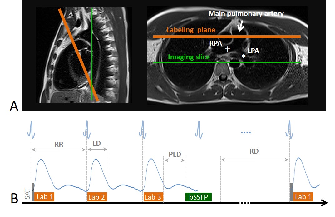
Figure 1: (A) Labeling plane (orange) was positioned nearly perpendicular to main pulmonary artery (arrow) and imaging slice (green) was in coronal orientation. LPA (asterisk) and RPA (cross) are the left and right pulmonary artery respectively. (B) Schema of PCASL sequence with series of three ECG-triggered labeling pulses (Lab 1-3) and bSSFP readout. (SAT: saturation pulse; RR: cardiac cycle; LD: labeling duration; PLD: post-labeling delay; RD: repetition delay).
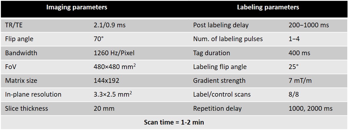
Table 1: Measurement parameters of multiple labeling PCASL sequence.
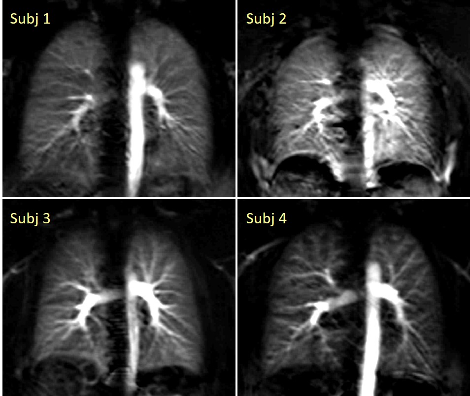
Figure 2: Exemplary PCASL perfusion-weighted images of the lungs in healthy subjects. Images were acquired using the proposed multiple labeling approach with NL=3, PLD=800 ms and RD=2000 ms. (NL – number of labeling pulses; PLD – post labeling delay; RD – repetition delay).
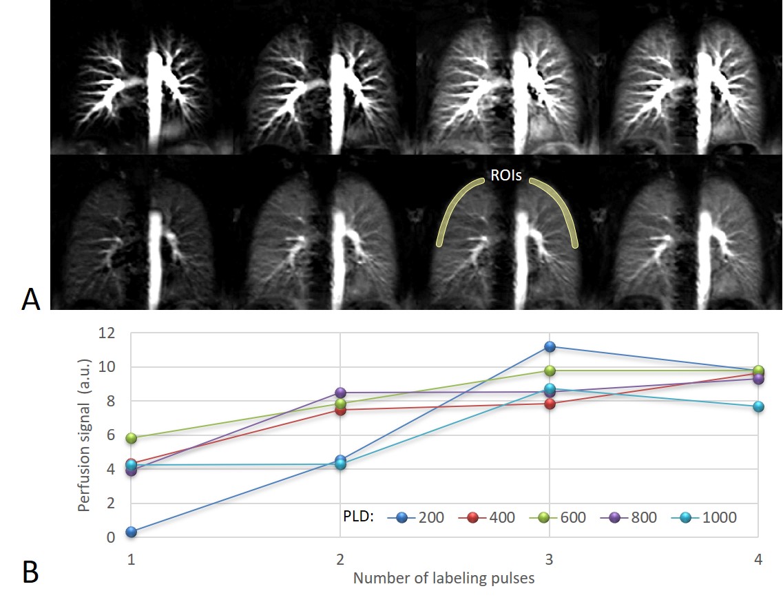
Figure 3: (A) Exemplary PCASL perfusion-weighted images of the lungs in a healthy subject. Images were acquired at a PLD of 200 ms (top) and 800 ms (bottom) and NL from 1 to 4 (left to right). RD was 2000 ms in all measurements. (B) Using multiple labeling, perfusion signal of the lung parenchyma (mean of ROIs) increases and reaches a maximum at NL of 3 to 4. (NL – number of labeling pulses; PLD – post labeling delay; RD – repetition delay).
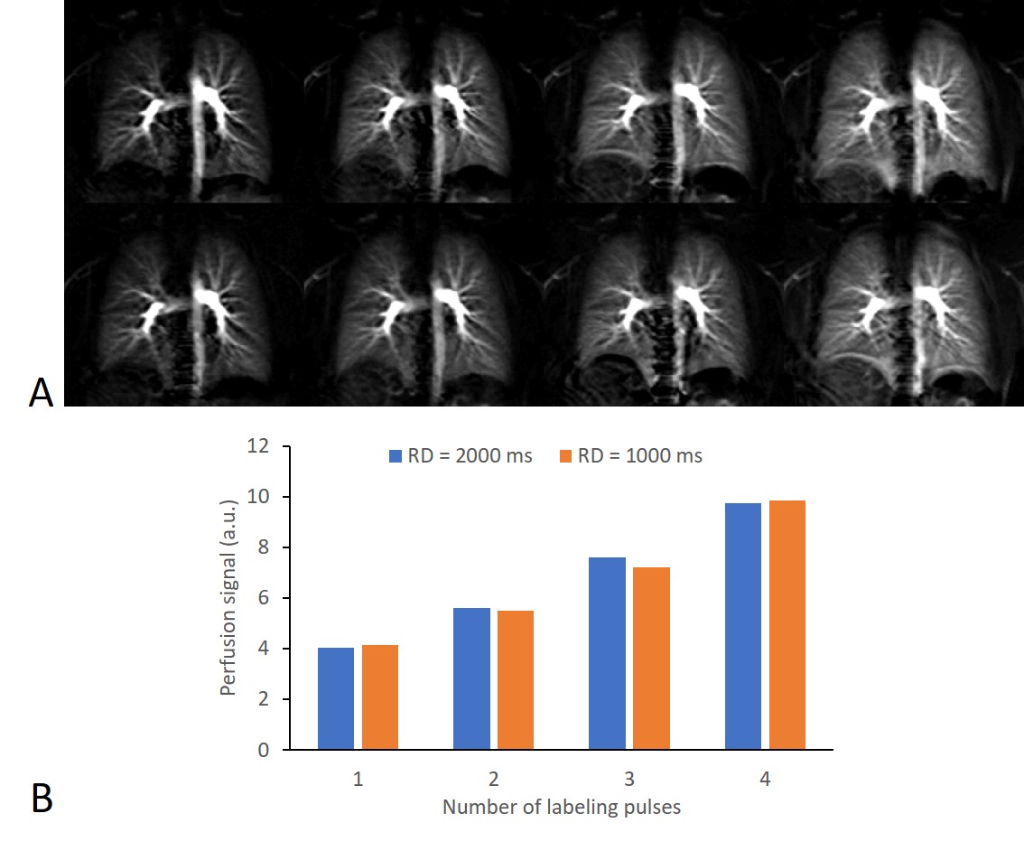
Figure 4: (A) PCASL perfusion-weighted images of the lungs in a healthy subject. Images were acquired at a RD of 2000 ms (top) and 1000 ms (bottom) and NL from 1 to 4 (left to right). PLD was 1000 ms in all measurements. (B) Only slight changes in the perfusion signal are observed between both RDs. (NL – number of labeling pulses; PLD – post labeling delay; RD – repetition delay).