1995
High fidelity, distortion-free brain diffusion-weighed imaging through Compressed SENSE combined Deep Learning reconstruction1MR R&D, Philips Healthcare, Suzhou, China, 2Philips Healthcare (China), Shanghai, China, 3MR Application, Philips Healthcare, Suzhou, China
Synopsis
Keywords: Head & Neck/ENT, Head & Neck/ENT
Motivation: Distortion-free brain diffusion-weighted imaging (DWI) remains a challenge due to susceptibility artifacts and low signal-to-noise ratio (SNR).
Goal(s): Assess the effectiveness of Compressed SENSE (CS) combined with deep learning (CS-DL) in improving brain DWI image quality.
Approach: We compared various acceleration schemes and reconstruction methods on TSE-DWI brain images.
Results: CS-DL with a factor of 4 improved image quality and SNR, while reducing scan time by 22%.
Impact: Implementation of CS-DL in TSE-DWI holds promise for high-fidelity, distortion-free imaging, facilitating detailed analysis of small brain abnormalities in regions affected by magnetic field inhomogeneity and susceptibility.
Introduction
Diffusion-weighted magnetic resonance imaging (DWI) and its apparent diffusion coefficient (ADC) have extensive clinical applications in acute cerebral infarction, tumor characterization, and the assessment of acute stroke, multiple sclerosis, and tumors. Single shot echo-planar imaging (ssEPI) often results in geometric distortion and susceptibility artifacts due to inhomogeneous magnetic field and line phase discrepancies in k-space, which hinders the identification of small lesions in such regions as inner ear, pituitary, oral cavity and orbit1,2. Turbo spin-echo (TSE)-DWI is less sensitive to susceptibility artifacts and decoupled from magnetic field homogeneity, thus eliminating image distortion and providing distortion-free images, however, it suffers from low signal-to-noise ratio (SNR) and consequently limited spatial resolution.Compressed SENSE (CS) has been demonstrated promising results in improving EPI based DWI3,4, and its integration with deep learning (CS-DL) allows for superior de-noising performance6. In this study, we aim to assess the impact of CS and CS-DL on TSE-DWI brain images.
Methods
Four volunteers were scanned on an Ingenia Elition 3.0 T MR system (Philips Healthcare, the Netherlands) under written informed consents. The 16-channel head array coil was used to acquire the anatomical T2-weighted (T2w) and single-shot TSE-based diffusion-weighted (TSE-DWI) images. The scanning parameters of each acquisition protocol were listed in Table 1. For the scan acceleration schemes, we used SENSE factor 2.5 as a baseline, and repeated the scans using different CS factors (listed in Table 1). The data acquired from SENSE and CS acquisitions were reconstructed using their default reconstruction methods. Additionally, the CS data was also reconstructed using the Adaptive-CS-Net neural network model described in [5].To quantitatively compare the image quality, six ROIs were manually delineated, covering white matter, gray matter and mixed regions at upper and lower brains on different subjects ( k=1,…,6). These ROIs were then mapped to the images obtained using different TSE-DWI scanning schemes. The signal-to-noise ratio (SNR) of each ROI ( ) was calculated as: , where SI stands for the signal intensity.
Results
Figure 1 displays representative TSE-DWI brain images using different acceleration and reconstruction schemes and the corresponding T2w image as anatomical reference. With the same acquisition time of TSE-DWIs (4m30s), the CS-DL approach notably improved the image quality, exhibiting superior de-noising capabilities. Figure 2 illustrates the SNR of the six ROIs for different scanning schemes. CS acquisitions obviously improved the SNR in most ROIs compared to conventional SENSE acceleration scans. Moreover, the CS-DL reconstruction outperformed CS reconstruction, particularly evident at CS-DL factor of 3, providing the highest SNR under the same acquisition time. Further investigation into the impact of increasing CS acceleration factors was conducted, as shown in Figure 3. While TSE-DWI with SENSE factor of 4 failed, the CS-DL with factor 4 yielded comparable results to the SENSE 2.5 images, with a reduced acquisition time of 3m33s compared to 4m30s for SENSE 2.5 images, reflecting a 22% time reduction.Discussion and Conclusion
In this work, we show that the distortion-free DWI can be achieved by TSE-DWI with improved SNR using CS-DL technique, and CS-DL with factor 4 provides improved image quality in terms of signal-to-noise ratio as well as reducing the scan time by 22%. TSE-DWI combined with CS acquisition and DL reconstruction can produce high-fidelity, distortion-free DW images, offering significant potential for the detailed visualization of small brain abnormalities in regions with magnetic field inhomogeneity and susceptibility, such as the orbit, inner ear, and pituitary.Acknowledgements
No acknowledgement found.References
1. Gumeler E, Parlak S, Yazici G, et al. Single shot echo planar imaging (ssEPI) vs single shot turbo spin echo (ssTSE) DWI of the orbit in patients with ocular melanoma. Br J Radiol. 2021 Feb 1;94(1118):20200825.
2. Hirata, Kenichiro; Nakaura, Takeshi; Okuaki, Tomoyuki, et al. Comparison of the image quality of turbo spin echo- and echo-planar diffusion-weighted images of the oral cavity. Medicine 97(19):p e0447, May 2018.
3. Kaga T, et al. Diffusion-weighted imaging of the abdomen using echo planar imaging with compressed SENSE: Feasibility, image quality, and ADC value evaluation. Eur J Radiol. 2021 Sep;142:109889.
4. Tamada T, et al. Clinical application of single-shot echo-planar diffusion-weighted imaging with compressed SENSE in prostate MRI at 3T: preliminary experience. MAGMA. 2022 Aug;35(4):549-556.
5. Pezzotti N, Yousefi S , MS Elmahdy, et al. An Adaptive Intelligence Algorithm for Undersampled Knee MRI Reconstruction: Application to the 2019 fastMRI Challenge[J]. 2020.
6. K. Yuda, et al. Myocardial T2-weighted black-blood imaging with a deep learning constrained Compressed SENSE reconstruction. ISMRM 2021.
Figures

Table 1 scanning parameters of T2w and TSE-DWI imaging with different acceleration and reconstruction schemes.
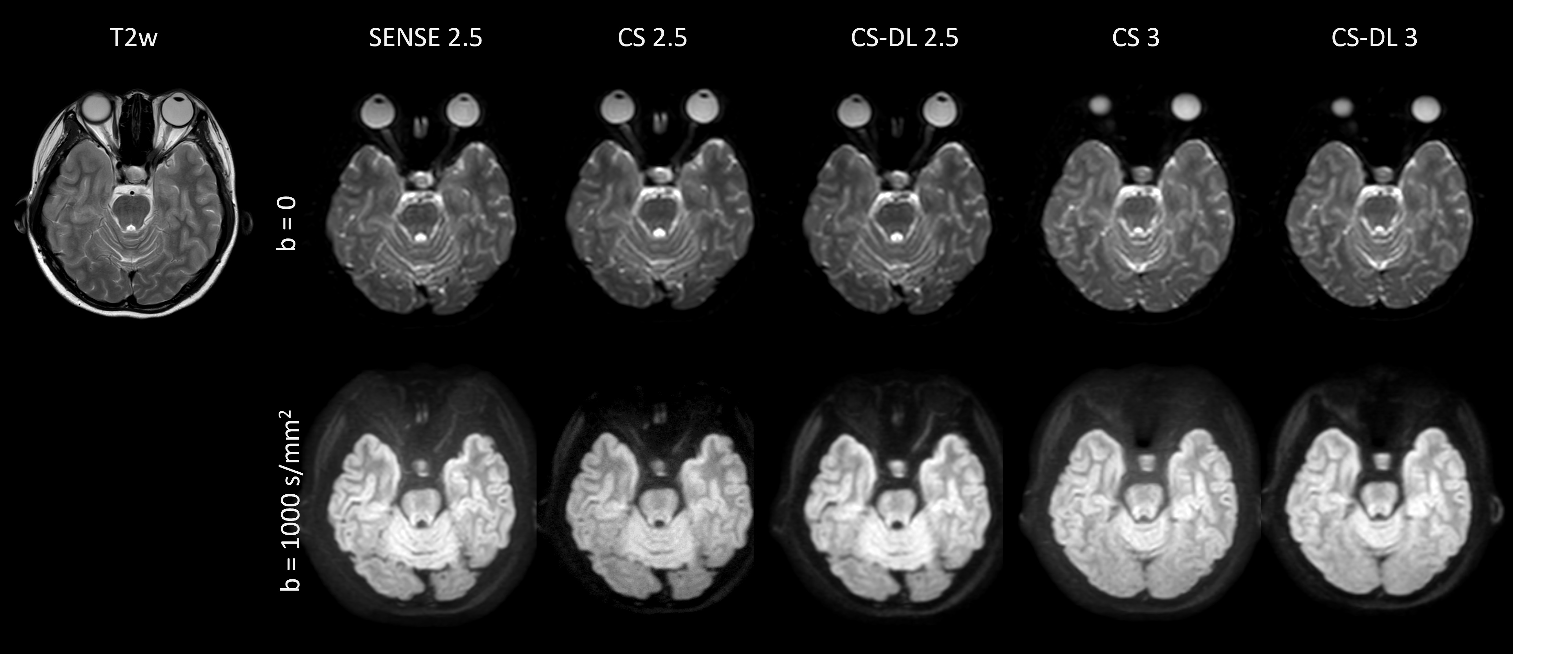
Fig.1 Brain TSE-DW images using different acceleration and reconstruction schemes were shown in axial orientation. The images of b=0 and b=1000 s/mm2 were shown with SENSE or Compressed SENSE (CS) with different acceleration factors. T2-weighted image was shown as anatomical reference. Acquisition time for all TSE-DW sequences were the same (4m30s).
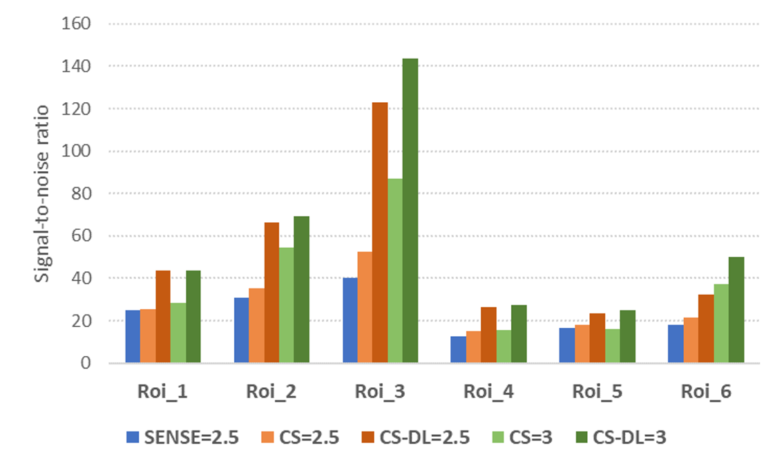
Fig.2 Comparison of signal-to-noise ratio for different acceleration and reconstruction schemes. Results from six ROIs were shown. Compared to conventional SENSE acceleration, CS acquisitions improved the SNR in most ROIs; the DL reconstruction combined with CS further de-noised the images compared to the CS wavelet-based reconstruction.
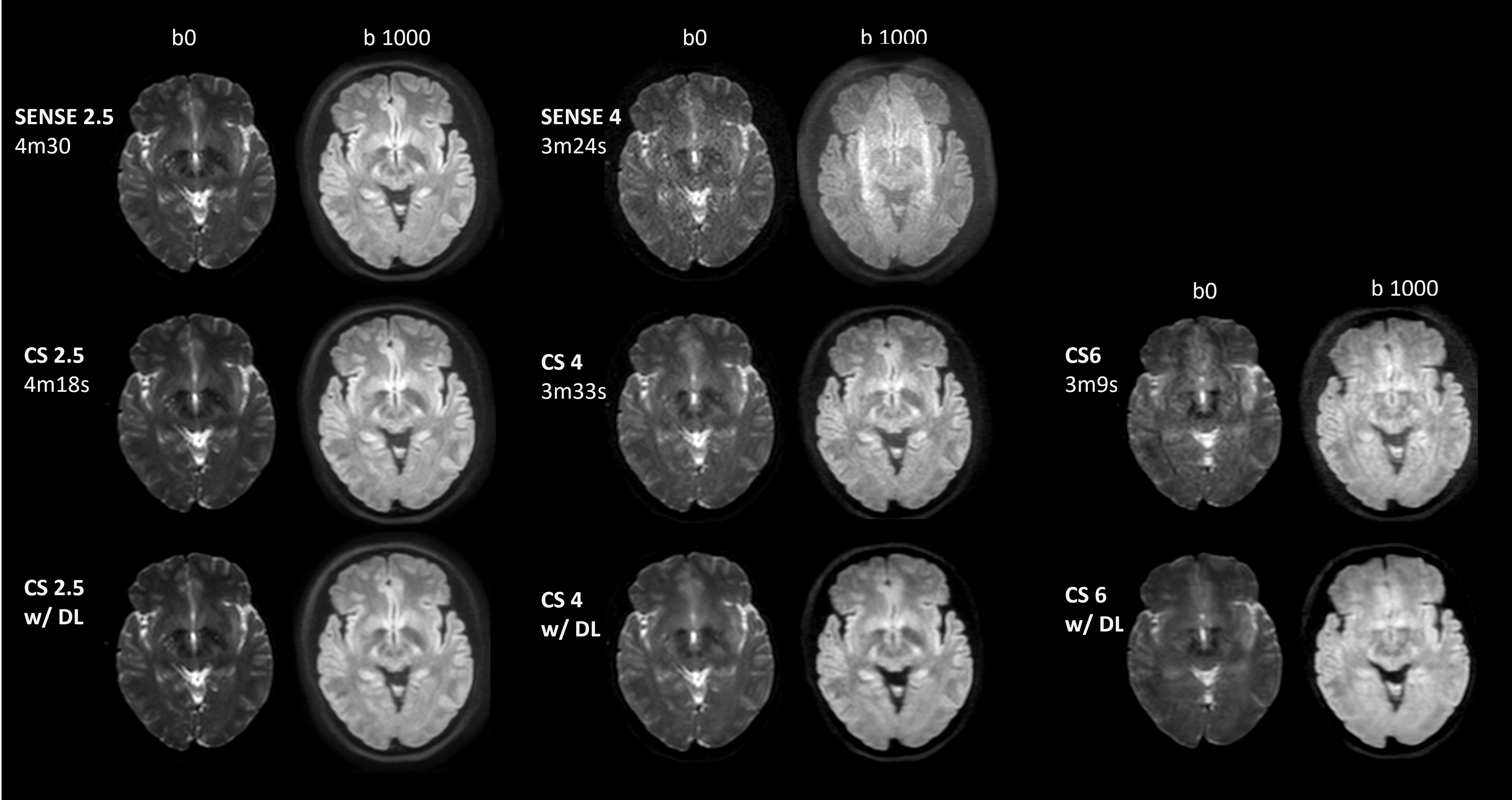
Fig. 3 Demonstration of the impact of increasing CS acceleration factors with regard to image quality.