1987
Application of deep learning–based image reconstruction in MRI of the temporomandibular joint to improve image quality and reduce scan time1Xuanwu Hospital Capital Medical University, Beijing, China, 2GE HealthCare, Beijing, China
Synopsis
Keywords: AI/ML Image Reconstruction, Machine Learning/Artificial Intelligence
Motivation: MRI examination of temporomandibular joint takes a long time to scan, especially when scanning in the open-mouth position, the patient cannot maintain the open-mouth state for a long time, it is easy to cause movements of the temporomandibular joint, resulting in motion artifacts in the scanned image and failure of the examination. On the premise of ensuring image quality, it is particularly important to reduce scan time.
Goal(s): Applying deep learning reconstruction to improve temporomandibular joint MRI image quality and reduce scanning time.
Approach: clinical experiments.
Results: Deep learning reconstruction can improve image quality and significantly shorten scanning time of temporomandibular joint MRI.
Impact: Deep learning reconstruction can significantly shorten scan time while improving image quality, helping radiologists to diagnose quickly and confidently,helping patients to spend less time feeling anxious in an MRI and more time living their life.
Purpose: To explore the application value of DLR in improving the quality of MRI imaging of the temporomandibular joint and shortening the scanning time.
Materials and Methods: 40 volunteers who agreed to undergo temporomandibular joint scanning from March 10, 2023 to April 30, 2023 were prospectively included, and each volunteer underwent temporomandibular joint MRI PDWI scan and DLR-PDWI scan. Two radiologists conducted qualitative and quantitative evaluations of the image quality of the two groups respectively. Qualitative evaluation: Likert scale (5 points scale) was used to qualitatively score the image anatomical structure clarity and overall image quality. Quantitative evaluation uses signal-to-noise ratio (SNR) and contrast-to-noise ratio (CNR) to quantitatively evaluate image quality. One-way analysis of variance and Friedman test were performed on normally distributed and non-normally distributed data respectively. The intra-class correlation coefficient (ICC) was used to compare the consistency of the subjective ratings of the two readers.
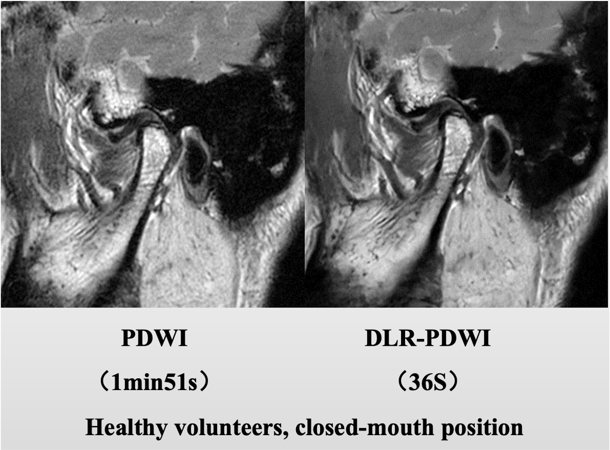
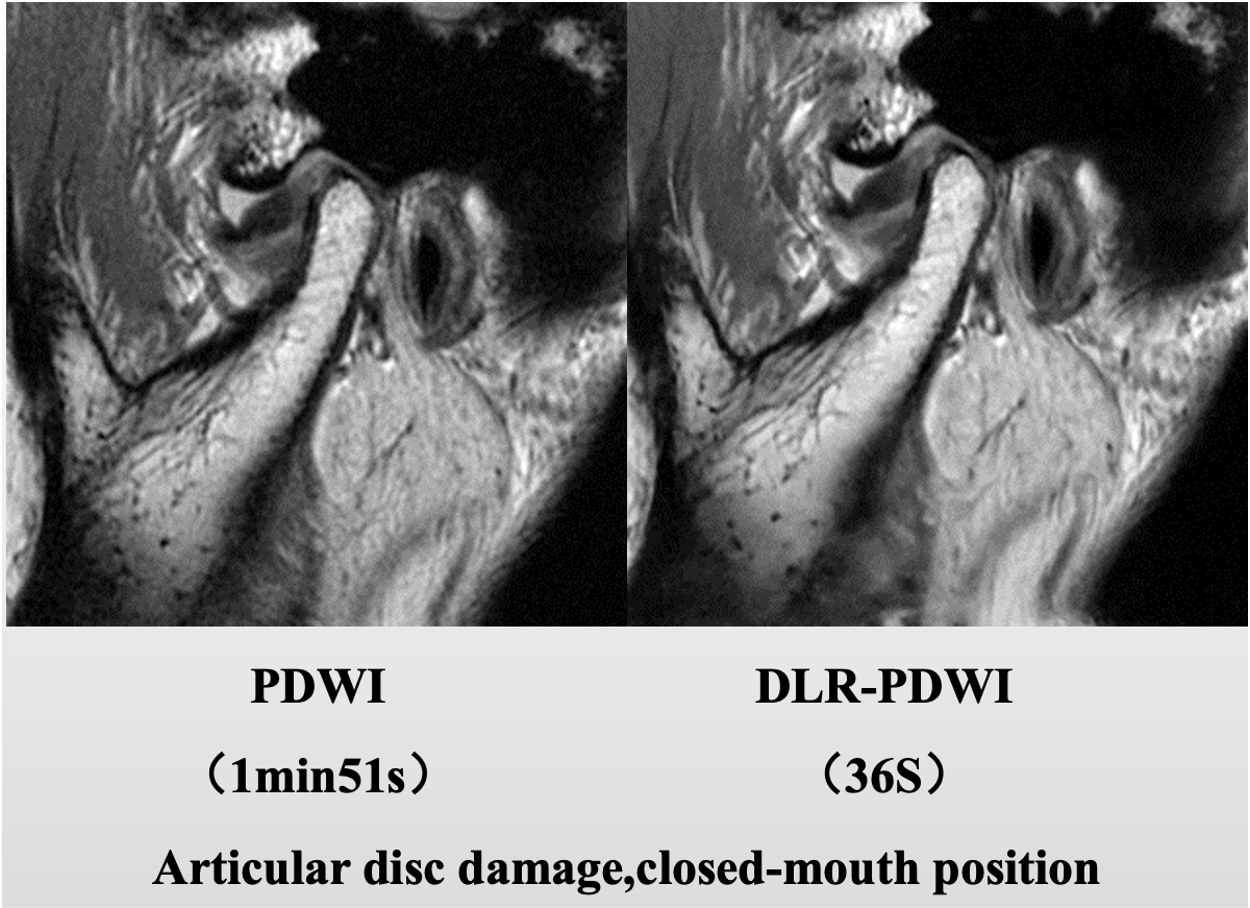
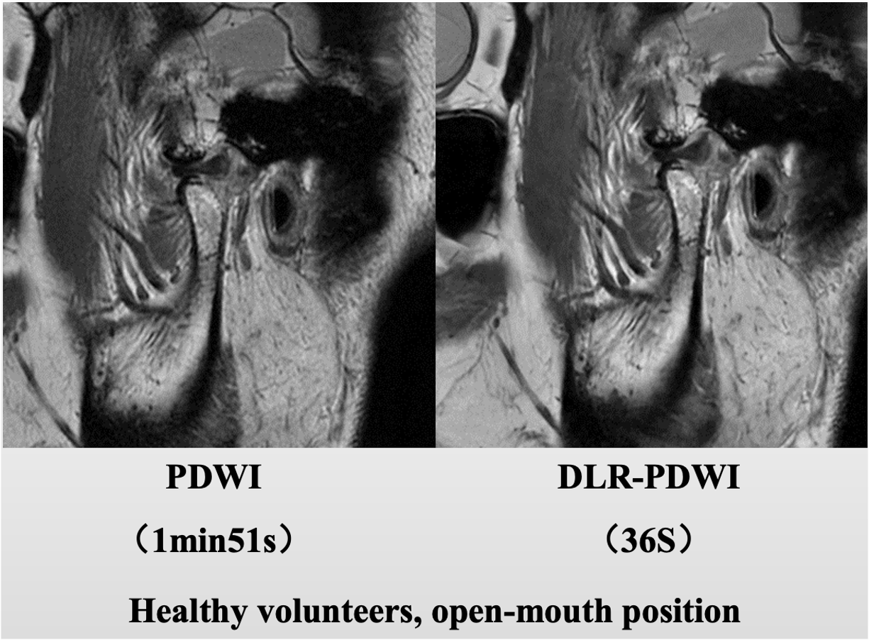
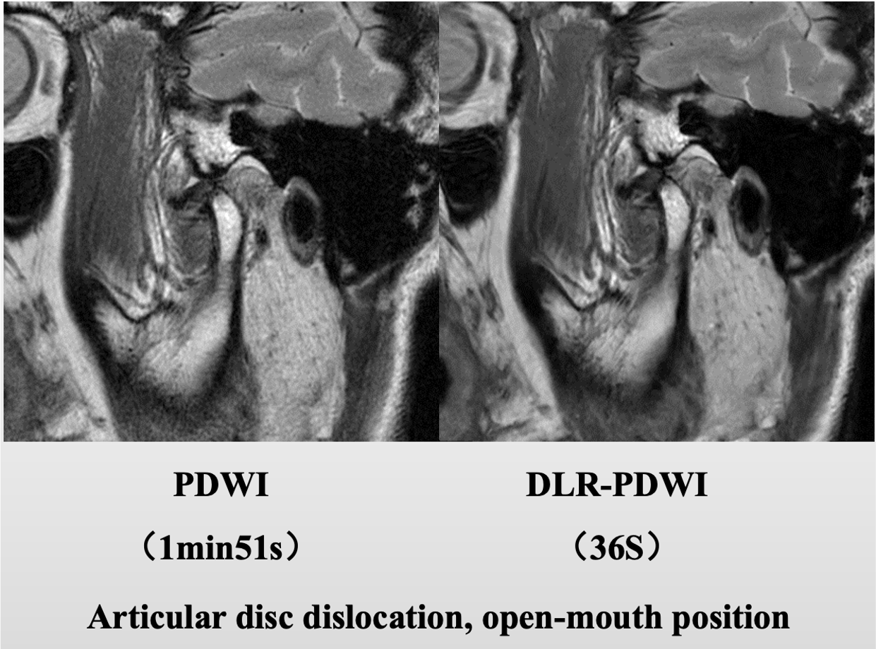
Results: Compared with the PDWI group, the scanning time of the DLR-PDWI group was shortened by 67%. The agreement between the two radiologists on the subjective ratings of image anatomical structure clarity and overall image quality was good (ICCs were 0.80 and 0.78, respectively). The scoring results of the two doctors showed that there were significant differences in the image anatomical structure clarity and overall image quality scores between the PDWI group and the DLR-PDWI group (P<0.05); The difference in SNR and CNR between the two groups of images is statistically significant (P<0.05); the qualitative and quantitative evaluation results of the DLR-PDWI group were significantly better than those of the PDWI group.
Conclusion: DLR can significantly shorten scanning time and improve image quality.
Acknowledgements
No acknowledgement found.References
1. Almansour H, Herrmann J, Gassenmaier S, Afat S, Jacoby J, Koerzdoerfer G, Nickel D, Mostapha M, Nadar M, Othman AE. Deep Learning Reconstruction for Accelerated Spine MRI: Prospective Analysis of Interchangeability. Radiology. 2023 Mar;306(3):e212922. doi: 10.1148/radiol.212922. Epub 2022 Nov 1. PMID: 36318032.
2. Johnson PM, Lin DJ, Zbontar J, Zitnick CL, Sriram A, Muckley M, Babb JS, Kline M, Ciavarra G, Alaia E, Samim M, Walter WR, Calderon L, Pock T, Sodickson DK, Recht MP, Knoll F. Deep Learning Reconstruction Enables Prospectively Accelerated Clinical Knee MRI. Radiology. 2023 Apr;307(2):e220425. doi: 10.1148/radiol.220425. Epub 2023 Jan 17. PMID: 36648347; PMCID: PMC10102623.
3. Kaniewska M, Deininger-Czermak E, Getzmann JM, Wang X, Lohezic M, Guggenberger R. Application of deep learning-based image reconstruction in MR imaging of the shoulder joint to improve image quality and reduce scan time. Eur Radiol. 2023 Mar;33(3):1513-1525. doi: 10.1007/s00330-022-09151-1. Epub 2022 Sep 27. PMID: 36166084; PMCID: PMC9935676.
4. Kim M, Kim HS, Kim HJ, Park JE, Park SY, Kim YH, Kim SJ, Lee J, Lebel MR. Thin-Slice Pituitary MRI with Deep Learning-based Reconstruction: Diagnostic Performance in a Postoperative Setting. Radiology. 2021 Jan;298(1):114-122. doi: 10.1148/radiol.2020200723. Epub 2020 Nov 3. PMID: 33141001.
5. Ueda T, Ohno Y, Yamamoto K, Murayama K, Ikedo M, Yui M, Hanamatsu S, Tanaka Y, Obama Y, Ikeda H, Toyama H. Deep Learning Reconstruction of Diffusion-weighted MRI Improves Image Quality for Prostatic Imaging. Radiology. 2022 May;303(2):373-381. doi: 10.1148/radiol.204097. Epub 2022 Feb 1. PMID: 35103536.
Figures