1984
Intracranial Vascular Segmentation in TOF-MRA Images Using Transfer Learning1Department of Medical Imaging, Henan Provincial People's Hospital & People's Hospital of Zhengzhou University, Zhengzhou, China, 2Institute for Integrated Medical Science and Engineering, Henan Academy of Sciences, Zhengzhou, China, 3School of Medicine, Tsinghua University, Beijing, China, 4Shukun Technology Co., Ltd, Beijing, China
Synopsis
Keywords: AI/ML Image Reconstruction, Vessels
Motivation: Addressing the challenge of segmenting cerebral vessels in TOF-MRA images, we explored transfer learning to overcome the need for large, annotated datasets.
Goal(s): Assess the feasibility of using a refined CTA-based 3D CNN model for MRA vascular segmentation with a limited dataset.
Approach: Implemented transfer learning on a ResU-Net3 model, initially trained on CTA scans, fine-tuned with a small MRA dataset.
Results: Post-transfer learning, the model's DSC improved dramatically, indicating effective MRA vessel segmentation with limited data.
Impact: This study benefits radiologists by streamlining the segmentation of cerebral vessels in MRA, reducing the workload associated with annotation. The method has the potential to be integrated into clinical workflows, enhancing the efficacy of vascular reconstruction in clinical settings.
Introduction
Time-of-flight magnetic resonance angiography (TOF-MRA) is an essential non-invasive imaging modality, playing a crucial role in visualizing and evaluating cerebral arteries for the diagnosis of cerebrovascular diseases1,2. However, segmenting vessels within 3D TOF-MRA volumes presents considerable challenges due to the brain's complex anatomy and visual resemblance of some background regions to cerebral vessels3.Deep learning models for vascular segmentation heavily depend on extensive, high-quality annotations by experts, a process that is laborious and time-consuming. Utilizing large datasets with tens of thousands of cases, pioneering studies in vascular imaging have implemented physiological anatomical-based 3D convolutional neural networks, enabling high-precision vessel segmentation in 3D computed tomography angiography (CTA) images4.
Transfer learning leverages knowledge from one domain to mitigate data scarcity in related tasks. Given its potential, this study aims to assess the feasibility of using transfer learning for segmenting vessels in MRA on small datasets.
Methods
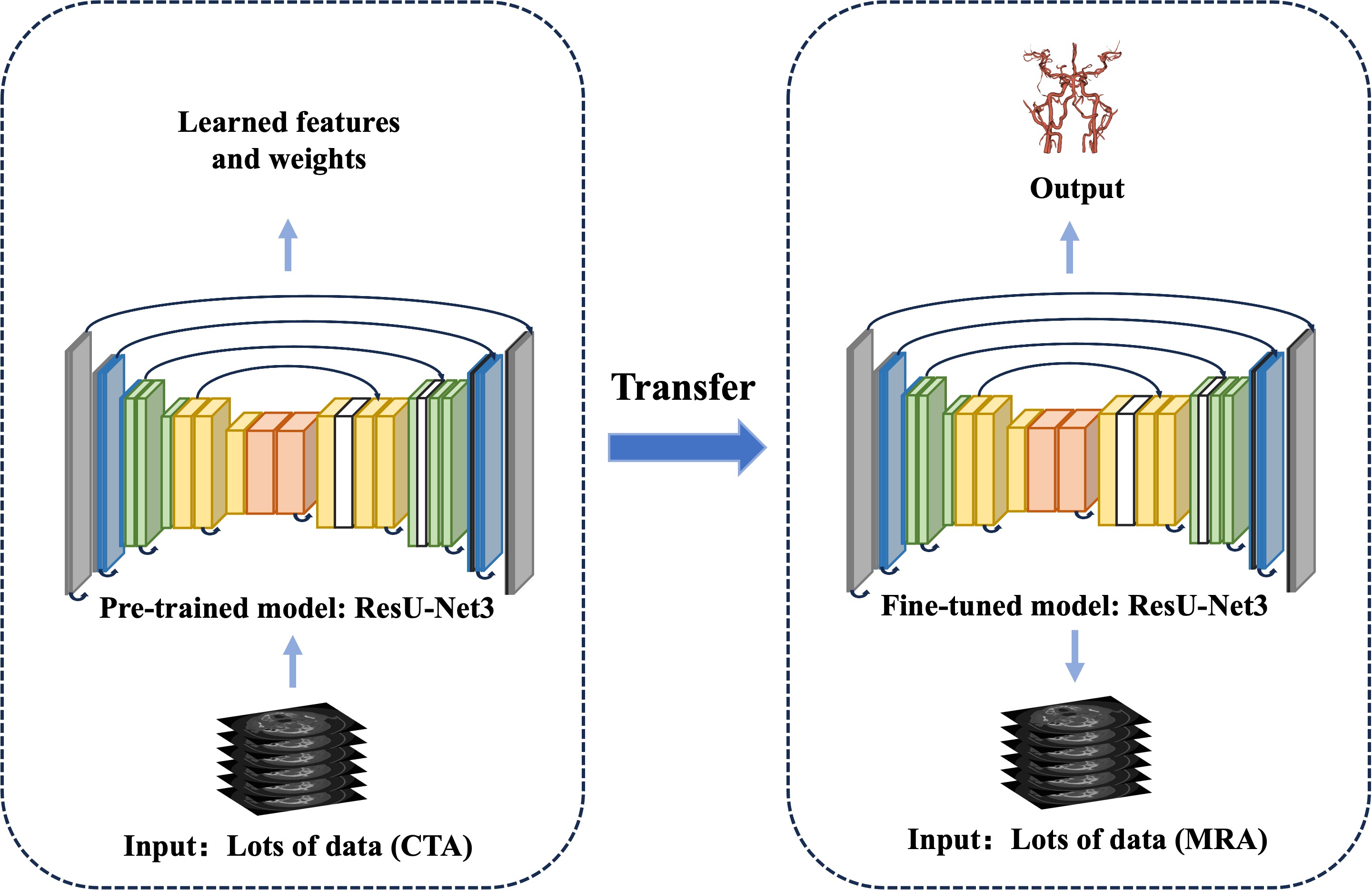
We utilized the MIDAS MRA TOF dataset of volunteers, imaged on a 3T scanner following standardized protocols, with matrix and voxel sizes of 448×448×128 and 0.51×0.51×0.8 mm³.5 Previous work supplied arterial labels for 20 samples from this collection2. With these labeled data, 15 and 5 patients were randomly chosen for the training and validation datasets, respectively, and we applied transfer learning to refine a CTA to MRA vascular segmentation model.As previously described, our proposed CTA reconstruction system utilizes an optimized 3D CNN, which has significantly lowered the labor intensity and error rate for technicians4. Trained and tested with 18,766 head and neck CTA scans, the system boasts an independent test set accuracy of 0.931 and a clinical qualification rate of 92.1%. Key components include the cascaded ResU-Net model and CGPM. Notably, the ResU-Net3 model precisely segments vascular structures based on anatomy, effectively identifying and differentiating major arteries such as the aorta, carotid, and intracranial arteries. The addition of CGPM corrects any segmentation errors, ensuring accurate morphological recognition.
Our research utilized transfer learning to fine-tune the ResU-Net3 model parameters, effectively bridging the distribution gap between source CTA data and target MRA data, thus achieving precise arterial segmentation transfer from CTA to MRA, overall framework as illustrated in Figure 1. The model was trained on a set of 15 MRA images, each paired with a corresponding vascular segmentation mask. Throughout the training process, we adjusted the model parameters through back-propagation and gradient descent algorithms to minimize the loss function, specifically employing the momentum stochastic gradient descent (momentum SGD) method for this optimization. The performance during training guided the fine-tuning of hyperparameters, including settings for learning rate, batch size, and the number of training iterations, all aimed at enhancing the model's efficacy. The accuracy of the vascular segmentation was quantified using the Dice Similarity Coefficient (DSC).
Results
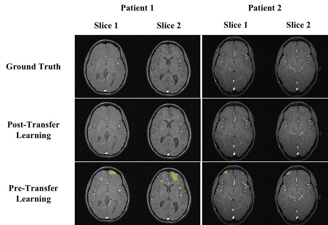
This study has achieved precise segmentation of intracranial vessels. Prior to transfer learning, the Dice Similarity Coefficient (DSC) for the model on the training and testing datasets was merely 0.145 and 0.172, respectively. After fine-tuning, the DSCs significantly improved to 0.766 for the training set and 0.732 for the testing set. Compared to the initial results, there was a substantial increase in both, amounting to 429.3% and 325.7%, respectively. The details are presented in Table 1. Typical segmentation results from the training and testing datasets are illustrated in Figure 2.Discussion
Previous studies have predominantly focused on the segmentation of intracranial vessels using CTA. However, MRA has significant advantages in clinical settings when compared to CTA. The main challenge with MRA is that image post-processing is labor-intensive and requires the specialized knowledge of radiologists.To overcome this challenge, we have introduced a novel and efficient method for the automatic segmentation of MRA images. Our research suggests that by leveraging an established CTA vessel segmentation network and applying transfer learning with fine-tuning on a small dataset, the predictive performance of the model can approximate that of a deep learning model specifically tailored for large MRA datasets6, 7.
In the future, we plan to expand our dataset and conduct more comprehensive studies and tests using data from multiple manufacturers. Our ongoing research will also focus on automating image reconstruction based on the arterial vessel segmentation and evaluating whether the reconstruction quality meets the diagnostic standards of radiology, as well as assessing the model’s efficiency for clinical application.
Conclusion
In conclusion, this study has developed an automated segmentation network for intracranial vessels, demonstrating the feasibility of employing transfer learning for segmenting vessels in MRA images using small datasets. This approach not only improves image segmentation but also advances MRA image analysis and its clinical applications.Acknowledgements
The authors declare that they have no conflicts of interest concerning this article.References
[1] Pal S C, Banerjee S, Toumpanakis D, et al. Segmentation of Major Cerebral Vessel from MRA images and Evaluation using U-Net Family[C]//2022 IEEE 6th International Conference on Condition Assessment Techniques in Electrical Systems (CATCON). IEEE, 2022: 235-238.
[2] Hilbert A, Madai V I, Akay E M, et al. BRAVE-NET: fully automated arterial brain vessel segmentation in patients with cerebrovascular disease[J]. Frontiers in artificial intelligence, 2020: 78.
[3] Chen Y, Jin D, Guo B, et al. Attention-assisted adversarial model for cerebrovascular segmentation in 3D TOF-MRA volumes[J]. IEEE Transactions on Medical Imaging, 2022, 41(12): 3520-3532.
[4] Fu F, Wei J, Zhang M, et al. Rapid vessel segmentation and reconstruction of head and neck angiograms using 3D convolutional neural network[J]. Nature communications, 2020, 11(1): 4829.
[5] Bullitt, Elizabeth, UNC. ITKTubeTK - Bullitt - Healthy MR Database. https://data.kitware.com/#collection/591086ee8d777f16d01e0724/folder/58a372e38d777f0721a64dc6. Accessed November 8, 2023.
[6] Alidoost M, Ghodrati V, Ahmadian A, et al. Model utility of a deep learning-based segmentation is not Dice coefficient dependent: A case study in volumetric brain blood vessel segmentation[J]. Intelligence-Based Medicine, 2023, 7: 100092.
[7] Pal S C, Toumpanakis D, Wikström J, et al. Multi-level Residual Dual Attention Network for Major Cerebral Arteries Segmentation in MRA towards Diagnosis of Cerebrovascular Disorders[J]. IEEE Transactions on NanoBioscience, 2023.
Figures