1974
CECNN-B1: Confidence-Enhanced CNN in B1 inhomogeneity Correction for Quantitative CEST MRI at 5 T1Paul C. Lauterbur Research Center for Biomedical Imaging, Shenzhen Institute of Advanced Technology, Chinese Academy of Sciences, Shenzhen, Guangdong, China
Synopsis
Keywords: Analysis/Processing, CEST & MT
Motivation: B1-correction is important in CEST MRI. Conventional correction methods require multiple B1 acquisitions, making them less practical in clinical adoptions.
Goal(s): This study proposed a CNN-based model that can correct B1 inhomogeneity from a single B1 acquisition.
Approach: A CECNN-B1 model incorporating B1 distribution was designed to enhance the confidence of CEST quantification, and its performance was evaluated on a creatine phantom at 5 T.
Results: Substantial variation of B1 distribution was observed, resulting in inhomogeneous CEST map. After correction, the B1-induced artifact was effectively alleviated. The image quality of the corrected CEST map was superior to that calculated with conventional interpolation method.
Impact: The proposed CECNN-B1 model enabled B1 inhomogeneity correction from single B1 acquisition. A creatine phantom study showed its superiority over the conventional interpolation methods requiring multiple B1 acquisitions, providing an efficient way for improved CEST MRI on high-field scanners.
INTRODUCTION
Chemical exchange saturation transfer (CEST) imaging can detect endogenous metabolites/compounds and exogenous CEST agents1. Its contrast depends on radiofrequency (RF) saturation level B1. Spatial variation of B1 would deviate CEST quantification, especially on high-field scanners2, 3. Conventional B1-corrected methods require multiple B1 acquisitions, which lengthened the scan time and are not desired in clinical settings4, 5.In this study, a deep learning-based B1 correction method was developed, and its feasibility in correcting B1 inhomogeneity in CEST MRI was evaluated on a creatine phantom from single B1 acquisition at 5 T.
MATERIALS AND METHODS
MRI study: A phantom consisting of two identical 60-mM creatine vials was prepared and imaged on a 5 T MR scanner (uMR Jupiter, Shanghai UIH, China) using a 48-channels head coil. Three slices of CEST images, relative B1-field (rB1) distributions, and WASSR6 maps were acquired at four B1 pulse amplitudes (0.75, 1.0, 1.5 and 2 µT), all with the same image resolution (2.08×2.08×10 mm3) and a matrix size of 96×96×3. The readout FSE sequence parameters for CEST images were Tsat = 3000 ms, TR = 6000 ms, TE = 6.72 ms and a flip angle of 110°, without acceleration. Z-spectra were acquired with 61 equidistant frequency offsets ranging from -3 to 3 ppm. An unsaturation scan also performed at +100 ppm before the offset. WASSR maps were collected from -0.5 to 0.5 ppm with intervals of 0.05 ppm using sequence of Tsat = 300 ms, TR = 1500 ms. rB1 distributions were acquired using a pre-conditioning RF pulse with TurboFLASH7 readout sequence (TR = 3.6 ms, TE = 1.8 ms, flip angle = 70°, NEX = 1). The scan times were 6.1 mins for CEST images, 32s for WASSR, and 16s for the rB1 map.Data analysis: Saturation scans was normalized by the unsaturation scan, interpolated, and corrected for B0 inhomogeneity using the WASSR approach6. Conventional interpolation-based B1 correction method employed four above B1 acquisitions was used to correct Z-spectra4, from which creatine CEST effect was quantified as $$$ MTR_{Rex}=1/Z(Δω)-1/Z(Δω)$$$8.
Model design: Figure 1 illustrates the CECNN-B1 structure. It consists a three CNN layers as the main body, an output tail for CEST effect $$$\widehat{y^1_\theta}$$$, and rB1 value $$$\widehat{y^2_\theta}$$$, and an uncertainty tail for standard derivation (SD) of $$$\sigma_{\theta}$$$. A combined negative Gaussian log-likelihood (GNLL) training loss was employed as
$$argmin_\theta{\left\{ -logp_\theta^1(y^1;\widehat{y^1_\theta},\sigma_{\theta}(\textbf{x})) -logp_\theta^2(y^2;\widehat{y^2_\theta},\sigma_{\theta}(\textbf{x})) \right\}}$$
where, $$$\textbf{x}$$$ is acquired Z-spectra, $$$y^1$$$ is the reference of $$$MTR_{Rex}$$$ measured from the conventional interpolation-based B1-correction method and $$$y^2$$$ is ground-truth (GT) rB1. $$$p_\theta$$$ is a Gaussian likelihood function with model weights $$$\theta$$$. The $$$\sigma_{\theta}$$$ was shared by both $$$p_\theta^1$$$ and $$$p_\theta^2$$$, thereby enhancing model confidence, especially with the experimentally acquired $$$rB_1$$$ map.
Pixel-wise Z-spectra under a representative nominal B1 of 0.75 µT from two slices were used as the training dataset, which was augmented with the addition of Gaussian noise levels ranging from 0.001 to 0.01, result in 14,610 Z-spectra. Respective rB1 and $$$MTR_{Rex}$$$ maps were used as GT and reference labels. Creatine CEST map on the other slice at the same B1 level was calculated to test the performance of the network in B1 correction. Model trained and tested on a NVIDIA RTX A2000 GPU.
RESULTS
Figure 2 shows the rB1 map and $$$ MTR_{Rex}$$$ images before and after conventional B1 correction. The notably inhomogeneous rB1 distribution led to spatially varied $$$ MTR_{Rex}$$$ values, which was corrected with the conventional interpolation-based B1-correction algorithm.The trained CECNN-B1 model weights saved at epoch 1000 was used for referencing, where validation Huber loss of rB1 was converged (Figure 3). Figure 4 shows the rB1 map obtained from the proposed CECNN-B1 method was similar with the acquired rB1 map. Comparable $$$MTR_{Rex}$$$ maps were obtained with B1 inhomogeneity-induced artifact mitigated between the conventional interpolation-based and the proposed CECNN-B1 methods. Moreover, the proposed CECNN-B1 model generated more homogeneous $$$ MTR_{Rex}$$$ map than the interpolation-based method, with narrower width histogram distribution and smaller SD of 0.42 % (Table 2).
DISCUSSION AND CONCLUSION
This study developed a deep learning-based CECNN-B1 model to correct B1-inhomogeneity artifact in CEST MRI, with the confidence enhanced by sharing uncertainty measurement for recovery of rB1 map. A creatine phantom study at 5 T demonstrated the ability of the model in reducing B1 inhomogeneity. The proposed model is advantageous to the conventional interpolation-based B1-correction method, in terms of less B1 acquisition and more homogeneous $$$ MTR_{Rex}$$$ map. The proposed CECNN-B1 provides a novel way to efficiently correct B1 inhomogeneity for improved CEST MRI, promising in clinical adoptions at high field strength.Acknowledgements
The financial support of the National Natural Science Foundation of China (81871348 and 82271976), and the Outstanding Scientific and Technological Innovation Talent Training Program of Shenzhen (RCJC20221008092809018) are gratefully acknowledged.References
1. Zaiss, M., & Bachert, P. (2013). Chemical exchange saturation transfer (CEST) and MR Z-spectroscopy in vivo: a review of theoretical approaches and methods. Physics in Medicine & Biology, 58(22), R221.
2. Zaiß, M., Schmitt, B., & Bachert, P. (2011). Quantitative separation of CEST effect from magnetization transfer and spillover effects by Lorentzian-line-fit analysis of z-spectra. Journal of magnetic resonance, 211(2), 149-155.
3. Setsompop, K., Feinberg, D. A., & Polimeni, J. R. (2016). Rapid brain MRI acquisition techniques at ultra‐high fields. NMR in Biomedicine, 29(9), 1198-1221.
4. Windschuh, J., Zaiss, M., Meissner, J. E., Paech, D., Radbruch, A., Ladd, M. E., & Bachert, P. (2015). Correction of B1‐inhomogeneities for relaxation‐compensated CEST imaging at 7 T. NMR in biomedicine, 28(5), 529-537.
5. Schuenke, P., Windschuh, J., Roeloffs, V., Ladd, M. E., Bachert, P., & Zaiss, M. (2017). Simultaneous mapping of water shift and B1 (WASABI)—application to field‐inhomogeneity correction of CEST MRI data. Magnetic resonance in medicine, 77(2), 571-580.
6. Kim, M., Gillen, J., Landman, B. A., Zhou, J., & Van Zijl, P. C. (2009). Water saturation shift referencing (WASSR) for chemical exchange saturation transfer (CEST) experiments. Magnetic Resonance in Medicine: An Official Journal of the International Society for Magnetic Resonance in Medicine, 61(6), 1441-1450.
7. Chung, S., Kim, D., Breton, E., & Axel, L. (2010). Rapid B1+ mapping using a preconditioning RF pulse with TurboFLASH readout. Magnetic resonance in medicine, 64(2), 439-446.
8. Zaiss, M., Xu, J., Goerke, S., Khan, I. S., Singer, R. J., Gore, J. C., ... & Bachert, P. (2014). Inverse Z‐spectrum analysis for spillover‐, MT‐, and T1‐corrected steady‐state pulsed CEST‐MRI–application to pH‐weighted MRI of acute stroke. NMR in biomedicine, 27(3), 240-252.
Figures
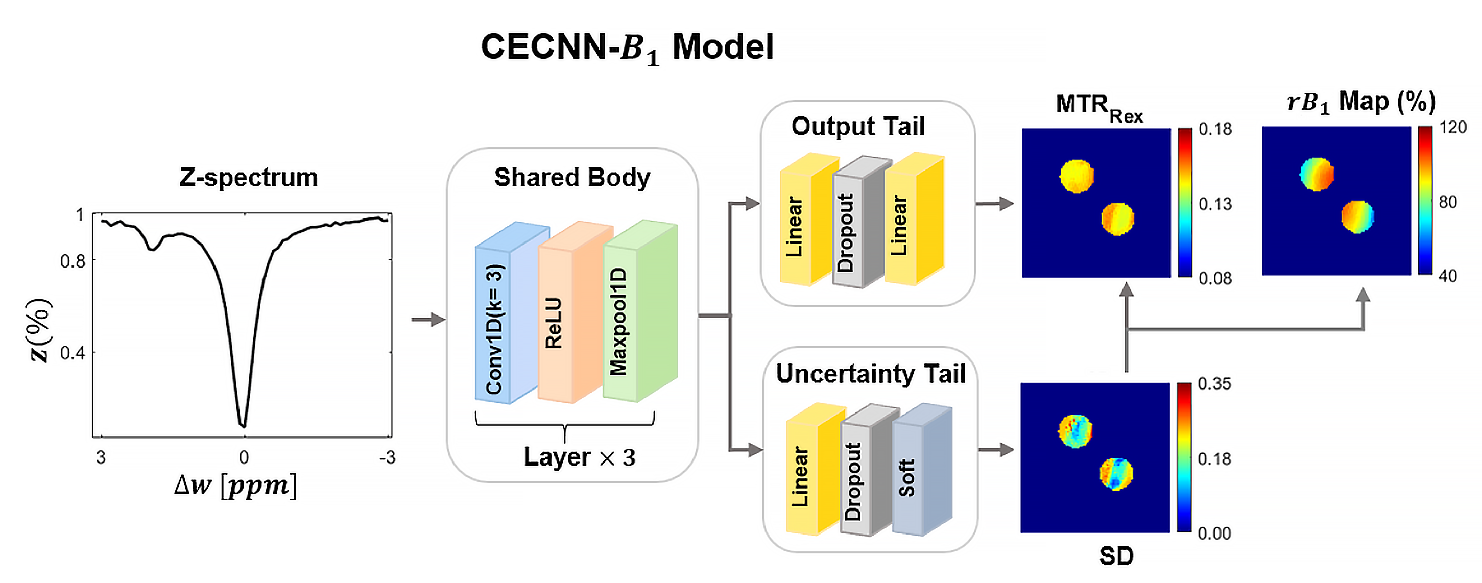
Figure 1. The structure of the confidence-enhanced CNN for B1-Correction (CECNN-B1).
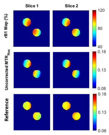
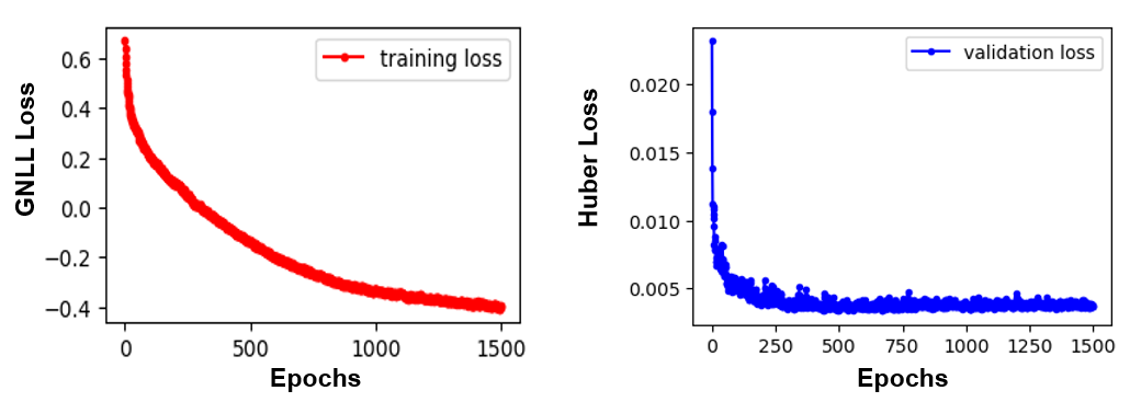
Figure 3. Gaussian negative log likelihood (GNLL) training loss and Huber validation loss of rB1.
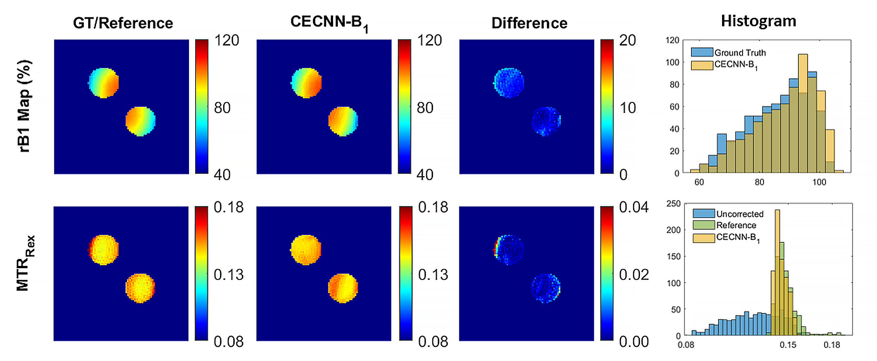
Figure 4. Ground truth of rB1, reference of $$$MTR_{Rex}$$$, their predictions from CECNN-B1 model, absolute differences and respective histograms of slice 3 under B1 level of 0.75 µ𝑇.
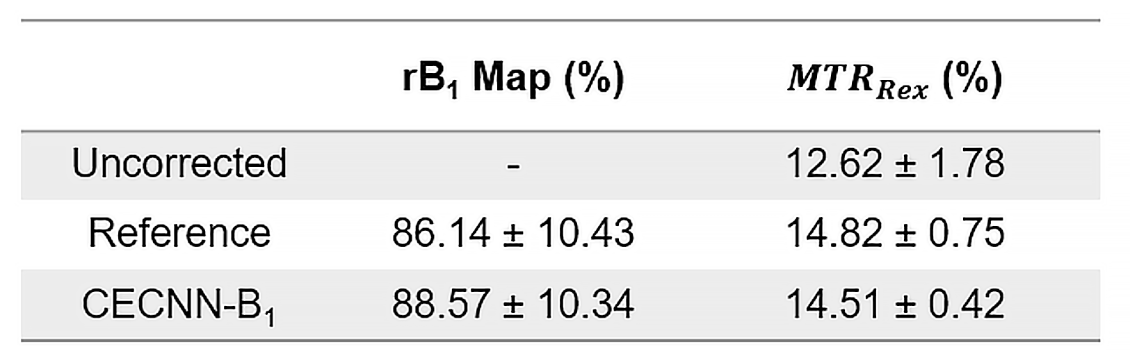
Table 1. Comparison of rB1 and $$$MTR_{Rex}$$$ values.