1969
Feasibility of thin-slice pituitary microadenoma MRI with super-resolution deep learning-constrained compressed sensing reconstruction1Department of Radiology, West China Hospital, Sichuan University, Chengdu, China, 2Clinical Science, Philips Healthcare, Chengdu, China
Synopsis
Keywords: AI/ML Image Reconstruction, Machine Learning/Artificial Intelligence, pituitary microadenoma
Motivation: The spatial resolution of MRI is still limited to the detection of pituitary microadenomas. CSAISR framework can reduce noise and improve image resolution.
Goal(s): To assess the image quality and pituitary microadenoma detection performance of the thin-slice MRI using CSAISR framework.
Approach: In this work, 1.5mm-CSAISR, 1.5mm-CSAI, 1.5mm-CS and 3mm-CSAISR images were obtained. These 1.5mm images were evaluated subjectively and objectively, and the detection rate of 1.5mm-CSAISR and 3mm-CSAISR were compared.
Results: Combined with subjective and objective evaluation, the image quality of 1.5mm-CSAISR images was the best. Meanwhile, the detection rate of 1.5mm-CSAISR reached 92.5%, which was significantly better than that of 3mm-CSAISR.
Impact: The results suggest that thin-slice MRI combined with CSAISR framework can balance the relationship between noise reduction and spatial resolution improvement, increase the detection rate of pituitary microadenoma and is meaningful for the diagnosis, follow-up and localization of this disease.
Introduction
Pituitary microadenomas are common in sellar region, and magnetic resonance imaging (MRI) plays an important role in the detection of pituitary microadenoma [1]. However, pituitary microadenomas have a high negative detection rate on conventional 3mm MRI, such as the 50% negative rate of adrenocorticotropic hormone-secreting tumors [2,3]. With the development of compressed sensing artificial intelligence (CSAI) framework in recent years, its effect of noise reduction has been confirmed in the studies of accelerated image acquisition [4-7]. Moreover, the compressed sensing artificial intelligence super-resolution (CSAISR) framework is further proposed to preserve image details while improving resolution, which may increase the detection rate of pituitary microadenomas [8]. In this study we aim to assess the image quality of 1.5mm-CSAISR MRI, comparing with 1.5mm-CSAI MRI and 1.5mm-compressed sensing (CS) MRI, and compared the detection performance of 1.5mm-CSAISR MRI with that of 3mm-CSAISR MRI.Methods
A total of 40 patients with pituitary microadenoma were enrolled from June 2023 to August 2023. All examinations were performed on a 3.0T MRI scanner (Ingenia Elition, Philips Healthcare) with a 32-channel head coil. All participants received the dynamic contrast-enhanced (DCE) coronal T1 weighted imaging based on CSAISR framework. The main parameters of DCE were as follows: TR = 513 ms, TE = 7.0 ms, flig angle = 90°, FOV = 160 × 160 mm2, slice thickness = 1.5 mm, gap = 0, acquired resolution = 0.65 × 0.65 × 1.5 mm, NEX = 1, scanning time = 1min15s/phase × 6 phases (including one unenhanced phase). The enhanced phase was started simultaneously with the injection, the gadolinium contrast agent was injected via the vein at a rate of 1-2ml/s by hand. It needs to be stressed that the raw data acquired under this framework can be reconstructed through K-space-based deep learning, and three groups of images can be obtained directly through one scan, which are 1.5mm-CSAISR, 1.5mm-CSAI and 1.5mm-CS. The reconstructed resolution for CSAISR framework is 0.312 × 0.312 × 1.5 mm using a super-resolution convolutional neural network [8]. Besides, the 3mm-CSAISR images were respectively reconstructed from the 1.5mm-CSAISR datasets.Image qualities of 1.5mm-CSAISR, 1.5mm-CSAI and 1.5mm-CS were evaluated subjectively and objectively. In the subjective double-blind evaluation, all of images were out of order, and two radiologists with more than 5 years of imaging diagnosis experience used the five-point scale to score the overall image quality, image noise, sharpness, contrast and structure conspicuity. When there was difference in scoring, two radiologists reached consensus after negotiation. In the objective evaluation, a radiologist with more than 10 years of imaging diagnosis experience used following formula to calculate the signal-to-noise ratio (SNR) and the contrast noise ratio (CNR) of normal pituitary glands and lesions by delineating the region of interest (ROI) to measure the signal intensity (SI) and standard deviation (SD). The ROIs were placed on the normal pituitary gland, the lesion, the brain parenchyma adjacent to the lateral ventricle and the air background.
$$SNR=\frac{SI_{interested\space tissue}}{SD_{background\space noise}}$$
$$CNR=\frac{|SI_{interested\space tissue}-SI_{contrast\space brain\space tisssue}|}{SD_{background\space noise}}$$
To evaluate the detection performance with 1.5mm-CSAISR and 3mm-CSAISR images, the detection rates of pituitary microadenoma were calculated. The Friedman test was used for subjective analysis and the One-Way ANOVA with a Least Significant Difference post hoc test was used for objective analysis. P<0.05 indicates that the difference is statistically significant.
Results
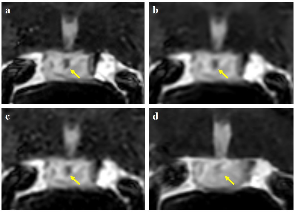
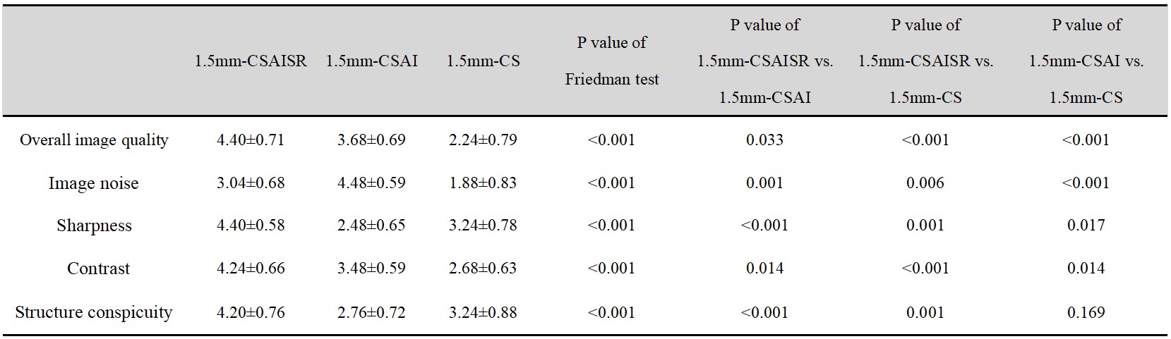
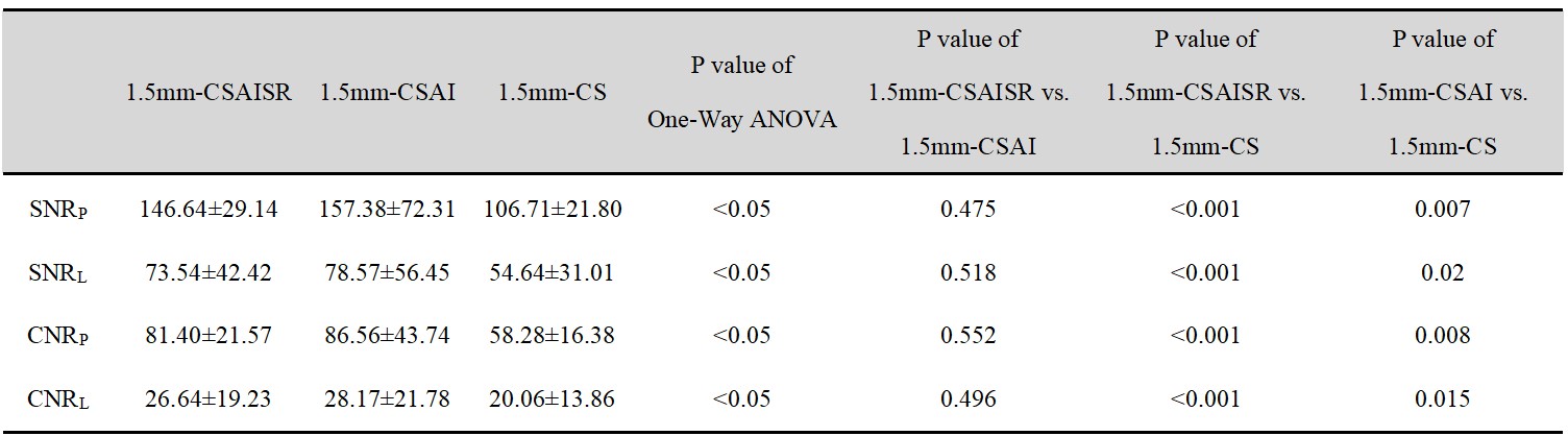
A typical set of 1.5mm-CSAISR, 1.5mm-CSAI, 1.5mm-CS and 3mm-CSAISR images from the same patient are shown in Figure 1. The subjective evaluation results are shown in Figure 2. and the objective evaluation results are shown in Figure 3. Moreover, the detection rate of 1.5mm-CSAISR has reached 92.5%, which is better than the 75% in 3mm-CSAISR.Discussion
The objective analysis demonstrates that both CSAISR and CSAI framework have good noise reduction effect when trying thin-slice pituitary microadenoma MRI and CSAI framework has the best performance. However, while using CSAI framework to reduce the scanning slice thickness, it can not balance SNR and spatial resolution, which lost the structural details and produced the worst sharpness. For small lesions such as pituitary microadenomas, it is necessary to retain anatomical details as much as possible to improve spatial resolution. In this regard, CSAISR framework makes up for the limitations of CSAI framework. Meanwhile, the improved detection rate comparing to thick-slice MRI further reveals the potential of thin-slice MRI with CSAISR framework in the detection of pituitary microadenoma.Conclusion
The CSAISR framework can reduce noise, and anatomical details are preserved as much as possible to improve resolution. With CSAISR framework, the thin-slice MRI can improve the detection rate of pituitary microadenoma, comparing to the conventional thick-slice MRI.Acknowledgements
NoneReferences
[1] Tritos NA, Miller KK. Diagnosis and Management of Pituitary Adenomas: A Review. JAMA. 2023;329(16):1386-1398.
[2] Molitch ME. Diagnosis and Treatment of Pituitary Adenomas: A Review. JAMA. 2017;317(5):516-524.
[3] Lee DH, Park JE, Nam YK, et al. Deep learning-based thin-section MRI reconstruction improves tumour detection and delineation in pre- and post-treatment pituitary adenoma. Sci Rep. 2021;11(1):21302.
[4] Hu SX, Xiao Y, Peng WL, et al. Accelerated 3D MR neurography of the brachial plexus using deep learning-constrained compressed sensing [published online ahead of print, 2023 Aug 22]. Eur Radiol. 2023;10.1007/s00330-023-09996-0.
[5] Zhang Y, Peng W, Xiao Y, et al. Rapid 3D breath-hold MR cholangiopancreatography using deep learning-constrained compressed sensing reconstruction. Eur Radiol. 2023;33(4):2500-2509.
[6] Wu X, Tang L, Li W, et al. Feasibility of accelerated non-contrast-enhanced whole-heart bSSFP coronary MR angiography by deep learning-constrained compressed sensing. Eur Radiol. 2023;33(11):8180-8190.
[7] Pezzotti N, Yousef S, Elmahdy MS et al. An adaptive intelligence algorithm for undersampled knee MRI reconstruction. IEEE Access. 2020;8:204825–204838
[8] Dong C, Loy CC, He K, Tang X. Image Super-Resolution Using Deep Convolutional Networks. IEEE Trans Pattern Anal Mach Intell. 2016;38(2):295-307.
Figures