1968
Usefulness of Super-Resolution Deep Learning-Based Reconstruction on High-Resolution 3D T2WI for Evaluation of the Internal Auditory Canal1Diagnostic Radiology, Kumamoto university, Kumamoto, Japan, 2Central Radiology section, Kumamoto University Hospital, Kumamoto, Japan, 3MRI sales department, sales engineer group, Canon Medical Systems Corporation, Kawasaki, Japan
Synopsis
Keywords: AI/ML Image Reconstruction, Image Reconstruction, deep learning; internal auditory canal; high resolution T2-weighted imaging; super-resolution.
Motivation: There are no reports on the usefulness of high-resolution three-dimensional T2-weighted imaging (HR-3D T2WI) using super-resolution deep learning-based reconstruction (SR-DLR) technique.
Goal(s): We aimed to determine whether a novel SR-DLR technique can improve the image quality of HR-3D T2WI for assessing the IAC and inner ear.
Approach: We qualitatively assessed the image quality of HR-3D T2WI with and without SR-DLR for the visualization of detailed structures of the IAC and inner ear.
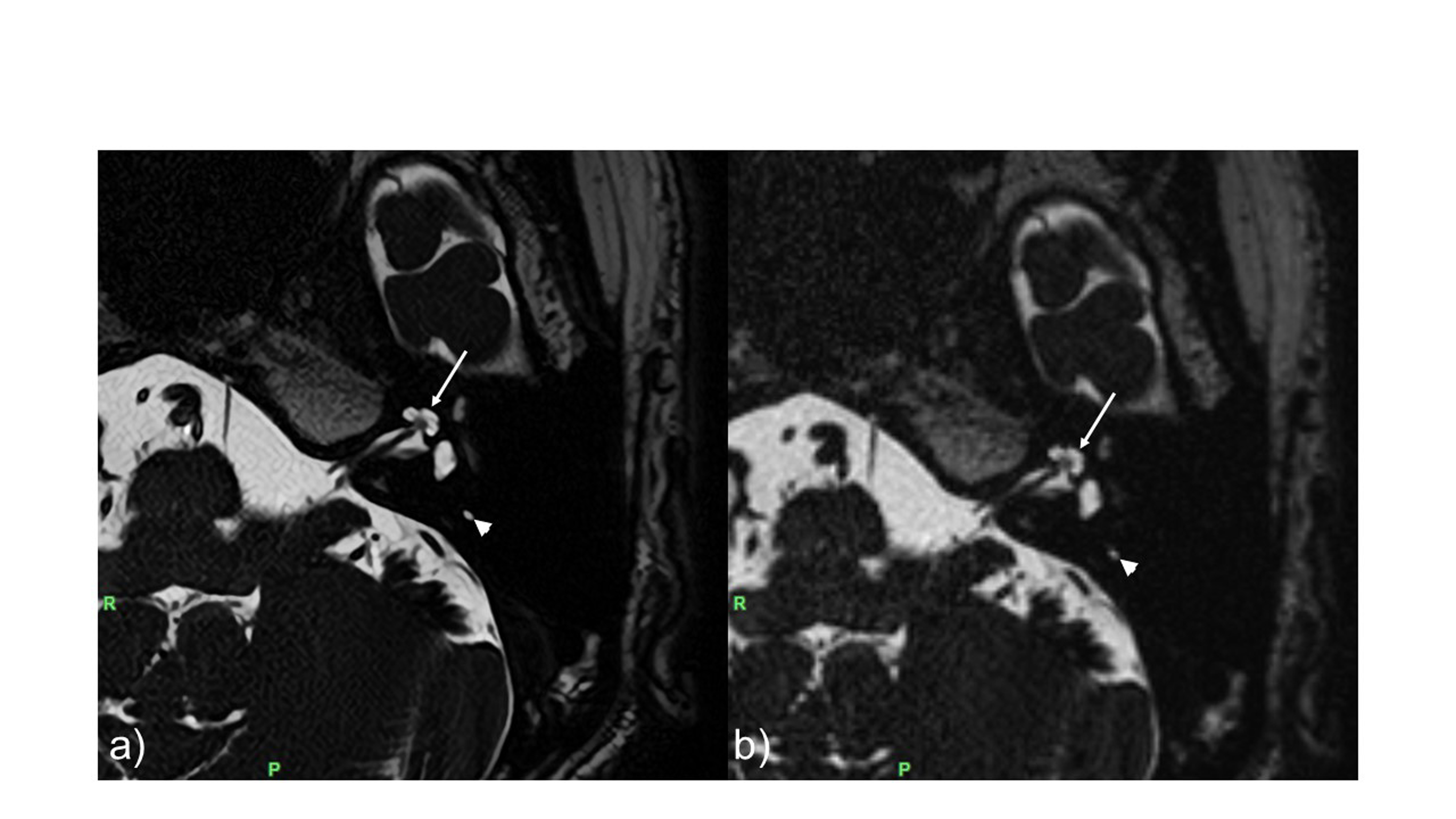
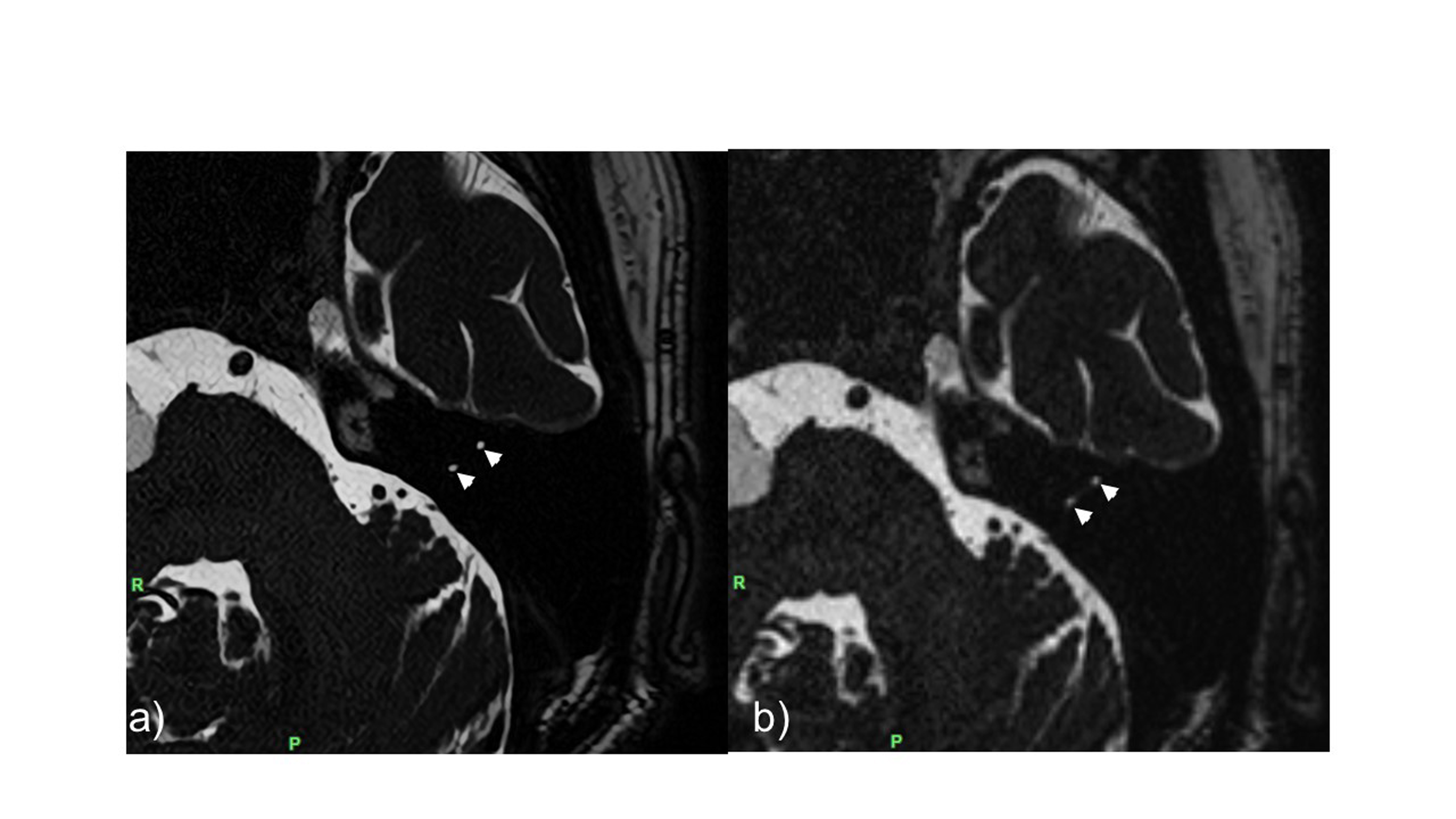
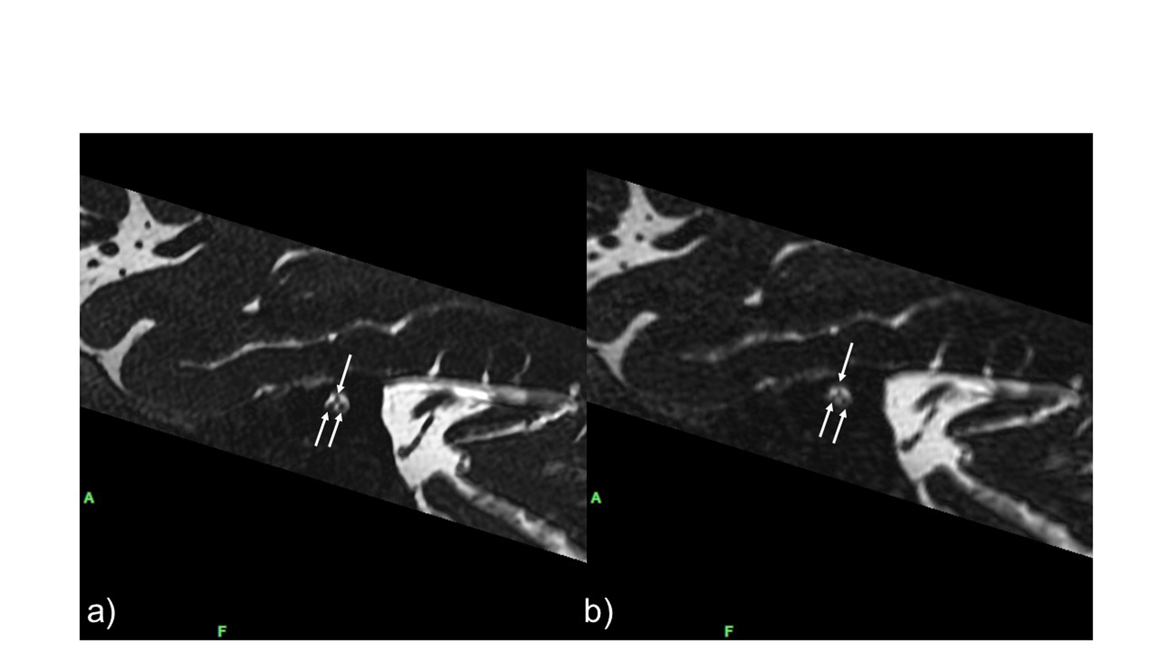
Results: SR-DLR was shown to significantly improve the image quality of HR-3D T2WI in visualizing nerves in the IAC and inner ear compared to conventional HR-3D T2WI without SR-DLR.
Impact: High-resolution three-dimensional T2-weighted imaging with super-resolution deep learning-based reconstruction can improve the visualization of detailed structures of the internal auditory canal and inner ear without extended acquisition time.
INTRODUCTION
High-resolution three-dimensional T2-weighted imaging (HR-3D T2WI) is commonly used to evaluate the internal auditory canal (IAC) and inner ear in patients with sensorineural hearing loss and dizziness, but this usually requires long acquisition times (1). To reduce acquisition time, many studies have reported the usefulness of a parallel imaging technique (2) and compressed sensing technique (1,3). The disadvantages of this approach are a decrease in signal-to-noise ratio (SNR) (4) or global ringing artifacts and blurring of fine details (5,6), making it difficult to depict small structures at high acceleration and denoising levels. Recently, the deep learning-based reconstruction (DLR) technique has been applied for MRI noise reduction (7-9). Several super-resolution techniques using DLR for MRI, called super-resolution DLR (SR-DLR), have emerged in recent years (10-12). A novel SR-DLR technique using zero-padding interpolation (ZIP) for the matrix enhancement within k-space and deep learning for noise and ringing artifact removal processing has been developed and reported to be useful in brain diffusion-weighted imaging (DWI)(13). To our knowledge, there are no reports on the usefulness of the SR-DLR technique for HR-3D T2WI. We aimed to determine whether the novel SR-DLR technique can improve the image quality of HR-3D T2WI for assessing the IAC and inner ear.METHODS
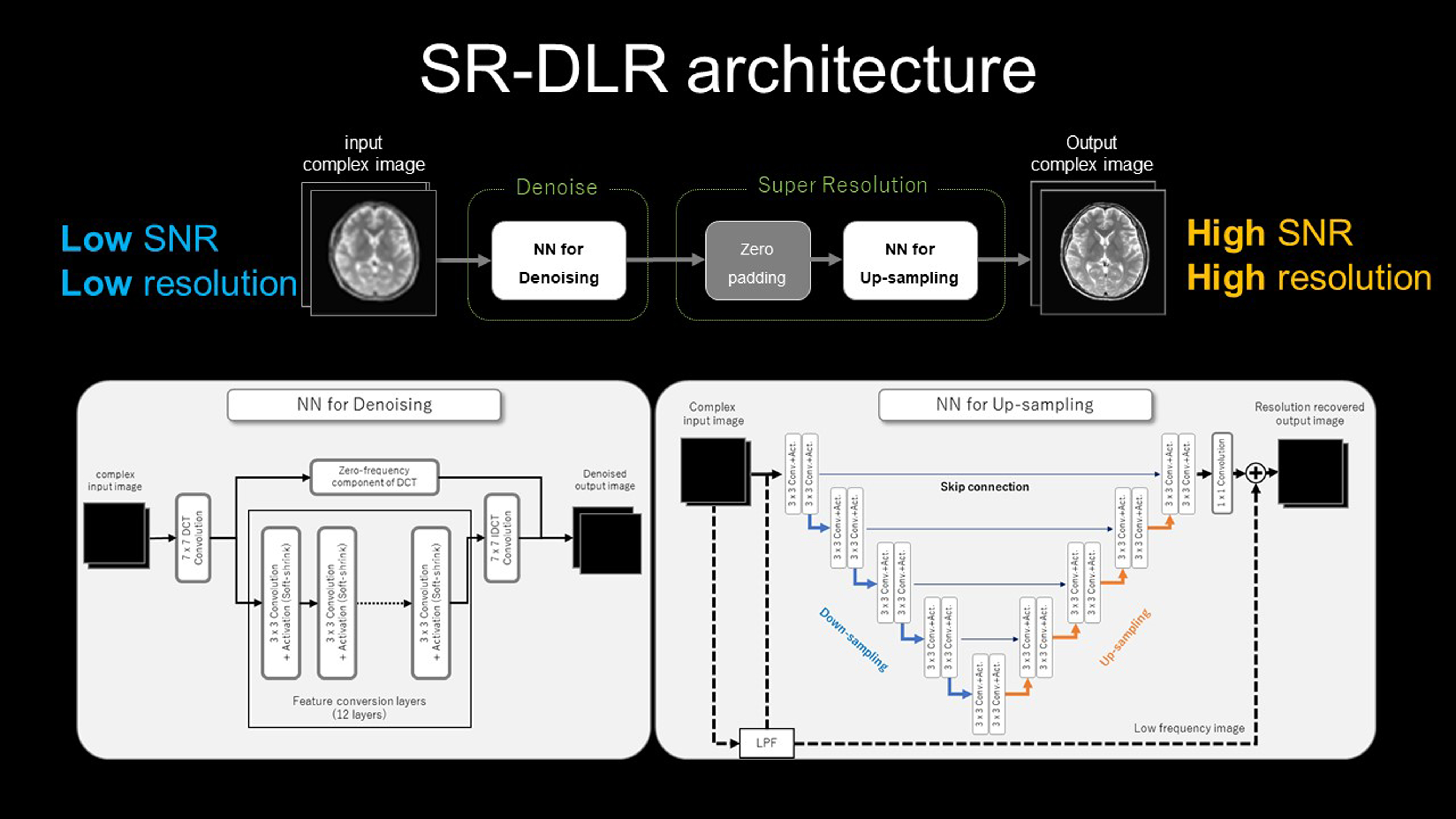
This retrospective study included 13 patients (six male, 64.1 ± 13.2 years) who underwent HR-3D T2WI using a 3T MRI system (Vantage Centurian; CANON Medical Systems) for assessing sensorineural hearing loss, dizziness, and IAC tumors. Scan parameters of HR-3D T2WI were as follows: TR/TE, 2200/227.5 ms; field of view, 180×180 mm; matrix, 320×320; slice thickness, 0.6 mm; parallel imaging factor, 3.0; and acquisition time, 3 min 45 sec. We reconstructed HR-3D T2W images with SR-DLR (matrix size, 960×960) and without SR-DLR (matrix size, 320×320). Figure 1 shows the reconstruction pipeline of the SR-DLR technique (13). Two neuroradiologists evaluated the visualization of the cochlea, vestibule, semicircular canal, anterior inferior cerebellar artery (AICA) on axial images, nerves within the IAC (facial nerve, cochlear nerve and superior and inferior vestibular nerve) on sagittal images, and overall image quality of the two types of images using a 5-point scale (from grade 1 [unacceptable] to grade 5 [excellent]) (1). The median scores between images with and without the SR-DLR were compared using the Wilcoxon signed-rank test.RESULTS
For the visualization of the cochlea, semicircular canal, AICA, nerves within the IAC, and overall image quality, the median score was significantly higher for images with SR-DLR than images without SR-DLR (the cochlea, 5.0 [interquartile range, 5.0-5.0] vs 3.0 [3.0-4.0], P <0.01; semicircular canal, 5.0 [5.0-5.0] vs 4.0 [4.0-4.0], P < 0.01; AICA, 5.0 [5.0-5.0] vs 4.0 [4.0-5.0], P = 0.018; IAC, 5.0 [5.0-5.0] vs 4.0 [3.75-4.0], P < 0.01 and overall image quality, 5.0 [5.0-5.0] vs 4.0 [4.0-4.0], P < 0.01).DISCUSSION
In our study, SR-DLR was shown to significantly improve the image quality of HR-3D T2WI in visualizing nerves in the IAC and inner ear structures compared to conventional HR-3D T2WI without SR-DLR. Our previous report suggested that SR-DLR was a useful tool to improve the SNR and contrast-to-noise ratio in DWI (13). This SR-DLR method integrates a ZIP technique and two types of DLR methods for noise reduction and ringing artifact removal to achieve a 3-fold scaling rate. High spatial resolution and noise reduction by the SR-DLR technique must have contributed to our results.CONCLUSION
The novel SR-DLR technique can enhance the image quality of HR-3D T2WI for assessing detailed structures in the IAC and inner ear.Acknowledgements
noneReferences
1. Yuhasz M, Hoch MJ, Hagiwara M, et al. Accelerated Internal Auditory Canal Screening Magnetic Resonance Imaging Protocol With Compressed Sensing 3-Dimensional T2-Weighted Sequence. Invest Radiol 2018;53(12):742-747.
2. Wang Y. Description of parallel imaging in MRI using multiple coils. Magnetic resonance in medicine 2000;44(3):495-499.
3. Zhou R, Huang W, Yang Y, et al. Simple motion correction strategy reduces respiratory-induced motion artifacts for k-t accelerated and compressed-sensing cardiovascular magnetic resonance perfusion imaging. J Cardiovasc Magn Reson 2018;20(1):6.
4. Ham CL, Engels JM, van de Wiel GT, Machielsen A. Peripheral nerve stimulation during MRI: effects of high gradient amplitudes and switching rates. J Magn Reson Imaging 1997;7(5):933-937.
5. Sharma SD, Fong CL, Tzung BS, Law M, Nayak KS. Clinical image quality assessment of accelerated magnetic resonance neuroimaging using compressed sensing. Invest Radiol 2013;48(9):638-645.
6. Zhang T, Chowdhury S, Lustig M, et al. Clinical performance of contrast enhanced abdominal pediatric MRI with fast combined parallel imaging compressed sensing reconstruction. J Magn Reson Imaging 2014;40(1):13-25.
7. Kidoh M, Shinoda K, Kitajima M, et al. Deep Learning Based Noise Reduction for Brain MR Imaging: Tests on Phantoms and Healthy Volunteers. Magnetic resonance in medical sciences : MRMS : an official journal of Japan Society of Magnetic Resonance in Medicine 2020;19(3):195-206.
8. Uetani H, Nakaura T, Kitajima M, et al. A preliminary study of deep learning-based reconstruction specialized for denoising in high-frequency domain: usefulness in high-resolution three-dimensional magnetic resonance cisternography of the cerebellopontine angle. Neuroradiology 2021;63(1):63-71.
9. Uetani H, Nakaura T, Kitajima M, et al. Hybrid deep-learning-based denoising method for compressed sensing in pituitary MRI: comparison with the conventional wavelet-based denoising method. Eur Radiol 2022;32(7):4527-4536.
10. Chaudhari AS, Fang Z, Kogan F, et al. Super-resolution musculoskeletal MRI using deep learning. Magnetic resonance in medicine 2018;80(5):2139-2154.
11. Pham CH, Tor-Diez C, Meunier H, et al. Multiscale brain MRI super-resolution using deep 3D convolutional networks. Comput Med Imaging Graph 2019;77:101647.
12. Chaudhari AS, Stevens KJ, Wood JP, et al. Utility of deep learning super-resolution in the context of osteoarthritis MRI biomarkers. J Magn Reson Imaging 2020;51(3):768-779.
13. Matsuo K, Nakaura T, Morita K, et al. Feasibility study of super-resolution deep learning-based reconstruction using k-space data in brain diffusion-weighted images. Neuroradiology 2023;65(11):1619-1629.
Figures