1965
Denoising AutoEncoder as a Pre-processor for Knee MRI Analysis1Center for Advanced Imaging Innovation and Research (CAI2R), Department of Radiology, New York University Grossman School of Medicine, New York, NY, United States, 2Bernard and Irene Schwartz Center for Biomedical Imaging, Department of Radiology, New York University Grossman School of Medicine, New York, NY, United States, 3Center for Data Science, New York University, New York, NY, United States
Synopsis
Keywords: Analysis/Processing, Data Processing
Motivation: Pre-processing MR images is a necessary step prior to image analysis due to variability of intensity scale in MR images.
Goal(s): To develop a deep learning algorithm for standardizing knee MR images prior to analysis.
Approach: We developed a denoising autoencoder with VNet architecture achieving on-the-fly image pre-processing (Bias field correction and intensity normalization) and denoising. Image quality was evaluated using SNR, NMSE, PSNR, and SSIM.
Results: Our approach achieved an improved SNR with an efficient runtime compared to conventional pre-processing methods.
Impact: Our DL-based knee MRI pre-processing tool generates standardized MRI outputs for image analysis and DL model development. This tool can be incorporated into a wide range of image analysis pipelines for the knee.
Introduction
In the rapidly advancing domain of medical imaging, artificial intelligence (AI), particularly deep learning (DL) models, has revolutionized clinical decision-making processes. These models effectively extract patterns from imaging data, thereby improving diagnostic accuracy in radiology. Compared to other imaging modalities, developing models for MR imaging contains additional challenges such as intensity scale variations and susceptibility to noise and motion artifacts1. These challenges are further exacerbated during transfer learning, where developed models often suffer performance degradation when used on different contrasts or out-of-distribution datasets due to domain shifts2. Recently, several studies have investigated solutions for these challenges3-8. In this study, we developed a DL-based pre-processing pipeline that integrates established MR image pre-processing techniques with a denoising autoencoder to generate standardized MRI outputs for analysis and improve model generalizability.Methods
DatasetWe used a subset of the Osteoarthritis Initiative (OAI) dataset9, comprising Sagittal intermediate-weighted turbo spin echo with fat suppression (SAG IW TSE FS) images (TE = 30 ms, TR = 3200 ms, FOV = 160 mm, Slice Thickness = 3.0 mm, Inplane Resolution = 0.36 mm × 0.51 mm, Bandwidth = 248 Hz/pixel, Matrix Size = 444 × 448 × 37). The images were centrally cropped into 384 × 384 × 32 matrices as DL model inputs. Our dataset includes 353 matched case-control pairs, divided as follows: 255 pairs for the training, 51 for the validation, and 47 for the test as previous study10,11.
Denoising AutoEncoder as Image Preprocessor
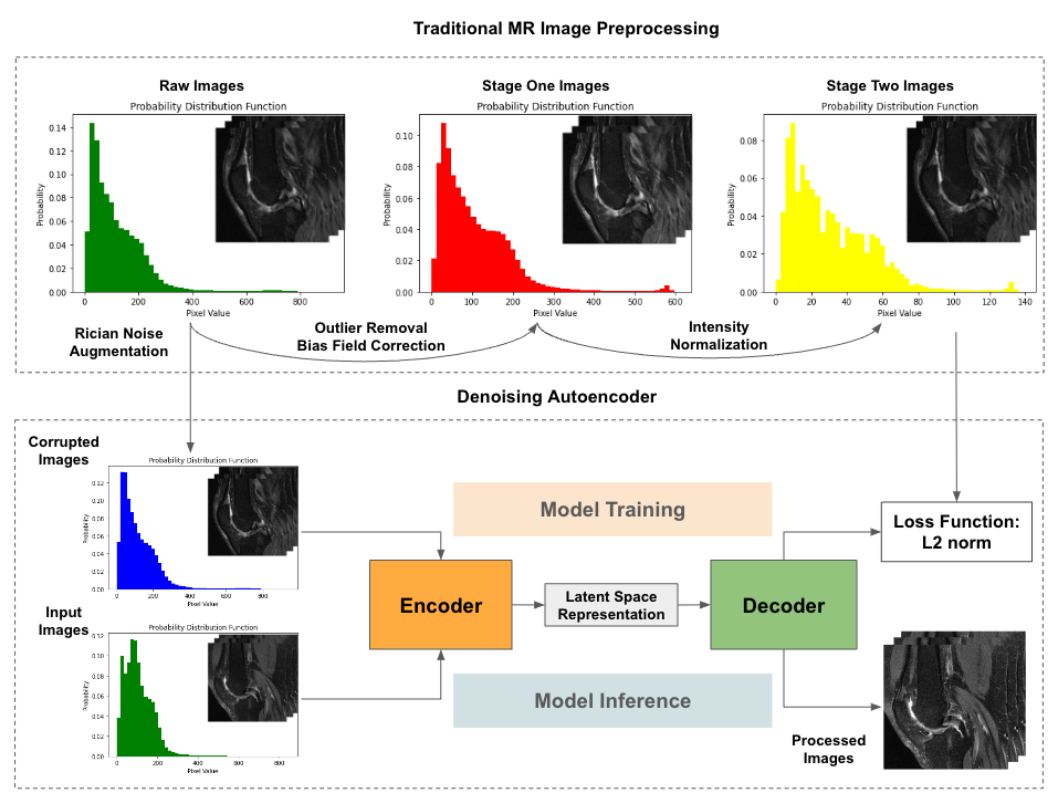
The preprocessing pipeline of knee MR images includes two conventional stages (Figure 1) adapted from a previous work12 and the last stage utilizing a denoising autoencoder (DAE) (Figure 2). Initially, outliers with very low occurrence were removed from probability distribution histograms. The bias field distortion, which causes the variability of MR intensity values13, was corrected using the N4 bias correction algorithm14. Subsequently, Nyul’s intensity standardization15 was employed to establish a consistent intensity scale for the entire dataset. 50 samples were randomly selected from the training set, and 10 landmarks were learned from them as an average reference to recalibrate the intensities of all images within the dataset.
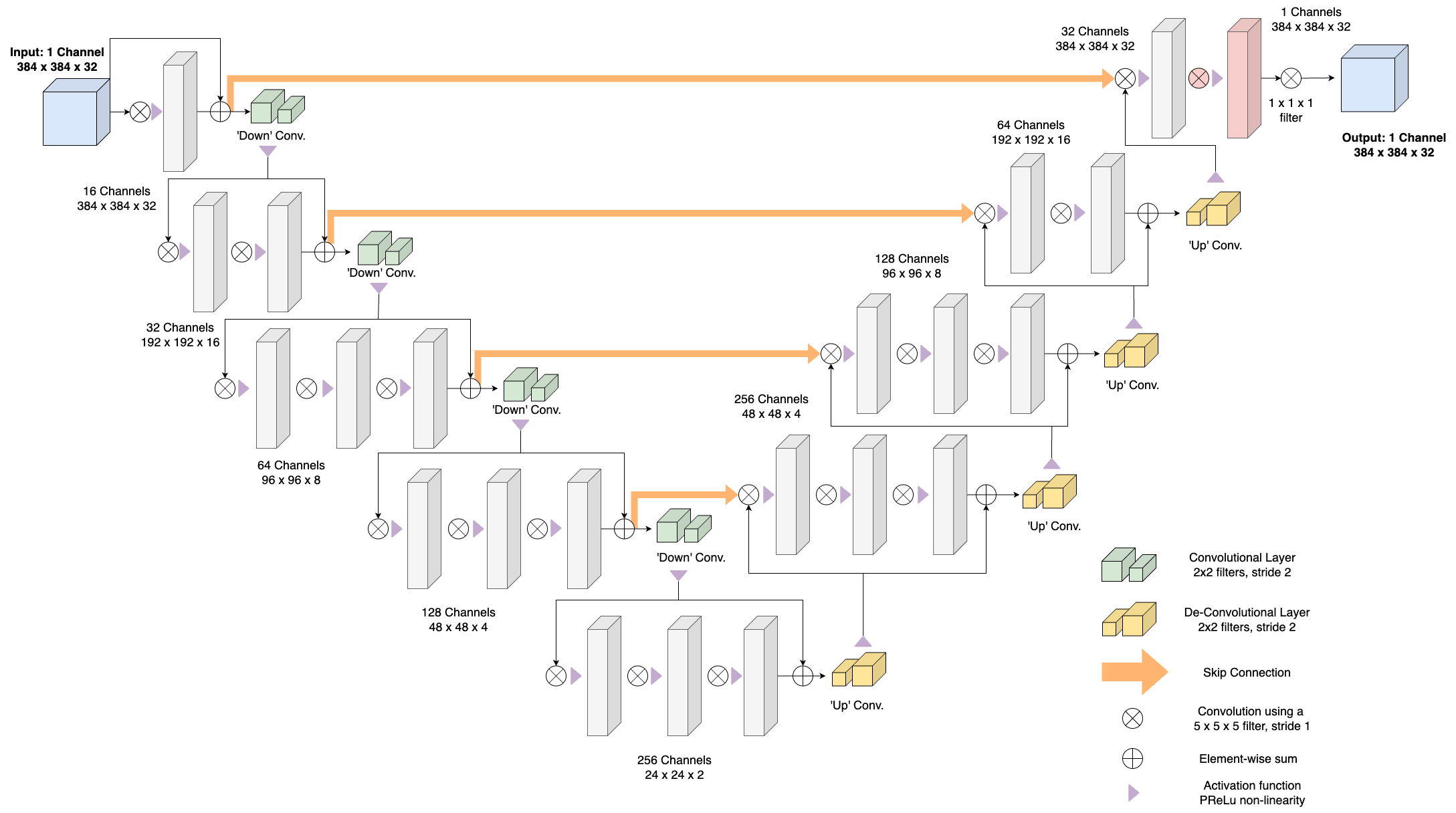
The DAE with a modified VNet architecture16 was employed in the final stage. The DAE took input images corrupted with Rician noise17 and produced denoised images that are aligned with the standardized images from stage two. A convolutional layer was added in the last up transition layer before the very last convolutional layer as detailed in Figure 1. The training of the autoencoder utilized the Adam optimizer with learning rate of 0.0001, adjusted by a ReduceLRPlateau schedule over 100 epochs. Data augmentation techniques such as flipping and rotation (with 10 degrees along the XY plane) were randomly applied to further improve the robustness of the model. The loss function was the L2 norm.
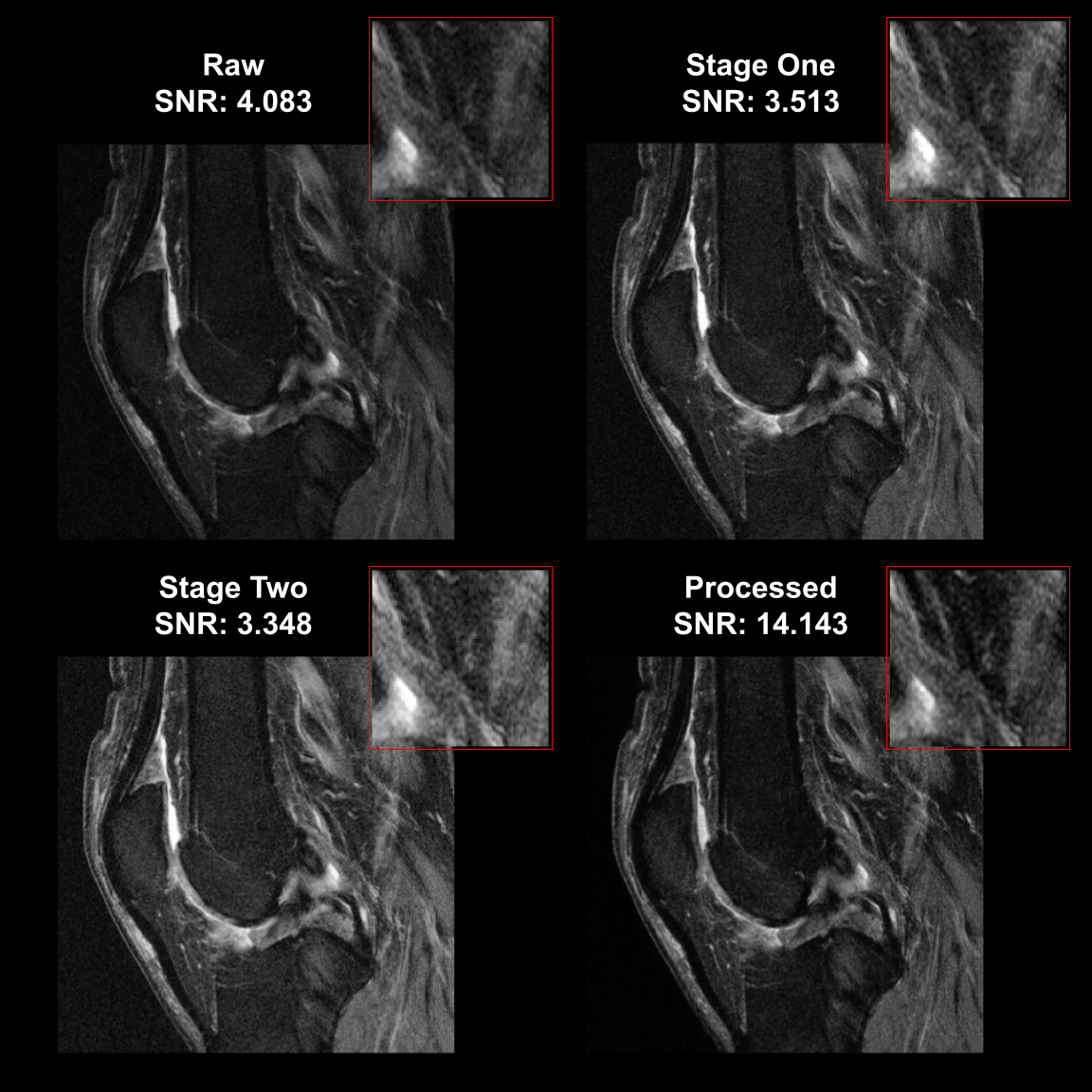
Quality assessment of the images includes calculations of Normalized Mean Squared Error (NMSE), Peak Signal-to-Noise Ratio (PSNR), and Structural Similarity Index (SSIM)18, comparing target (stage two) and processed images. Additionally, the signal-to-noise ratio (SNR) was calculated by measuring the mean pixel intensity within a box placed in the region of interest over the standard deviation of pixel values in a corresponding background region as seen in Figure 3.
Results
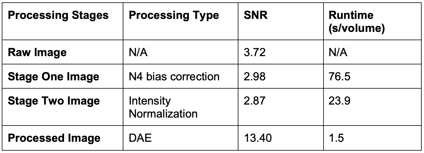
Figure 3 shows a sequential illustration of the image processing workflow, indicating the transition from raw images through three processed stages. The SNR is quantitatively evaluated at each stage from a central slice in a random volume. The SNR initially decreases through Stage One (3.51) and Stage Two (3.35). The output of DAE exhibits an enhanced SNR of 14.14. The zoomed-in regions provide finer details. Noise reduction is also observed within the image background.Table 1 shows a comparison of SNR and processing runtime across all stages of pre-processing on the test set. The processed image shows a substantial improvement in SNR to an average of 13.40 and the runtime is greatly reduced to 1.46 seconds for running inference on raw data to get processed images. When compared with target images, processed images have an average NMSE of 0.0373 (0.006), PSNR of 30.35 (2.19), and SSIM of 0.86 (0.03).
Conclusion
Our study introduces a DAE integrating traditional MRI preprocessing techniques to enhance the quality of knee MR images and to generate standardized MRI outputs for analysis. The quantitative analysis indicates that our DAE not only improves the SNR but also achieves this while substantially reducing the inference time. Importantly, the improved image quality would be helpful to enhance the performance of downstream image analysis tasks, showing promise for clinical deployment and the advancement of diagnostic procedures.Acknowledgements
This work was supported in part by the NIH R01 AR074453, and was performed under the rubric of the Center for Advanced Imaging Innovation and Research (CAI2R, www.cai2r.net), an NIBIB National Center for Biomedical Imaging and Bioengineering (NIH P41 EB017183).References
- Moradmand, Hajar, Seyed Mahmoud Reza Aghamiri, and Reza Ghaderi. "Impact of image preprocessing methods on reproducibility of radiomic features in multimodal magnetic resonance imaging in glioblastoma." Journal of applied clinical medical physics 21.1 (2020): 179-190.
- Yoon, Jee Seok, et al. "Domain Generalization for Medical Image Analysis: A Survey." arXiv preprint arXiv:2310.08598 (2023).
- Madaan, Divyam, et al. "On Sensitivity and Robustness of Normalization Schemes to Input Distribution Shifts in Automatic MR Image Diagnosis." arXiv preprint arXiv:2306.13276 (2023).
- Liu, Hong, et al. "Denoising 3D MR images by the enhanced non-local means filter for Rician noise." Magnetic resonance imaging 28.10 (2010): 1485-1496.
- Yasenko, Lev, Yaroslav Klyatchenko, and Oksana Tarasenko-Klyatchenko. "Image noise reduction by denoising autoencoder." 2020 IEEE 11th International Conference on Dependable Systems, Services and Technologies (DESSERT). IEEE, 2020.
- Carré, Alexandre, et al. "Standardization of brain MR images across machines and protocols: bridging the gap for MRI-based radiomics." Scientific reports 10.1 (2020): 12340.
- He, Xinzi, Alan Q. Wang, and Mert R. Sabuncu. "Neural Pre-processing: A Learning Framework for End-to-End Brain MRI Pre-processing." International Conference on Medical Image Computing and Computer-Assisted Intervention. Cham: Springer Nature Switzerland, 2023.
- Drozdzal, Michal, et al. "Learning normalized inputs for iterative estimation in medical image segmentation." Medical image analysis 44 (2018): 1-13.
- Peterfy, C.G., M.D., Ph.D, Schneider E, Ph.D, Nevitt M, Ph.D. The osteoarthritis initiative: Report on the design rationale for the magnetic resonance imaging protocol for the knee. Osteoarthritis and cartilage. 2008;16(12):1433-1441.
- Rajamohan, H.R., Wang, T., Leung, K. et al. Prediction of total knee replacement using deep learning analysis of knee MRI. Sci Rep 13, 6922 (2023).
- Leung, Kevin, et al. "Prediction of total knee replacement and diagnosis of osteoarthritis by using deep learning on knee radiographs: data from the osteoarthritis initiative." Radiology 296.3 (2020): 584-593.
- Masoudi, Samira, et al. "Quick guide on radiology image pre-processing for deep learning applications in prostate cancer research." Journal of Medical Imaging 8.1 (2021): 010901-010901.
- Guillemaud, Régis, and Michael Brady. "Estimating the bias field of MR images." IEEE Transactions on Medical imaging 16.3 (1997): 238-251.
- Tustison, Nicholas J., et al. "N4ITK: improved N3 bias correction." IEEE transactions on medical imaging 29.6 (2010): 1310-1320.
- Nyúl, László G., and Jayaram K. Udupa. "On standardizing the MR image intensity scale." Magnetic Resonance in Medicine: An Official Journal of the International Society for Magnetic Resonance in Medicine 42.6 (1999): 1072-1081.
- Milletari, Fausto, Nassir Navab, and Seyed-Ahmad Ahmadi. "V-net: Fully convolutional neural networks for volumetric medical image segmentation." 2016 fourth international conference on 3D vision (3DV). Ieee, 2016.
- Gudbjartsson, Hákon, and Samuel Patz. "The Rician distribution of noisy MRI data." Magnetic resonance in medicine 34.6 (1995): 910-914.
- Wang, Z., Bovik, A. C., Sheikh, H. R., & Simoncelli, E. P. (2004). Image quality assessment: from error visibility to structural similarity. IEEE transactions on image processing, 13(4), 600-612.
Figures