1959
Deep Learning Classification of Muscular Dystrophy from MR Images using Swin Transformer1Istituto di Tecnologie Biomediche, Consiglio Nazionale delle Ricerche, Segrate (MI), Italy, 2Istituto di Sistemi e Tecnologie Industriali Intelligenti per il Manifatturiero Avanzato, Consiglio Nazionale delle Ricerche, Milano, Italy, 3Unit of rehabilitation of rare diseases of the central and peripherical nervous system, Scientific Institute IRCCS "Eugenio Medea", Bosisio Parini (LC), Italy, 4Neuroimaging Unit, Scientific Institute IRCCS “Eugenio Medea”, Bosisio Parini (LC), Italy
Synopsis
Keywords: Diagnosis/Prediction, Muscle, Deep Learning; Classification; MRI; Swin Transformer; Generative AI; Dystrophy
Motivation: This study aims to improve the accuracy of muscular dystrophy (MD) diagnosis by applying AI and multiparametric MRI to distinguish subtypes with similar muscle involvement patterns.
Goal(s): The primary goal is to develop a Swin Transformer (SwinT) AI-based classification approach for BMD, LGMD2, and healthy subjects using muscle MR images and identify the optimal MRI contrast for accurate classification.
Approach: In a retrospective study, we utilized SwinT and VGG19 AI models with various MRI contrasts in a 10-fold cross-validation setup.
Results: SwinT outperformed VGG19, with the Fat Fraction contrast delivering the highest accuracy of 89.3%±4.9%, highlighting the potential for more accurate MD diagnosis.
Impact: This work could improve muscular dystrophy diagnosis, offering clinicians a more objective and accurate tool. Patients may benefit from earlier and more precise interventions, while scientists can explore novel research avenues in AI-driven medical diagnostics, ultimately reducing healthcare disparities.
Introduction
Muscular dystrophies (MD) pose challenges for accurate diagnosis, with reliance on methods like muscle biopsy and genetic testing, which sometimes have limitations in accuracy1,2. Magnetic Resonance Imaging (MRI) can aid but often depends on individual expertise, potentially leading to inaccuracies3. Artificial Intelligence (AI) offers a path to more objective and precise diagnostics, with Swin Transformer (SwinT) emerging as an innovative AI tool adept at capturing long-range dependencies in medical images4,5. However, there is limited research on the application of SwinT in this context. Additionally, there is a lack of research on automatic detection and differential diagnosis among MD subtypes with overlapping muscle involvement patterns like Becker muscular dystrophy (BMD) and Limb-girdle muscular dystrophies (LGMDs)6. This study's primary goal is to develop a novel classification approach using SwinT for patients with BMD, LGMD2 and healthy control based on muscle MR images. Considering the intrinsic multiparametric nature of MRI protocols, the study also aims to determine the best MRI contrast not only for accurate disease classification but also to extend its utility beyond diagnosis, offering insights into treatment responses in clinical trials and practice7.Methods
In a retrospective study, MRI data from 54 subjects were divided into three groups: 17 healthy volunteers, 17 BMD patients, and 20 LGMD2 patients (10 LGMD2A and 10 LGMD2B), see Figure 1. Fifteen individuals were part of a longitudinal study with multiple scans, resulting in a total of 75 MRI scans, with varying numbers of images in each scan. For the analytical phase, a selection of 46 subjects, contributing a total of 2036 2D images, was chosen for training and subjected to a 10-fold cross-validation approach. The remaining 8 subjects, yielding 260 2D images were reserved for the test. As a first step, experiments involving AI architectures were conducted with the specific aim to compare the VGG19, a Convolutional Neural Network (CNN), and the SwinT, both initially pre-trained on the ImageNet-1k. Independent training and testing of the AI models were conducted using four distinct MRI contrasts: T1-weighted (T1w), Fat Fraction (FF), Fat, and Water (Wat) images. A two-way ANOVA was performed to determine the significance of architecture and contrast on classification performance. Pairwise Wilcoxon tests were employed to compare the classification results among the various AI models and contrasts. This analysis aimed to uncover the most effective combination of AI model and MRI contrast for accurate classification of muscular dystrophy subtypes. Classification performance was evaluated calculating the accuracy for the overall performance and the F-Score for the class-specific performance.Results
The study demonstrates that both DL architectures and image contrast significantly impact the classification performance, with p-values of 5.6*10^-40 and 2.3*10^-08, respectively. SwinT outperforms VGG19 in all image contrasts except T1w, highlighting the superior performance of SwinT over CNN, as illustrated in Figure 2. The evaluation of image contrast reveals that FF consistently produces significantly better results, particularly when compared with T1w images. Notably, the highest accuracy of 89.3%±4.9% was achieved with the Fat Fraction contrast when using SwinT (Figure 3). Additionally, F-Score values for each class (99.8%±0.4% for healthy, 86.3%±5.8% for BMD and 83.4%±8.5% for LGMD2) highlight the method’s ability in distinguishing overlapping disease patterns within MD.Discussion
This research introduced a novel DL method for the three-class classification among Healthy subjects, BDM, and LGMD2 muscular dystrophies from muscle MR images. It identified the most effective MRI contrast, with the FF proving to be the best choice, achieving the highest accuracy of 89.3%. The study emphasized SwinT superiority over traditional CNNs, thanks to its ability to capture long-range dependencies through self-attention mechanisms and its efficient, compact architecture5. The use of FF maps as an image-based biomarker for muscular dystrophy diagnosis proved effective, enhancing sensitivity for an effective screening via MRI8. With respect to literature, this study stands out by using the SwinT architecture for a three-class classification task, addressing the challenge of similar disease patterns within MD.Moreover, the research extends the potential of MRI and AI integration in detecting MD, making a cost-effective diagnostic tool and reducing biases in interpretations3.
Conclusion
This study highlights the potential of AI and multiparametric MRI to improve muscular dystrophy diagnosis. SwinT, combined with the FF contrast, achieves the best accuracy. This research contributes to more objective disease classification, extending MRI's cost-effective diagnostic application. Further research and data standardization will enhance AI-driven medical diagnostics, offering more precise and objective disease classification and evolution.Acknowledgements
The work was in part funded by Fondazione Cariplo and Regione Lombardia, Progetto "Active3 - Everyone, Everywhere, Everyday", Ref. (2021-0612) and by “Ricerca Corrente 2023” fund provided by the Italian Ministry of Health.
References
1. Nigro V, Savarese M. Next-generation sequencing approaches for the diagnosis of skeletal muscle disorders. Curr Opin Neurol. 2016 Oct;29(5):621-7. doi: 10.1097/WCO.0000000000000371. PMID: 27454578.
2. Ghaoui R, Cooper ST, Lek M, Jones K, Corbett A, Reddel SW, Needham M, Liang C, Waddell LB, Nicholson G, O'Grady G, Kaur S, Ong R, Davis M, Sue CM, Laing NG, North KN, MacArthur DG, Clarke NF. Use of Whole-Exome Sequencing for Diagnosis of Limb-Girdle Muscular Dystrophy: Outcomes and Lessons Learned. JAMA Neurol. 2015 Dec;72(12):1424-32. doi: 10.1001/jamaneurol.2015.2274. PMID: 26436962.
3. Venturelli N, Tordjman M, Ammar A, Chetrit A, Renault V, Carlier R. Contribution of muscle MRI for diagnosis of myopathy. Revue Neurologique. 2023 Jan;179(1-2):61-80. doi: 10.1016/j.neurol.2022.12.002.
4. Kim M, Yun J, Cho Y, Shin K, Jang R, Bae HJ, Kim N. Deep Learning in Medical Imaging. Neurospine. 2020 Jun;17(2):471-472. doi: 10.14245/ns.1938396.198.c1. PMID: 32615703.
5. Liu Z, Lin Y, Cao Y, Hu H, Wei Y, Zhang Z, Lin S, Guo B. Swin Transformer: Hierarchical Vision Transformer using Shifted Windows. 2021 Aug. arXiv: 2103.14030.
6. Lovering RM, Porter NC, Bloch RJ. The muscular dystrophies: from genes to therapies. Phys Ther. 2005 Dec;85(12):1372-88. PMID: 16305275; PMCID: PMC4496952.
7. Nicolau S, Naddaf E. Muscle MRI for Neuromuscular Disorders Using muscle MRI to diagnose neuromuscular conditions requires awareness of different patterns of muscle involvement. Practical Neurology. 2020 Jul.
8. Gaeta M, Messina S, Mileto A, Vita GL, Ascenti G, Vinci S, Bottari A, Vita G, Settineri N, Bruschetta D, Racchiusa S, Minutoli F. Muscle fat-fraction and mapping in Duchenne muscular dystrophy: evaluation of disease distribution and correlation with clinical assessments. Preliminary experience. Skeletal Radiol. 2012 Aug;41(8):955-61. doi: 10.1007/s00256-011-1301-5. PMID: 22069033.
Figures
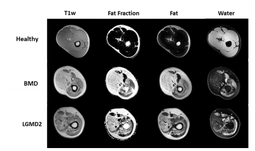
Figure 1: Example of dataset images representing the three classes, each with their corresponding image contrasts. The MRI protocol included both T1w and DIXON sequences, with the latter providing the Fat Fraction, the Fat and the Water quantitative maps.
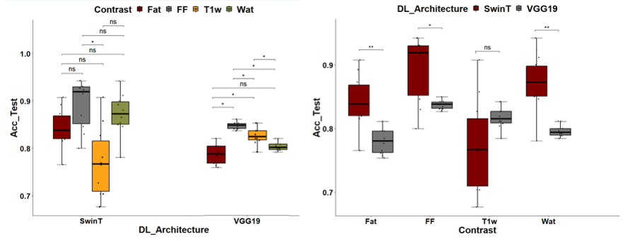
Figure 2: Classification performances associated with the different contrasts and DL architectures. The FF contrast provides the best performances independently of the DL architecture (Left panel). On the other hand, SwinT classifier outperforms the VGG16 classifier in each contrast, except for T1w. (*: significative difference according to the Wilcoxon pairwise test).
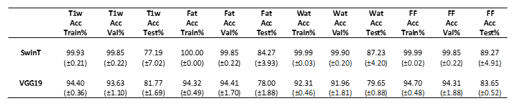
Figure 3: Accuracy values for T1w, Fat, Wat and FF averaged over the 10-fold cross-validation. FF shows the lowest performance drop between the training and the testing sets.