1948
Enhancing Eye Diagnostic Precision: Deep Learning Reconstruction Improves MRI Image Quality for Extraocular Rectus Muscle Assessment1Department of MRI, the First Affiliated Hospital of Zhengzhou University, Zhengzhou, China, 2MR Research China, GE Healthcare, Beijing, China
Synopsis
Keywords: Head & Neck/ENT, Image Reconstruction, deep learning, reconstruction, magnetic resonance imaging, eye, image quality
Motivation: Extraocular rectus muscles (ERMs) are essential for precise eye movement control, and their assessment is vital for diagnosing eye conditions. Magnetic Resonance Imaging (MRI) is preferred due to its superior soft-tissue resolution, but it has limitations.
Goal(s): To determine whether Deep Learning Reconstruction (DLR) can enhance eye image quality and diagnosis.
Approach: A study with 28 patients used DLR on ocular MRI. Two readers evaluated the images independently, and statistical analyses were conducted.
Results: DLR significantly improved ERM depiction and overall image quality compared to conventional MRI. Interobserver agreement was good, especially for structural depiction. DLR produced clear, detailed images, enhancing diagnostic potential.
Impact: This study demonstrates that DLR improves MRI image quality for assessing eye conditions, potentially leading to more accurate diagnoses, reduced repeat examinations, and enhanced patient care.
Introduction
The extraocular rectus muscles (ERMs) are vital for fine eye movement control, with each rectus muscle serving different functions. The Medial Rectus Muscle (MR) primarily acts as an eye adductor, while the Lateral Rectus Muscle (LR) is responsible for lateral eye movement [1,2]. Both the MR and LR play a role in the horizontal vestibulo-ocular reflex. The Superior Rectus Muscle (SR) aids in elevating the eye and contributes to adduction and medial rotation, while the Inferior Rectus Muscle (IR) causes depression, external rotation, and adduction of the eyeball. Enlargement of extraocular muscles is associated with various diseases, particularly thyroid-related ophthalmopathies, and can lead to a range of symptoms and signs.Magnetic Resonance Imaging (MRI) is preferred over CT scans for assessing disease activity due to its superior soft-tissue resolution and absence of ionizing radiation risk. However, conventional MRI has limitations, such as lower resolution, sensitivity to motion artifacts, and reduced sensitivity to specific lesions. Deep learning reconstruction (DLR) [3, 4] has emerged as a promising approach to improve MRI image quality, addressing these limitations. DLR, utilizing convolutional neural networks, has shown substantial potential in enhancing image quality and diagnostic performance.The study's goal is to investigate whether DLR can enhance the image quality of eyes in clinical practice, recognizing that its effectiveness may vary depending on clinical requirements and equipment capabilities. Consequently, DLR holds promise as a tool to improve the evaluation and diagnosis of eye conditions through improved MRI imaging.Methods
Between December 2022 and April 2023, a total of 28 patients (14 males and 14 females) with a mean age of 40.3 ± 17.3 years underwent ocular MRI at the First Affiliated Hospital of Zhengzhou University. The study included ten cases of eye-tubercle, four cases of eyelid edema, three cases without obvious abnormalities, and eleven other cases. The study was approved by the Institutional Review Board, and informed written consent was obtained from all participants.Data were acquired using a 3T GE MRI scanner (SIGNATM Premier, GE Healthcare, Waukesha, WI), with specific imaging parameters for axial T1WI and T2WI sequences. The study involved qualitative analysis, with three experienced radiologists independently assessing image sets for various factors, including structural clarity, overall image quality, and the presence of artifacts.Statistical analyses included the use of Wilcoxon signed-rank tests to compare differences between two image sets and intraclass correlations (ICC) to assess agreement between observers. Data were presented as median (interquartile range), and statistical significance was defined as p < 0.05. The analyses were conducted using SPSS version 27.Results and discussion
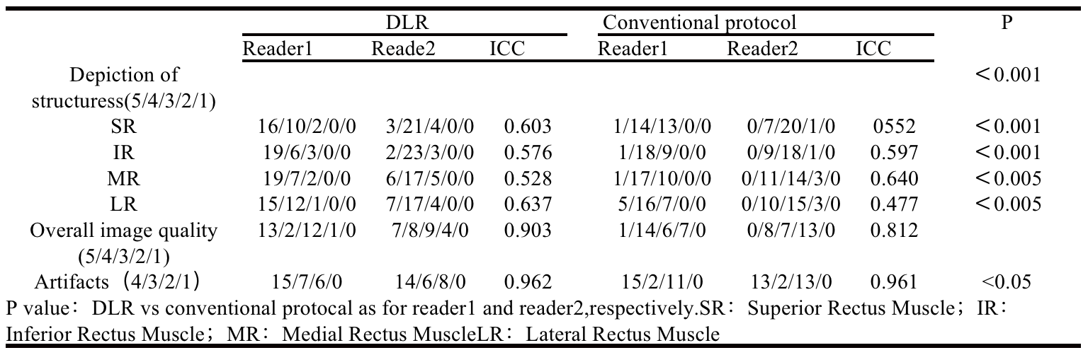
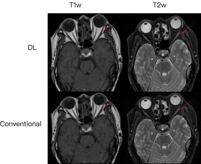
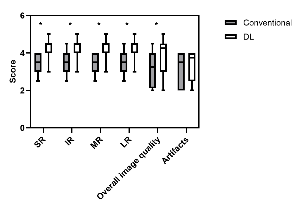
The conventional method had an average axial scan time of 1 minute and 12 seconds, whereas our Deep Learning Reconstruction (DLR) protocol reduced this to 33 seconds. Both readers unanimously agreed that DLR images significantly outperformed conventional protocol images in depicting Extraocular Muscles (ERMs) (p < 0.003 for both readers). The overall image quality of DLR images was also superior (p < 0.03 for both readers), although no substantial difference was observed between them (refer to Table 1 and Figure 2). The interobserver agreement was moderate to good when assessing structural depiction (ICC values of 0.603, 0.576, 0.528, and 0.637 for SR, IR, MR, and LR, respectively) and excellent for overall image quality assessment (ICC value = 0.903). The DL protocols produced clear and detailed images, as depicted in Figures 1. The structures were distinctly visible, highlighting the efficacy of the DLR method in enhancing image quality and providing a comprehensive view of the examined anatomical features.Conclusion
In summary, the intricate structure of the eye makes MRI a crucial tool for extraocular rectus muscle examination. It is imperative to emphasize the diagnostic significance of evaluating these muscles to minimize the need for repeat examinations and enhance clinical outcomes for patients. In this context, the utilization of Deep Learning Reconstruction (DLR) in the image reconstruction process should be seriously considered as a viable option. This approach holds the potential to significantly improve image quality and diagnostic performance, ultimately contributing to more effective and efficient patient care.Acknowledgements
No acknowledgment.References
[1] S. Farzavandi, “Surgical anatomy,” in Color Atlas of Strabis-mus Surgery, S. Farzavandi, Ed., pp. 91–101, Springer, New York, NY, USA, 2007.
[2] M. Peng, V. Poukens, R. M. da Silva Costa, L. Yoo, L. Tychsen, and J. L. Demer, “Compartmentalized innervation of primate lateral rectus muscle,” Investigative Opthalmology & Visual Science, vol. 51, no. 9, pp. 4612–4617, 2010.
[3] Lebel RM. Performance characterization of a novel deep learning-based MR image reconstruction pipeline. arXiv website. arxiv.org/abs/2008.06559.Published August 14, 2020.
[4] Kim M, Kim HS, Kim HJ, et al. Thin-slice pituitary MRI with deep learning–based reconstruction: diagnostic performance in a postoperative setting.Radiology 2021; 298:114–122.
Figures