1947
Utilizing Synthetic MRI and Amide Proton Transfer-Weighted MRI to Distinguish Malignant from Benign Sinonasal Lesions1Radiology, Second Affiliated Hospital of Xi’an Jiaotong University, Xi'an,Shaanxi, China, 2GE Healthcare, MR Research, Beijing, China
Synopsis
Keywords: Head & Neck/ENT, CEST & MT, Synthetic imaging, Sinonasal Lesions, APT
Motivation: Distinguishing between benign and malignant sinonasal lesions is crucial for determining the appropriate treatment regimen and predicting patient outcomes.
Goal(s): To evaluate the performance of synthetic MRI (SyMRI), combined with amide proton transfer (APT)-weighted MRI (APTw) in differentiating between malignant and benign sinonasal lesions.
Approach: Eighty patients with sinonasal lesions were underwent the SyMRI and ATPw scan and the quantitative parameters were analyzed.
Results: T1, T2, PD and APT values were significant between the benign and malignant groups. Combined SyMRI and APTw had the best diagnostic efficiency.
Impact: Combined the SyMRI with APTw would function as a quantitative and contrast-free approach, significantly enhancing the differentiation of benign and malignant sinonasal lesions. This method also helps overcome the limitations associated with the superficial nature of nasal endoscopic sampling.
Introduction
A broad spectrum of tumors and tumor-like lesions is common in the sinonasal region, composing approximately 3 % of all head and neck tumors[1-2]. The clinical presentations of sinonasal lesions are always nonspecific[3],as well as there was different prognosis and therapy strategy between benign and malignant entities[4-5]. Thus, the identification of benign and malignant sinonasal lesions is of great importance for therapeutic decisions and prognosis. Various MR techniques have been investigated on this topic[6-8]. SyMRI, also known as Magnetic Resonance Image Compilation (MAGiC), represents a relatively recent quantitative MR technique. By employing a multi-echo and multi-delay acquisition approach, SyMRI simultaneously yields a range of relaxometry mappings, including longitudinal relaxation time (T1), transverse relaxation time (T2), and proton density (PD). These mappings provide insights into microstructural distinctions at the cellular level within lesions[9]. Meanwhile, another novel molecular contrast imaging technique termed as amide proton transfer (APTw) could be used for detecting and quantifying endogenous cytoplasmic protein without exogenous contrast agent[10]. At present, there were no relevant reported studies of sinonasal masses concerning the utilization of the SyMRI and APTw imaging. Given that inherent microstructural features may be reflected through relaxation and APT rate, this study aims to assess the diagnostic capabilities of MAGiC and APTw imaging in distinguishing between benign and malignant sinonasal lesions.Methods
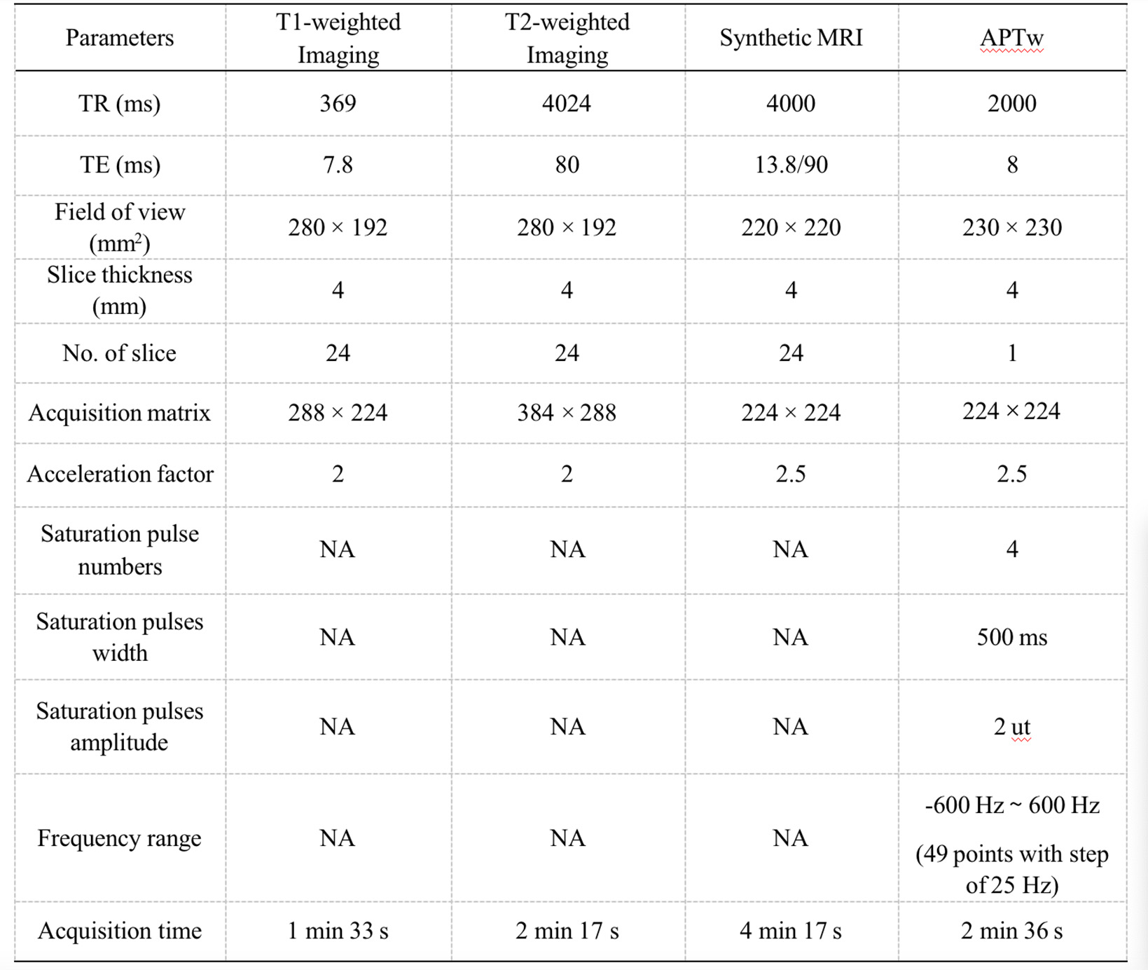
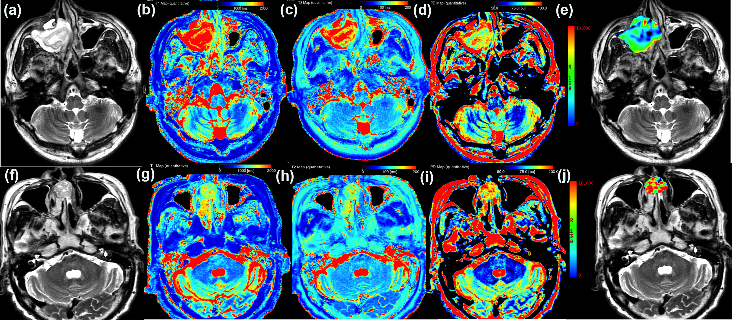
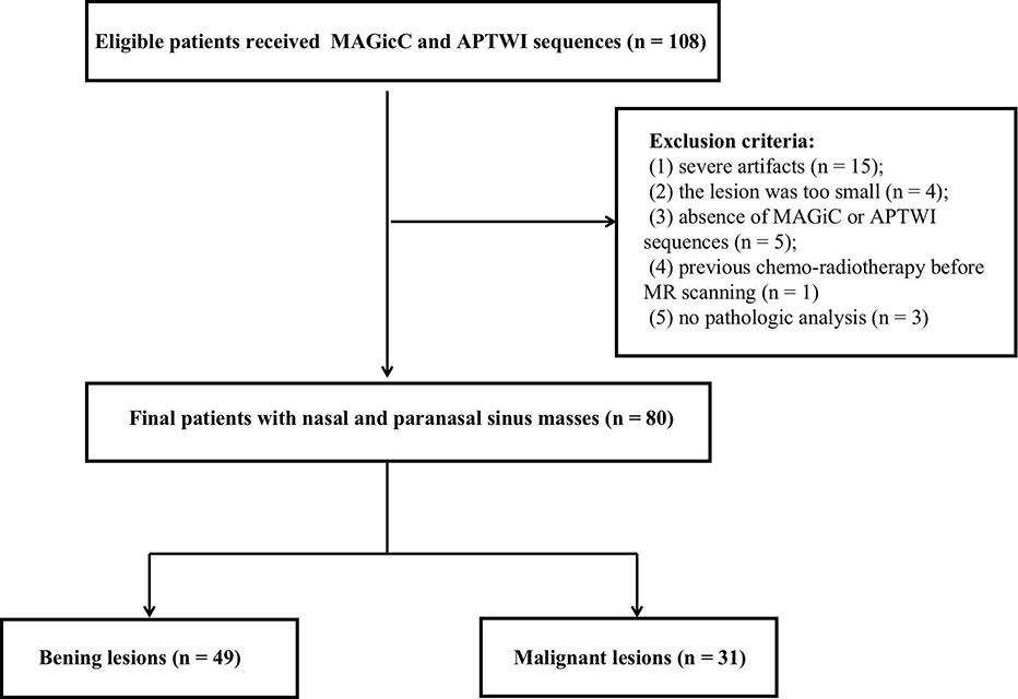
Our institutional review board approved this study, and the informed consent was waived due to the retrospective nature. From March 2021 to March 2023, a total of 80 patients (31 malignant and 49 benign) who were underwent the syMRI and APTTw examination with sinonasal lesions were selected for the study. All of the examinations were performed using a 3-T MRI system (Architect, GE Healthcare) equipped with a 16-channel head coil (scan parameters were detailed in Table 1). After scan, data were digitally transferred to the dedicated Advantage Windows workstation (GE Healthcare, AW 4.4 and 4.7) and vendor provided post-processing programs (MAGiC, v. 100.1.1 and Functool 9.4) were adopted to generate T1, T2 and PD maps from MAGiC sequence, as well as APT asymmetry maps (APT%) from APTw sequence, respectively. Subsequently, a region of interest (ROI) was manually delineated to encompass the maximum cross-sectional tumor areas, as illustrated in Figure 2. Diagnostic efficacy of single and combined parameters for differentiating malignant and benign lesions was evaluated with receiver operating characteristic (ROC) and regression analyses.Results
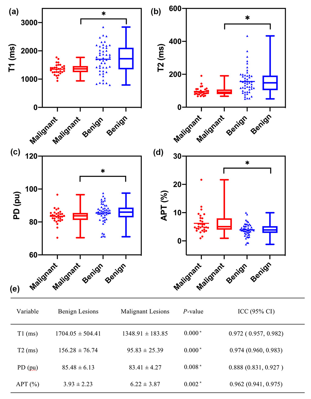
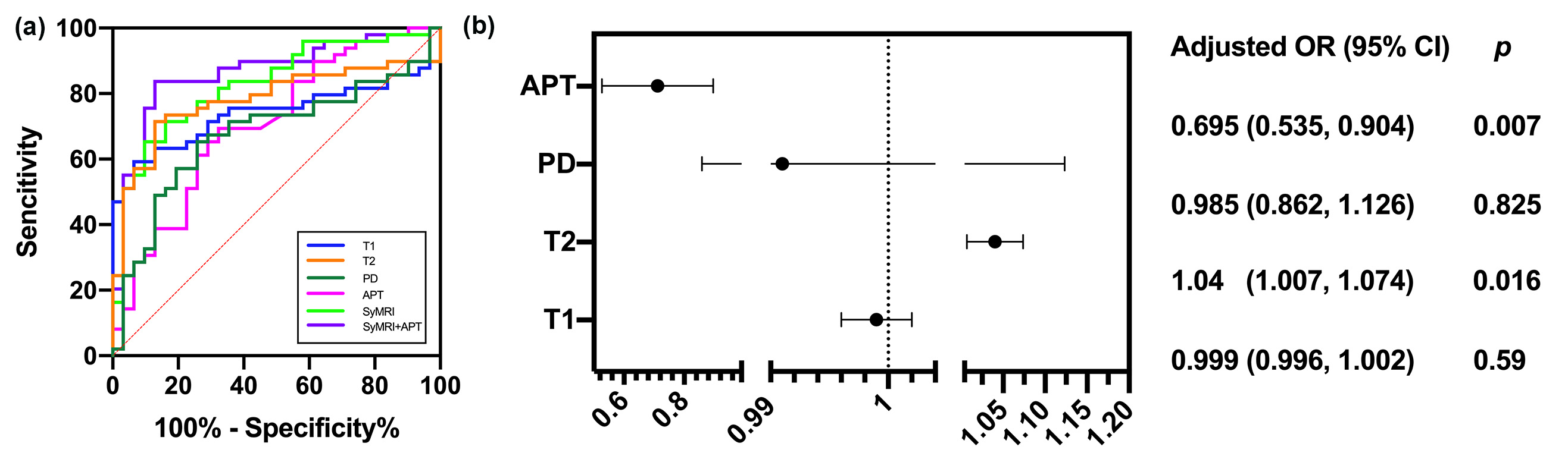
All of 80 patients with 31 malignant and 49 benign lesions were included (Table 3.). There were no significant differences in gender, age and diameters between the two groups (P < 0.05 ). As presented in Figure 4, syMR-derived parameters’ values were significantly higher and APT values was lower in benign than malignant lesions (P < 0.05). The ROC analysis showed that the AUCs of the syMRI-derived quantitative values and APT % ranged from 0.677 to 0.739 for differential diagnosis between benign and malignant sinonasal lesions. The AUC of combined SyMRI-derived multiple parameters (T1, T2, PD ) and APT were higher than that of any single parameter, which was significantly improved(Figure 5a). The SyMRI-derived multiple parameters and APT % were included in the regression analysis. The multivariate analysis revealed T2 and APT values were independent predictors for evaluating benign and malignant groups (Figure 5b).Discussion
The present study explored the clinical value of synthetic MRI–derived quantitative mapping and its combination with APT in distinguishing benign from malignant sinonasal entities. The results revealed that, on average, T1, T2, and PD values were higher in benign lesions compared to malignant ones. This disparity can be attributed to the elevated cellularity and nuclear-to-cytoplasmic ratios typically found in malignant lesions, which, in turn, result in a corresponding reduction in the extracellular fluid space and free water content. Consequently, this leads to lower T1, T2, and PD values in malignant lesions[9]. Meanwhile, since APTw is achieved by detection of the chemical exchange rate between amide proton and bulk water, it tends to exhibit higher values in cases of malignant tumors characterized by heightened cellular proliferation activity and protein synthesis relative to benign lesions. Notably, the optimal diagnostic performance was attained when SyMRI and APTw were employed in combination, surpassing the diagnostic capabilities of individual parameters.Conclusion
The findings indicate that synthetic MRI and APTw-derived quantitative parameters were served as a noninvasive means for distinguishing between benign and malignant sinonasal lesions. This approach holds potential value in preoperative treatment planning and prognostic assessments for patients, all without the need for exogenous contrast agents.Acknowledgements
No acknowledgement found.References
1. Sen S, Chandra A, Mukhopadhyay S, et al. Sinonasal tumors: computed tomography and MR imaging features. Neuroimaging Clin N Am 2015; 25(4): 595–618.
2. Eggesbø HB. Imaging of sinonasal tumours. Cancer Imaging 2012; 12(7):136–523.
3.Wang XY, Yan F, Hao H, et al. Improved performance in differentiating benign from malignant sinonasal tumors using diffusionweighted combined with dynamic contrast-enhanced magnetic resonance imaging. Chin Med J (Engl) 2015;128(5):586–924.
4.Je´goux F, Metreau A, Louvel G, et al. Paranasal sinus cancer. Eur Ann Otorhinolaryngol Head Neck Dis 2013;130(6):327–355.
5.Carta F, Blancal JP, Verillaud B, et al. Surgical management of inverted papilloma: approaching a new standard for surgery. Head Neck 2013;35(10):1415–206.
6.Zhao Z, Tang Z, Qiang J, et al. Intravoxel Incoherent Motion MR Imaging in the Differentiation of Benign and Malignant Sinonasal Lesions: Comparison with Conventional Diffusion-Weighted MR Imaging. AJNR Am J Neuroradiol 2018; 39(3):538-467.
7.Jiang JX, Tang ZH, Zhong YF, et al. Diffusion Kurtosis Imaging for Differentiating Between the Benign and Malignant Sinonasal Lesions. J Magn Reson Imaging 2017;45(5):1446-548.
8.Zhong Y, Xiao Z, Tang Z,et al. Intravoxel incoherent motion MRI for differentiating sinonasal small round cell malignant tumours (SRCMTs) from Non-SRCMTs: comparison and correlation with dynamic contrast-enhanced MRI. Clin Radiol 2018;73(11):966-74 9.
9.Jung Y, Gho SM, Back SN, et al. The feasibility of synthetic MRI in breast cancer patients: comparison of T2 relaxation time with multiecho spin echo T2 mapping method.Br J Radiol 2018;92(1093):2018047910.
10. Peng Y, Zou XL, Chen G, et al. Chemical Shift-Encoded Sequence (IDEAL-IQ) and Amide Proton Transfer (APT) MRI for Prediction of Histopathological Factors of Rectal Cancer. Bioengineering 2023;10(6):720
Figures