1942
A comprehensive MRI-based human craniofacial atlas1Institute of Science and Technology for Brain-Inspired Intelligence, Fudan University, ShangHai City, China, 2CAS Key Laboratory of Computational Biology, Shanghai Institute of Nutrition and Health, University of Chinese Academy of Sciences, Chinese Academy of Sciences, Shanghai, China, ShangHai City, China, 3Center for Excellence in Animal Evolution and Genetics, Chinese Academy of Sciences, Kunming, China, KunMing, China
Synopsis
Keywords: Head & Neck/ENT, Neuro, facial morphology, craniofacial atlas
Motivation: Current research lacks an understanding of the relationship between the craniofacial structure and the brain, and this connection is of significant importance for pre-diagnosis and neuroscience research.
Goal(s): Develop a MRI-based craniofacial atlas to support clinical-neuroscience research, particularly in understanding the relationship between the brain and craniofacial structure.
Approach: Gathered and processed MRI scans from 148 subjects, employing specialized algorithms for detailed craniofacial and skull mapping, aiding neuroscience research.
Results: The study revealed variations in skull thickness, particularly in the occipital bones, and a significant negative correlation between cortical and skull thickness in specific regions. A comprehensive facial atlas was also successfully developed.
Impact: Our research provides a clear research framework and atlas that can be used to explore the relationship between neurological disorders and craniofacial morphological changes in future studies. Additionally, it offers a potential approach for non-invasive and rapid preclinical diagnosis.
Introduction
MRI effectively captures the soft tissues of the human body, including muscles, fat, and craniofacial morphology. Although 3D optical camera systems can image facial features and textures, their integration into clinical practice remains complex, especially when it comes to merging their point cloud data with MRI imagery. CT scans, another approach for craniofacial mapping, are limited in routine use due to the associated radiation risks. Moreover, exploring the interplay between craniofacial structures and the brain is key for the development of innovative, non-invasive, facial scan-based diagnostic tools[1]. These advancements could revolutionize predictive healthcare. In response to these challenges and opportunities, our study has pioneered an MRI-aided atlas of the human face and cranial vault, an initiative increasingly crucial in the light of recent studies underscoring the connection between craniofacial anatomy and neurological conditions [2, 3]. This atlas not only aids in deeper understanding of this relationship but also opens up new avenues for personalized medical approaches in neuroscience and craniofacial research.Methods
We acquired T1-weighted and T2-FLAIR images (voxel size: 0.8x0.8x0.8mm³) from 148 healthy young Chinese participants, all of whom provided informed consent. The group had a mean age of 41±3.5 years and was gender balanced. Adhering to a comprehensive preprocessing regimen consistent with the Human Connectome Project (HCP) pipeline, which included denoising, registration, and surface reconstruction, we employed the Charm[4] algorithm to segment the skull from these MRI images, deliberately omitting the facial skull due to its intricate complexity. The inner and outer portions of the skull were meticulously extracted and reconstructed into detailed mesh structures using the advanced isosurface algorithm. In the process of parcellating the skull surface, vertices located on the superficial pial surface were projected onto the nearest point of the inner skull surface, then accurately transferred to the outer skull. Ensuring consistency in anatomical correlation, all surfaces were normalized to the Montreal Neurological Institute (MNI) space, employing previously established normalization warp fields. Subsequent to these steps, we utilized spherical registration to iteratively develop a population-averaged cranial atlas.In addressing facial morphology, we removed brain tissues from the images and conducted a voxel-based general Procrustes analysis on all T1-weighted images[5]. This analysis led to the creation of an initial facial template, derived by averaging these images. Utilizing Advanced Normalization Tools (ANTs), we developed a comprehensive population facial template. Following this, we reconstructed the facial mesh, aligning and registering it with an established East Asian facial mesh atlas. This meticulous process enabled the creation of a detailed, MRI-based facial atlas, contributing significantly to our understanding of craniofacial morphology in this demographic.Results
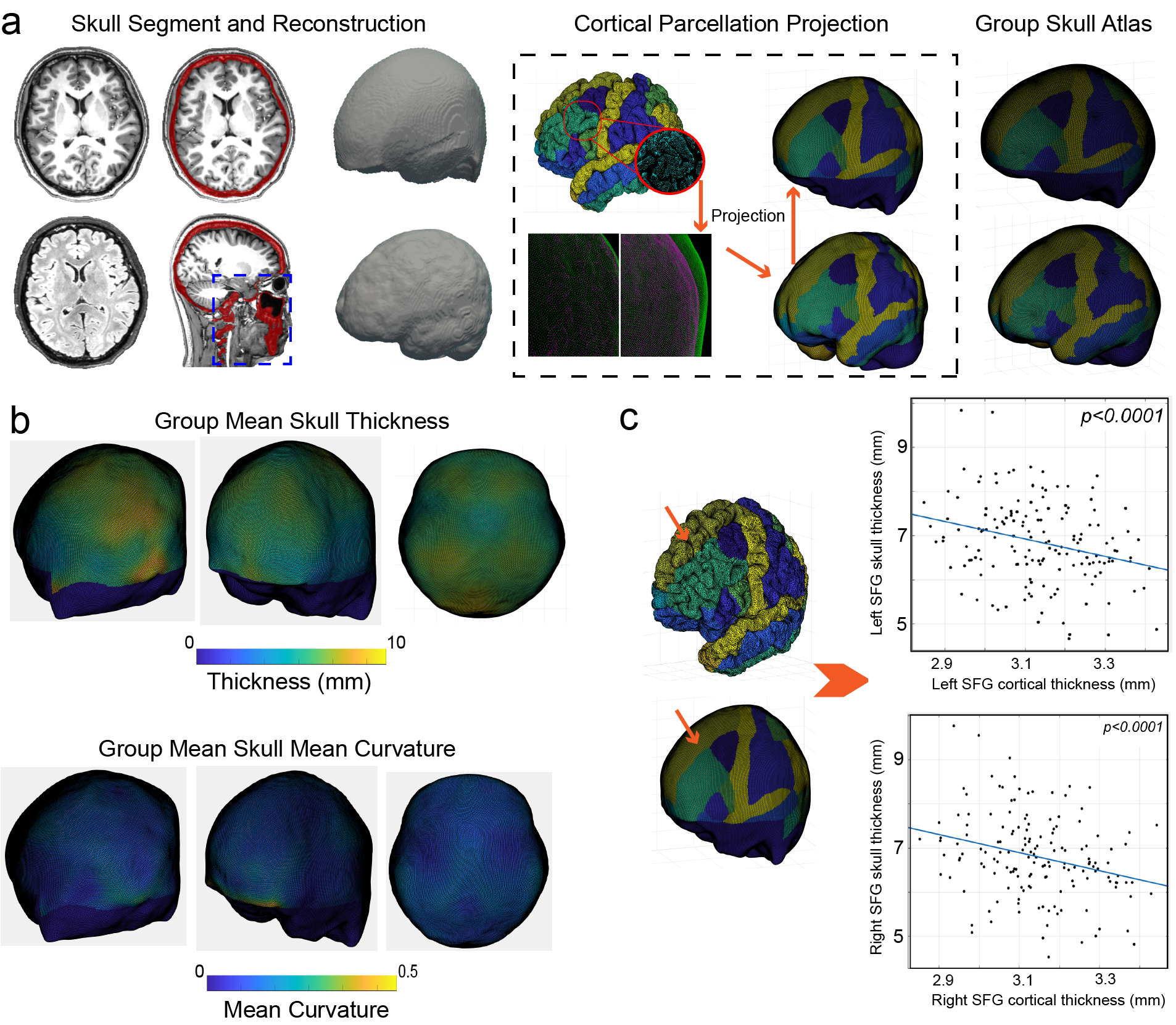
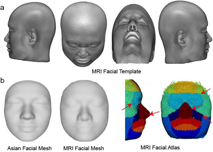
Figure 1a illustrates the pipeline for constructing the skull atlas[6]. The skull structure inside the red dotted box was removed. We computed skull thickness between the inner and outer surfaces, finding greater thickness in the occipital bones, particularly around the occipital protuberance. The skull mean curvature was also high near the eye socket and occipital protuberance (Figure 1b). Our correlation analysis (Figure 1c) between brain cortex and corresponding skull regions revealed a significant negative correlation between the cortical thickness of the bilateral superior frontal gyrus and corresponding skull regions after correcting for multiple comparisons (p < 0.0001).In Figure 2a, our voxel-based facial template is showcased, while Figure 2b present the facial mesh template with an existing Asian facial template for registration and segmentation [7]. Employing both rigid and non-rigid iterative closest point registration techniques, we have registered our facial mesh to the Asian facial template. Totally 7906 vertices were existed after registration, and all of the vertices were assigned with a unique label value. Finally, our facial atlas included 16 distinct regions, such as mouth, nose, bilateral eyes, bilateral nostril, and bilateral forehead. The red arrows in the figures highlight crucial landmarks determined by regional boundaries. These landmarks could be used to compute landmark distance and principle components of coordinates. This detailed mapping enhances our understanding of facial structure variations and supports further advances in facial analysis and reconstruction.
Conclusions
Our study presents a groundbreaking MRI-aided atlas of the human face and cranial vault, overcoming limitations of traditional imaging methods. Utilizing T1-weighted and T2-FLAIR MRI images from 148 individuals, we meticulously mapped craniofacial structures, revealing notable anatomical details, especially in the occipital region. The resulting skull and facial atlases show significant advancements in craniofacial analysis. The facial atlas, segmented into 16 distinct regions, enhances our understanding of structural variations and supports innovative approaches in facial analysis and reconstruction. Our present study marks a significant stride in clinical and neuroscientific research, aiding in the development of non-invasive diagnostics.Acknowledgements
This work was supported by the National Natural Science Foundation of China (No. 81971583), Shanghai Municipal Science and Technology Major Project (No. 2018SHZDZX01), National Key R&D Program of China (No. 2018YFC1312900), Shanghai Natural Science Foundation (No. 20ZR1406400).
This work was also supported by Shanghai Municipal Science and Technology Major Project (2017SHZDZX01 to L.J. and S.Wang, 2018SHZDZX01 to W.Q.); National Key Research and Development Project (2018YFC0910403 to S.Wang); CAS Interdisciplinary Innovation Team Project (to S.Wang); "Strategic Priority Research Program" of the Chinese Academy of Sciences (XDB38020400 to S.Wang); Max Planck-CAS Paul Gerson Unna Independent Research Group Leadership Award (to S.Wang); National Natural Science Foundation of China (31521003 to L.J., 31900408 to M.Z.); National Science & Technology Basic Research Project (2015FY111700 to L.J.); CAMS Innovation Fund for Medical Sciences (2019-I2M-5-066 to L.J.); The 111 Project (B13016 to L.J.); China Postdoctoral Science Foundation (2019M651352 to M.Z., 2020M670984 to W.Q.)
References
1. White, J.D., et al., Insights into the genetic architecture of the human face. Nature genetics, 2021. 53(1): p. 45-53.
2. Naqvi, S., et al., Shared heritability of human face and brain shape. Nature Genetics, 2021. 53(6): p. 830-839.
3. Kolabas, Z.I., et al., Distinct molecular profiles of skull bone marrow in health and neurological disorders. Cell, 2023. 186(17): p. 3706-3725. e29.
4. Saturnino, G.B., et al., SimNIBS 2.1: a comprehensive pipeline for individualized electric field modelling for transcranial brain stimulation. Brain and human body modeling: computational human modeling at EMBC 2018, 2019: p. 3-25.
5. Chakravarty, M.M., et al., Automated analysis of craniofacial morphology using magnetic resonance images. PLoS One, 2011. 6(5): p. e20241.
6. Lillie, E.M., et al., Evaluation of skull cortical thickness changes with age and sex from computed tomography scans. Journal of bone and mineral research, 2016. 31(2): p. 299-307.
7. Zhang, M., et al., Genetic variants underlying differences in facial morphology in East Asian and European populations. Nature Genetics, 2022. 54(4): p. 403-411.
Figures