1940
Brain Alterations in Nasopharyngeal Carcinoma patients after Chemotherapy based Multiplex MR imaging: A longitudinal MRI study1Hubei Cancer Hospital, Tongji Medical College, Huazhong University of Science and Technology, Wuhan, China, 2United Imaging Healthcare, Shanghai, China
Synopsis
Keywords: Head & Neck/ENT, Quantitative Imaging
Motivation: The objective of this study was to examine the potential impact of chemotherapy on the brain in patients with nasopharyngeal carcinoma (NPC) using Multiplex (MTP) MR imaging sequence, which generate multiple contrasts with one-time scan.
Goal(s): We aimed to investigate the altered MTP parameters and their correlation with clinical cognitive scores before and after chemotherapy.
Approach: All patients underwent MTP imaging, and the data were analyzed using BrainTool software.
Results: The findings revealed that patients who underwent chemotherapy exhibited reduced brain volume and elongated T2Star in some brain regions, although no significant correlation was observed with clinical cognitive scores.
Impact: The newly MTP MR sequence can evaluate brain alterations in NPC patients following chemotherapy. The observed decrease in volume values and increase in T2Star properties within brain regions may suggest neuron and myelin sheath damages in NPC patients after chemotherapy.
Introduction
Pre-irradiation induction chemotherapy has emerged as the primary treatment option for patients with NPC. Prior research has provided the direct influence of chemotherapy on the structural integrity of both white matter1 and gray matter2 within the central nervous system. Investigations3-6have revealed that prior to the observable changes in brain regions, there are already subtle structural and functional abnormalities in the brains of NPC following radiation. However, alterations in brain region properties and cognitive changes in NPC after chemotherapy remains limited. The MRI sequences employed in the aforementioned studies usually could generate only one kind of contrast with one scan and may only provide limited imaging information. Multiplex (MTP) MRI technology enables the acquisition of multiple contrasts and quantitative parameters from a single scan. It is necessary to explore its application of brain cognitive function abnormalities after chemotherapy in NPC. We hypothesized that patients with NPC would exhibit altered MTP properties before and after chemotherapy. The alterations could be correlated with potential cognitive changes after chemotherapy.Method
This study included a cohort of twenty-three patients with pathologically confirmed NPC who underwent MTP imaging before and after receiving 2-cycle chemotherapy. Montreal Cognitive Assessment (MoCA, Beijing Version) tests were administered on the same MRI scan day. All MRI data were acquired using a 3.0 T MRI scanner (uMRI 790, United Imaging Healthcare, Shanghai, China) equipped with a 32-channel phased-array head coil. A single MTP scan allowed for the acquisition of multiple contrasts, including aT1W, SWI, cPDW, cT1w, T2Star, T1Map, PDMap, R2Star, and QSM. The data were acquired using a MTP imaging sequence with the following parameters: repetition time (TR) = 35 ms, echo time (TE) = 3.01/7.02/9.68/13.69/16.35/20.36/23.02 ms, voxel = 1 × 1 × 2 mm3, slice thickness/ intersection gap = 1/0 mm, field of view (FOV) = 224 × 224 mm2, matrix size = 224 × 224, scan time = 6.21 min.MTP data were analyzed utilizing BrainTool software (uMRI 790, United Imaging Healthcare, Shanghai, China). The brain image was segmented into 106 bilateral brain regions based on deep learning techniques. Segmented MTP images underwent normalization, multimodal fusion, quantification analysis. Properties were obtained for each of the bilateral 106 brain regions. Some properties of the whole-brain white matter, grey matter, cerebrospinal fluid, and choroid plexus, were regressed out from the dataset as covariates. The normality of the remaining 89 brain regions' data was assessed using the Kolmogorov-Smirnov test in SPSS software version 22 (http://www.spss.com). A single-group paired t-test (https://fsl.fmrib.ox.ac.uk/fsl/fslwiki/PALM) was utilized to analyse the differences in properties before and after the 2-cycle chemotherapy(family-wise error (FWE) threshold of p < 0.05). Pearson correlations were computed using SPSS v.22.0 to examine the relationship between altered regional values and MoCA scores in NPC. Linear regression analysis was utilized to predict changes in MoCA scores following 2-cycle chemotherapy in NPC.
Result
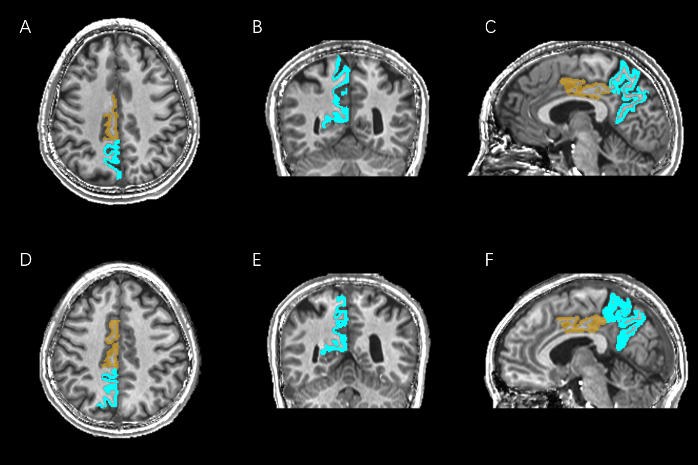
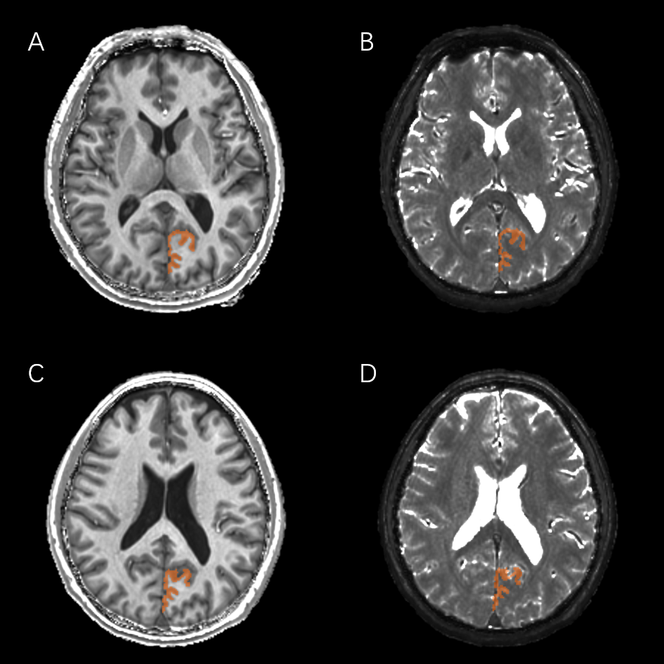
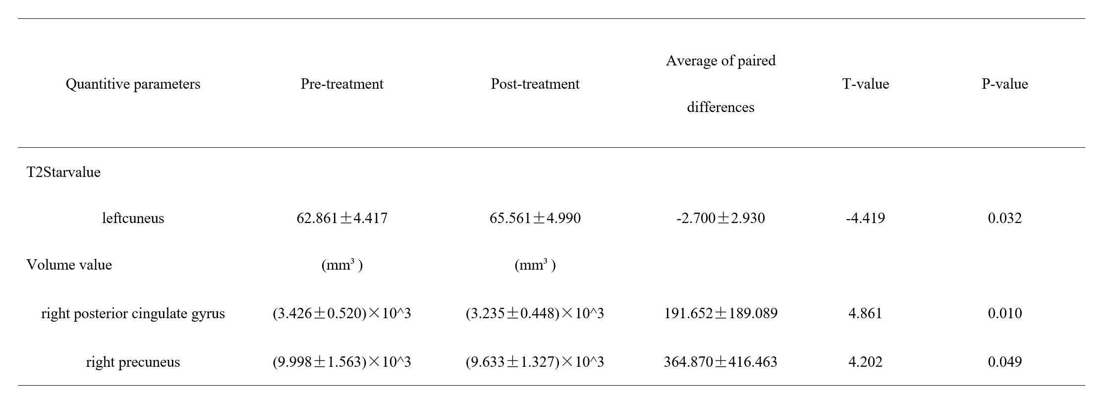
Compared to NPC patients prior to chemotherapy, NPC patients who underwent 2-cycle chemotherapy exhibited a significant decrease in volume in the right posterior cingulate gyrus (p = 0.010), a decrease in volume in the right precuneus (p = 0.049), and an increase in T2Star value in the left cuneus (p = 0.032) (Table 1). Figure 1 and 2 illustrate the altered brain regions in a patient before and after chemotherapy. The changes in the volume of the right posterior cingulate gyrus and right precuneus, as well as the T2Star value of the left cuneus, did not demonstrate a noticeable correlation with the change in the MoCA score (p > 0.05).Discussion
The MTP sequence can evaluate the potential alterations in brain regions of patients with NPC both prior to and following chemotherapy. This study indicates a decrease in volume in the right posterior cingulate gyrus and precuneus after chemotherapy, suggesting potential neuron impairment and loss of neuronal cells in visual space imagery and higher cognitive functions. The T2Star value of the left cuneus increased in the post group, possibly indicating myelin sheath damages in visual space information. The Pearson correlation analysis showed no significant correlation between brain volume reductions or T2Star alterations and the MoCA scores. Regression analysis showed these values did not predict the changes in MoCA scores in a statistically significant manner. This may be attributed to the limited sample size and the relatively short duration of chemotherapy treatment.Conclusion
MTP sequence can evaluate the changes in NPC patients pre and post chemotherapy. This study suggest that diminished brain volume in visual space imagery and higher cognitive regions was accompanied by alterations in the mean T2Star characteristics of visual space information.Acknowledgements
NoneReferences
[1] Matsos A , Loomes M , Zhou I ,et al. Chemotherapy-induced cognitive impairments: White matter pathologies[J].Cancer treatment reviews, 2017, 61:6-14.
[2] Niu R , Du M , Ren J ,et al.Chemotherapy-induced grey matter abnormalities in cancer survivors: a voxel-wise neuroimaging meta-analysis[J].Brain Imaging and Behavior, 2020(Suppl).
[3]Leng, Peng, Fang,et al. Structural MRI research in patients with nasopharyngeal carcinoma following radiotherapy: A DTI and VBM study.[J].Oncology letters, 2017.
[4] Yang Y , Lin X , Li J ,et al. Aberrant Brain Activity at Early Delay Stage Post-radiotherapy as a Biomarker for Predicting Neurocognitive Dysfunction Late-Delayed in Patients With Nasopharyngeal Carcinoma[J].Frontiers in Neurology, 2019, 10.
[5] Zhang Xinyuan, Pan Jie, Lin Yuhao et al. Structural network alterations in patients with nasopharyngeal carcinoma after radiotherapy: A 1-year longitudinal study.[J] .Front Neurosci, 2022, 16: 1059320.
[6] Kang Y F , Chen R T , Ding H ,et al. Structure-Function Decoupling: A Novel Perspective for Understanding the Radiation-Induced Brain Injury in Patients With Nasopharyngeal Carcinoma[J].Frontiers in neuroscience, 2022, 16:915164.
[7]Ye Y, Lyu J, Hu Y, Zhang Z, Xu J, Zhang W. MULTI-parametric MR imaging with fLEXible design (MULTIPLEX). Magn Reson Med. 2022 Feb;87(2):658-673. doi: 10.1002/mrm.28999. Epub 2021 Aug 31. Erratum in: Magn Reson Med. 2023 May;89(5):2145. PMID: 34464011.
Figures