1939
The Feasibility of Synthetic MRI in Monitoring NPC with Clival Invasion Changes During Radiation Treatment1Department of Radiology, The First Affiliated Hospital of Guangxi Medical University, Nanning, China, 2GE Healthcare, MR Research China, Beijing, China
Synopsis
Keywords: Head & Neck/ENT, Cancer, Nasopharyngeal Carcinoma
Motivation: Complications from radiotherapy in nasopharyngeal carcinoma impact patient quality of life, necessitating methods to determine the optimal radiation dose for patients.
Goal(s): To evaluate Synthetic MRI's ability to monitor clival invasion and its changes during treatment in nasopharyngeal carcinoma patients.
Approach: T1, T2 and PD values of clivus were acquired before treatment, at radiation doses of 6420cGy and 7062cGy, and were compared with each other.
Results: Prior to treatment, the clival T1 values in clival invasion patients were higher than those without clival invasion. No difference was found in Quantitative values between the radiation dose reached 6420cGy and 7062cGy.
Impact: Synthetic MRI can assess clival invasion in nasopharyngeal carcinoma and its changes during radiation treatment, potentially enabling the evaluation of the optimal radiation dose for patient.
Introduction
Nasopharyngeal carcinoma (NPC) is a common malignancy of the nasopharyngeal region in Southeast Asia1. Induction chemotherapy combined with intensity-modulated radiation therapy is the primary treatment for advanced stages of the disease2. However, radiation therapy can potentially lead to necrosis of the clivus and subsequent complications, which are contributors to poor patient prognosis and a decline in quality of life3,4. Therefore, reducing the radiation dose to the osseous region of the skull base, while ensuring effective treatment of the lesion, is crucial for minimizing radiogenic damage to the clivus. The quantitative parameters, acquired with Synthetic MRI (SyMRI) technique, have been widely used for the diagnosis and therapeutic efficacy prediction in various tumors5. Thus, the study aims to evaluate the feasibility of SyMRI in assessing the therapeutic effects of treatments at varying radiation doses in patients with nasopharyngeal carcinoma and bone invasion of the clivus.Materials and Methods
A prospective cohort of 7 patients with nasopharyngeal carcinoma staged T3 or higher were recruited from First Affiliated Hospital of Guangxi Medical University. The patients underwent induction chemotherapy followed by intensity-modulated radiation therapy. Among these, 4 patients presented with clival invasion before treatment, and 3 did not. Additionally, according to RECIST criteria, four patients experienced complete recovery after treatment with a 6420cGy radiation dose, while the remaining three patients were still in partial remission after treatment with a 7062cGy radiation dose.For each patient, MRI examinations were conducted at three time points using a 3.0 Tesla SIGNA Premier scanner (GE Healthcare, WI, USA): before clinical intervention, when the radiation dose reached approximately 6420cGy, and again at approximately 7062cGy. To collect images for the SyMRI technique, a 2D multiple-dynamic multiple-echo (MDME) sequence was utilized. Key parameters of the MDME sequence included an in-plane voxel size of 2.0 mm by 2.0 mm, a slice thickness of 6 mm with no interslice gap.
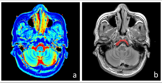
T1 mapping, T2 mapping, PD mapping and synthetic T1w images were generated with the vendor-provided postprocessing software (SyntheticMR, v11.2.2). Two experienced radiologists manually delineated the volume of interest (VOI) of the clivus on synthetic T1w images, using ITK-SNAP software. The mean values of T1, T2, and PD within the VOI were extracted and compared across different data acquisition time points.
Results
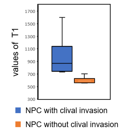
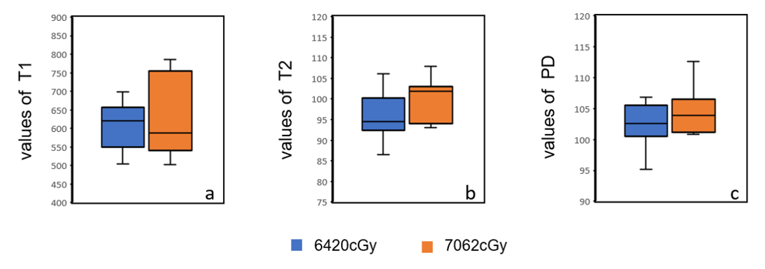
Fig. 2 The mean values of T1 is more sensitive than the mean values of T2 and PD in reflecting the clival invasion before clinical intervention. The mean values of T1 in the group with the clival invasion are higher than those in the group without the clival invasion.Fig. 3 When the radiation dose for nasopharyngeal cancer patients reaches approximately 6420 cGy compared to around 7062 cGy (at the end of treatment), there are no significant differences in the mean values of T1, T2, and PD.
Discussion
This study was designed to investigate the application of SyMRI technique for evaluating changes in the clivus during the treatment of nasopharyngeal carcinoma. Our results demonstrate that T1 mapping can accurately reflect the extent of tumor invasion in the clivus, likely due to factors such as bone marrow edema and transformation resulting from tumor encroachment. No significant difference was observed in the clivus T1 values between patients treated with a radiation dose of 6420cGy and those receiving 7062cGy, indicating that additional radiation therapy does not lead to further benefit for patients in terms of bone invasion at the clivus. Moreover, this finding is consistent with clinical outcomes: patients showing partial remission at a radiation dose of 6420cGy remained in partial remission even when the dose was increased to 7062cGy. Consequently, our findings suggest that SyMRI technique could serve as a non-invasive monitoring approach for detecting changes in the clivus during the treatment of nasopharyngeal carcinoma patients.Conclusions
Quantitative synthetic MRI technique can effectively assess the involvement of the clivus in nasopharyngeal carcinoma and monitor its changes throughout the treatment process.Acknowledgements
We sincerely thank the participants in this study.References
1. Chua MLK, Wee JTS, Hui EP, Chan ATC. Nasopharyngeal carcinoma. Lancet Lond Engl. 2016;387(10022):1012-1024. doi:10.1016/S0140-6736(15)00055-0
2. Ll T, Yp C, Cb C, et al. The Chinese Society of Clinical Oncology (CSCO) clinical guidelines for the diagnosis and treatment of nasopharyngeal carcinoma. Cancer Commun Lond Engl. 2021;41(11). doi:10.1002/cac2.12218
3. Han P, Wang X, Liang F, et al. Osteoradionecrosis of the Skull Base in Nasopharyngeal Carcinoma: Incidence and Risk Factors. Int J Radiat Oncol Biol Phys. 2018;102(3):552-555. doi:10.1016/j.ijrobp.2018.06.027
4. Li S, Luo C, Huang W, et al. Value of skull base invasion subclassification in nasopharyngeal carcinoma: implication for prognostic stratification and use of induction chemotherapy. Eur Radiol. 2022;32(11):7767-7777. doi:10.1007/s00330-022-08864-7
5. Ji S, Yang D, Lee J, Choi SH, Kim H, Kang KM. Synthetic MRI: Technologies and Applications in Neuroradiology. J Magn Reson Imaging JMRI. 2022;55(4):1013-1025. doi:10.1002/jmri.27440
Figures