1938
Interleaved HYDROPS: towards motion robust imaging of endolymphatic hydrops after intravenous administration of gadolinium1Philips Japan, Tokyo, Japan, 2Department of Radiology, Saitama Medical University Hospital, Saitama, Japan, 3Philips Healthcare, Best, Netherlands
Synopsis
Keywords: Head & Neck/ENT, Head & Neck/ENT
Motivation: HYDROS is useful method for detection of endolymphatic hydrops in patients with Meniere’s disease, but it has a potential risk for motion of patients during the acquisition because of its long scan time, resulting in misalignments of subtraction images.
Goal(s): To develop a new sequence, called interleaved HYDROPS, which acquires PPI and PEI images with an interleaved manner in one single scan.
Approach: We developed the interleaved 3D IR-prepared TSE with variable inversion delays. Interleaved HYDROPS were compared with conventional HYDROPS for image quality, particularly in the motion simulated situation.
Results: Initial findings indicated good feasibility of interleaved HYDROPS, encouraging further clinical evaluation.
Impact: Interleaved HYDROPS could provide PPI and PEI images with minimized motion effects during scan, compared with conventional separately acquired sequence. It holds promise for increasing robustness for motion-induced subtraction errors, but further studies are warranted to confirm its full potential.
INTRODUCTION
For detecting endolymphatic hydrops in patients with Meniere’s disease, MR imaging at 4 hours after intravenous administration of a single dose of gadolinium-based contrast agent (GBCA) has become popular in clinical practice1,2. For assessment of endolymphatic hydrops after contrast enhancement, the subtraction of two images obtained with different inversion times, so called HYbriD of Reversed image Of Positive endolymph signal and native image of positive perilymph Signal (HYDROPS) sequence1 is used. HYDROPS image is the subtraction of a positive endolymph image (PEI) from a positive perilymph image (PPI). Obtaining these images requires long scan time because it is 3D high resolution acquisition with heavily T2 weighted FLAIR contrast, which is quite sensitive to low concentrations of GBCA in fluid, but these images are essentially low SNR. Hence, HYDROPS has a potential risk for motion of patients during the acquisition of both images because of its long scan time, resulting in misalignments of subtraction images.To solve this problem, we developed a new sequence, called interleaved HYDROPS, which acquires PPI and PEI images with an interleaved manner in one single scan. The purpose of this study was to evaluate the feasibility of interleaved HYDROPS in comparison with conventional separated method.
METHODS
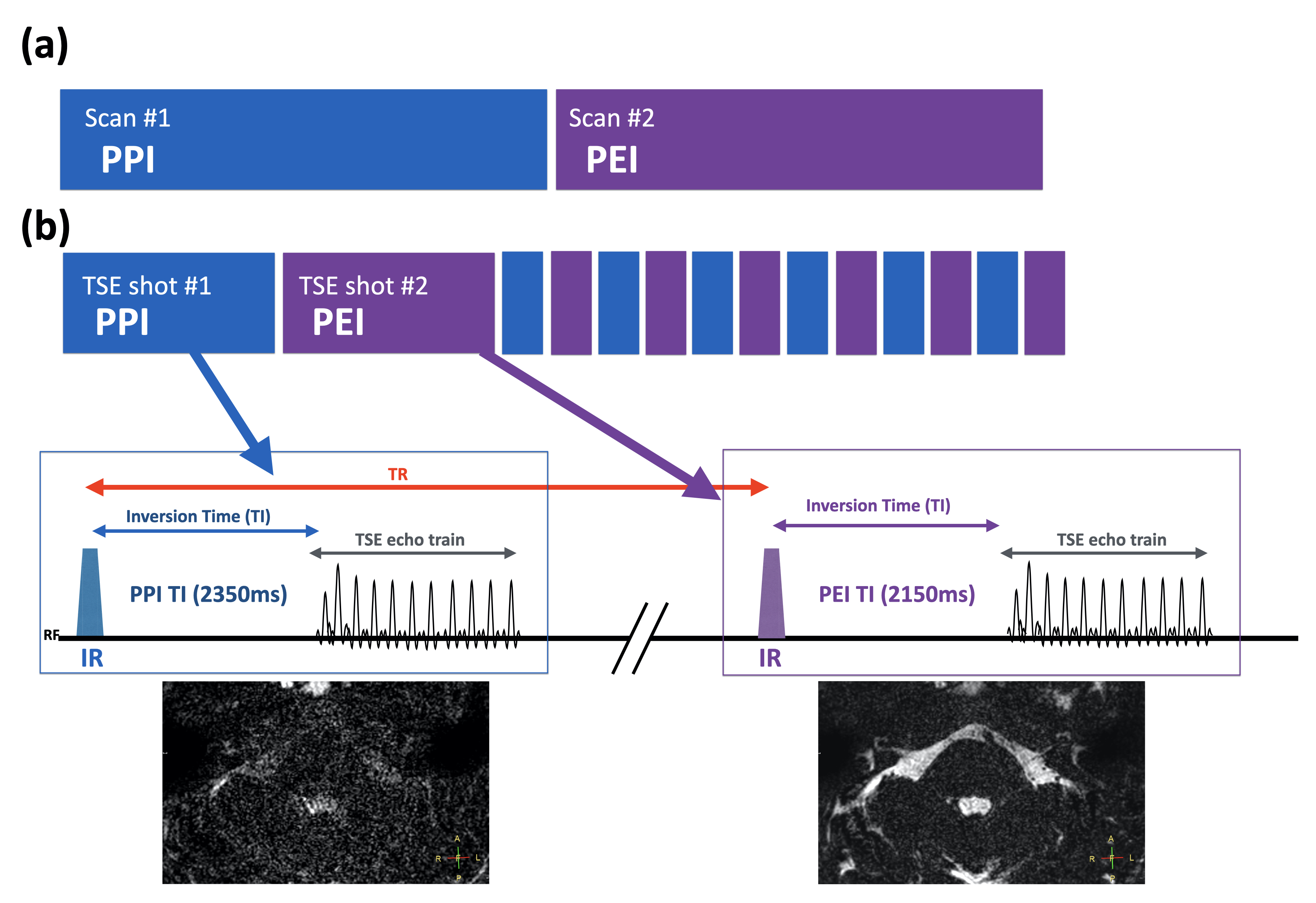
A schematic overview of the interleaved HYDROPS sequence is shown in Figure 1. It basically consists of two signal acquisition modules. Each module contains inversion recovery pre-pulse with different inversion delays followed by 3D turbo spin-echo (TSE) readouts. First module provides PPI images with the TI of 2350ms. After the repetition time (TR) period (9000ms in this study), the second signal acquisition is carried out preceded by the IR pulse with TI of 2150ms for obtaining PEI images. As all datasets including PPI and PEI images are inherently spatially aligned from one single scan, no post-processing for misalignment is needed.A total of 5 volunteers and 2 patients were examined on a 3.0T whole-body clinical system (Ingenia Elition X, Philips Healthcare, Best, the Netherlands). The study was approved by the local IRB, and written informed consent was obtained from all subjects.
Interleaved HYDROPS images were compared with conventional HYDROPS images for image quality, particularly in the motion simulated situation.
Imaging parameters for separated HYDROPS were; Axial, voxel size=0.6x0.6x2.0(1.0)mm3, 104 slices, TR=9000ms, TE=540ms, Refocusing flip angle=120°, TSE factor=256, TI02350(PPI)/2150(PEI)ms, SENSE reduction factor=2.2, and total acquisition time=7m48s for each.
Imaging parameters for interleaved HYDROPS were; Axial, voxel size=0.6x0.6x2.0(1.0)mm3, 104 slices, TR=9000ms, TE=540ms, Refocusing flip angle=120°, TSE factor=256, TI=2350(PPI)/2150(PEI)ms, SENSE reduction factor=2.2, and total acquisition time=15m18s.
RESULTS and DISCUSSION
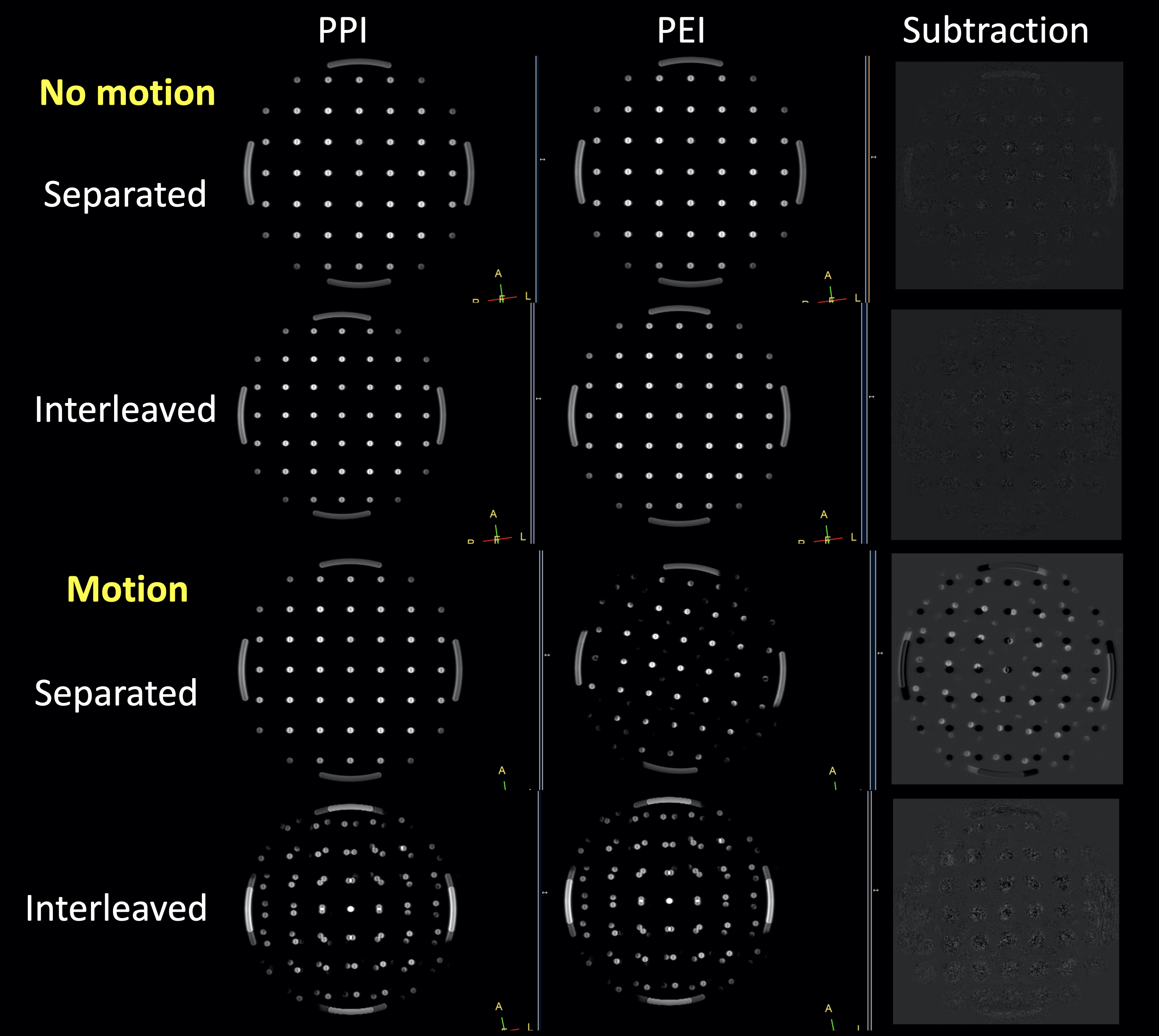
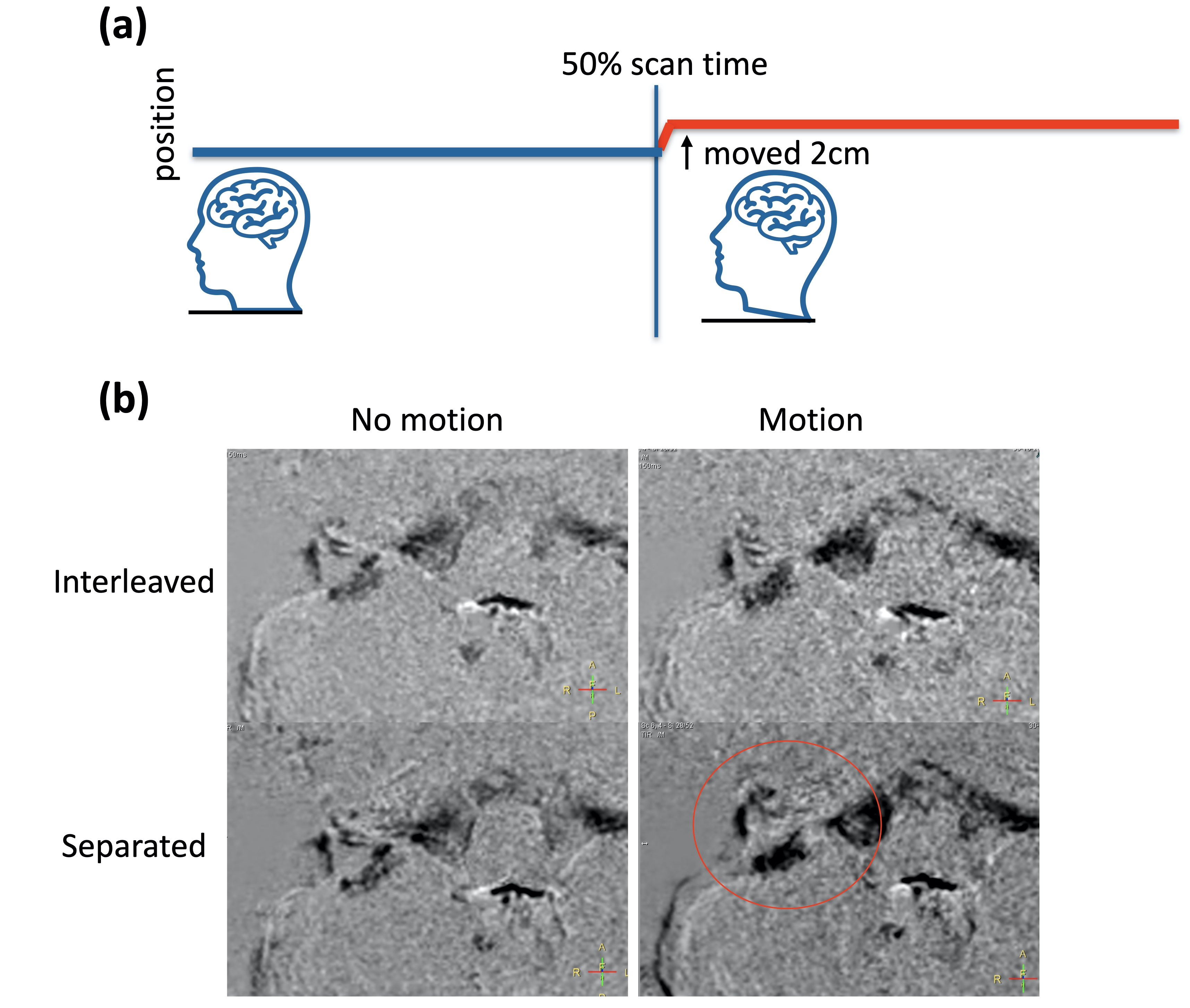
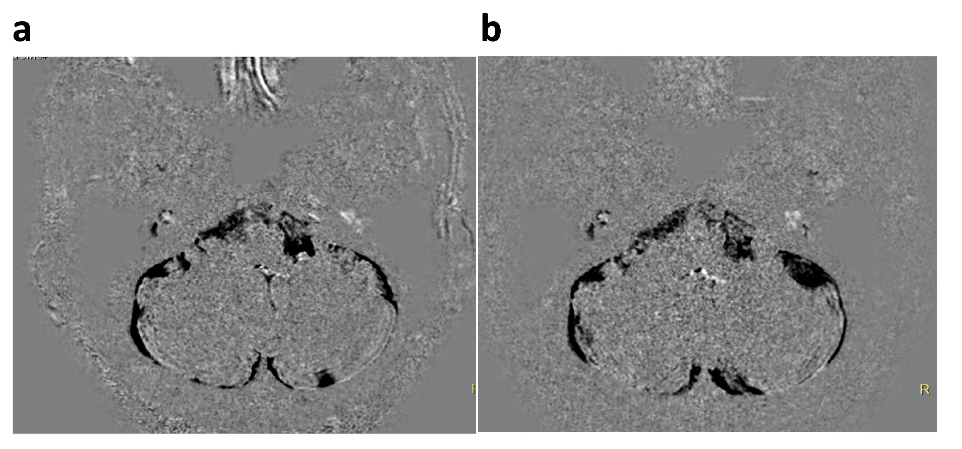
Figure 2 shows the results of phantom experiment without and with reproduction of motion during the scan. We manually moved the phantom about 2cm at the 50% of the total scan time. Interleaved HYDROPS showed robust subtraction images even phantom was moved during the scan. Also Figure 3 shows the representative results of volunteer experiment without and with motion during the scan. The volunteer moved about 2cm at the 50% of the total scan time and kept the same position after moving. Interleaved HYDROPS demonstrated robust subtraction images even the volunteer moved during the scan, whereas conventional separately acquired HYDROPS provided subtraction errors, some structures were blurred and shifted, due to the misalignment.One clinical comparison with a patient after GBCA administration is shown in Figure 4. Interleaved HYDROPS provided similar image information to conventional speared PPI/PEI acquisition.
Although further clinical investigation is needed, this method can be extended for the variants of HYDROPS, such as HYDROPS23, HYDROPS-Mi24 and HYDROPS2-Mi25, with different combination methods of PPI, PEI and/or MR cisternography images.
CONCLUSION
Interleaved HYDROPS enables to obtain both PPI and PEI images with minimized motion effects during scan by interleaved acquisition compared with the conventional separately acquired sequence. It holds promise for increasing robustness for motion-induced subtraction errors, but further clinical studies are warranted.Acknowledgements
No acknowledgement found.References
1. Nakashima T, et al. Meniere’s disease. Nat Rev Dis Primers 2016; 2:16028.
2. Naganawa S, et al. Improved HYDROPS: Imaging of Endolymphatic Hydrops after Intravenous Administration of Gadolinium. Magn Reson Med Sci. 2017 Oct 10;16(4):357-361. doi: 10.2463/mrms.tn.2016-0126.
3. Naganawa S, et al. Imaging of Ménière's disease by subtraction of MR cisternography from positive perilymph image. Magn Reson Med Sci. 2012;11(4):303-9. doi: 10.2463/mrms.11.303.
4. Naganawa S, et al Imaging of Ménière’s disease after intravenous administration of single-dose gadodiamide: utility of multiplication of MR cisternography and HYDROPS image. Magn Reson Med Sci 2013; 12:63–68.
5. Naganawa S, et al. MR Imaging of Endolymphatic Hydrops in Five Minutes. Magn Reson Med Sci. 2022 Jul 1;21(3):401-405. doi: 10.2463/mrms.ici.2021-0022. Epub 2021 Apr 24. PMID: 33896892; PMCID: PMC9316136.
Figures