1926
Deuterium labeling kinetics in rat cerebral cortex using ex-vivo 2H NMR spectroscopy1NMR Microimaging and Spectroscopy, Centre for Cellular and Molecular Biology, Hyderabad, India, 2Academy of Scientific and Innovative Research, Ghaziabad, India
Synopsis
Keywords: Neurotransmission, Deuterium, Neurometabolism, brain, Neuroscience, Spectroscopy
Motivation: Lower sensitivity of 13C NMR spectroscopy requires longer acquisition time while quantifying brain metabolites. An alternative technique is essential to aid faster detection.
Goal(s): To evaluate the kinetics of 2H labeling brain metabolites from [6,6’-2H2]glucose using 2H NMR spectroscopy.
Approach: The cortical extracts of rats infused with [6,6’-2H2]glucose were analyzed using 2H NMR spectroscopy.
Results: The signals of deuterated GlcC6, GluC4, LacC3, GABAC2and GlnC4 are seen in the 2H NMR spectrum of cortical extract obtained after 90 min of [6,6’-2H2]glucose infusion. The resonances of AspC2, GluC2, and GluC3 are absent suggesting complete loss/dilution of 2H from the TCA cycle intermediates beyond α-ketoglutarate.
Impact: Judicious use of 2H NMR together with suitable deuterated substrates may an alternative for neurometabolic analysis in neurological disorders.
INTRODUCTION
13C NMR spectroscopy is considered to be the gold standard for neurometabolic measurements. However, lower sensitivity of 13C NMR requires longer acquisition time. Recently, 2H NMR based neurometabolic measurements together with an infusion of deuterated glucose has been proposed.1This approach requires dedicated 2H Radio Frequency transmitters in the NMR spectrometer, and has lower sensitivity. Therefore, an indirect detection (subtraction) of deuterium by 1H NMR spectroscopy has been proposed very recently.2 However, unlike 13C-glucose metabolism where 13C labeled is lost only as CO2 in the tricarboxylic acid cycle (TCA), there is differential dilution/loss of 2H from the glycolytic and TCA cycle intermediates in case of [6,6’-2H2]glucose. It is not very clear, if the 2H label is incorporated in intermediates beyond α-ketoglutarate, and/or in molecules like glutamate and aspartate that are in the fast exchange with TCA cycle intermediates. Hence, the objective of the current study is to evaluate the kinetics of 2H labeling at different positions of aspartate, glutamate, and glutamine from [6,6’-2H2]glucose using 2H NMR spectroscopy.METHODS
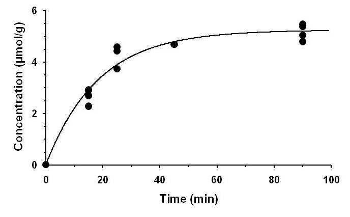
All animal experiments were performed under protocols approved by the Institutional Animal Ethics Committee of CCMB. Four weeks old SD (n=14) rats were used in the study. Rats were anesthetized with urethane (1.5 g/kg, intraperitoneal), and infused with [6,6’-2H2]glucose for 15, 25, 45 and 90 min using a bolus variable infusion rate.3 Blood was withdrawn from retro-orbital sinus, and brain metabolism was arrested by in situ freezing in liquid nitrogen. The brain was removed from the head, and was dissected in a cryostat (-20°C). Metabolites were extracted from the rat cerebral cortex using ethanol-extraction protocol.4 The lyophilized extracts were dissolved in a phosphate buffer (50 mM) containing D2O (1%) and TSP (0.25 mM).The 2H NMR spectroscopy of cortical extract from [6,6’-2H2]glucose infused rat was obtained with a zg2h pulse program using a triple resonance probe with following parameters: repetition time, 0.5 s; spectral width, 1197 Hz; and number of averages: 16384 in the block of 16 with 1024 scans each. An estimate of rate of glucose oxidation was obtained by the least square exponential fitting of the GluD4 concentration with time measured by 2H NMR.
RESULTS
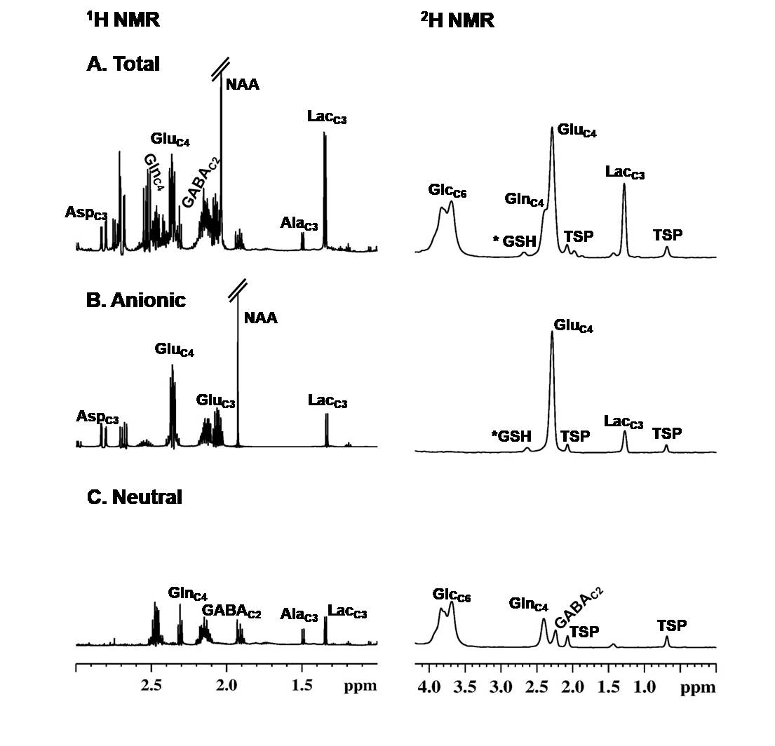
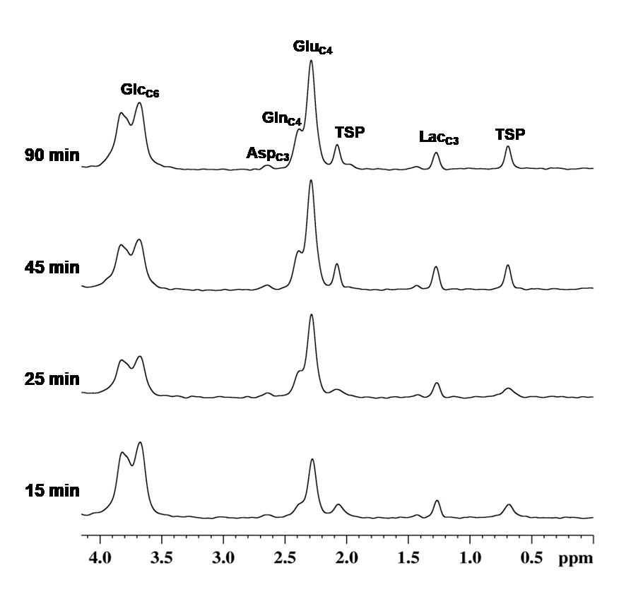
The 2H NMR and 1H spectra of cerebral cortex of rat infused with [6,6’-2H2]glucose for 90 min is shown in Fig.1. The presence of deuterium is seen in LacC3, GlcC6, GluC4, GlnC4and TSP. To gain further insight about labeling in other molecules, the neutral and anionic molecules were separated by passing the extract through anion exchange column. The signals of deuterated GlcC6, GABAC2 and GlnC4 are seen in the neutral fraction, while GluC4, LacC3 and TSP are seen in the anionic fraction. The resonances of AspC2 (~3.90 ppm), GluC2 (~3.76 ppm), and GluC3 (~2.09 ppm) are absent in the anionic fraction suggesting complete loss/dilution of 2H from the TCA cycle intermediates beyond α-ketoglutarate. The small signal at ~2.6 ppm may be of glutamate moiety of glutathione (GSH). The kinetics of GluD4 follows an exponential rise that saturates between 25 to 45 min (Fig. 2). The estimated glucose oxidation by exponential fitting was found to be 0.29±0.24 µmol/g/min, which is very much similar to that obtained by 1H-[13C]-NMR measurement in rats maintained under similar condition.5DISCUSSION
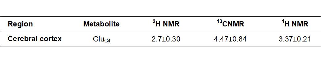
The findings of the study suggests that 2H from [6,6’-2H2]glucose is seen in metabolites till α-ketoglutarate(α-KG)/glutamate, and complete absence of 2H in TCA intermediates beyond α-KG. The labeling information of the second turn of the TCA cycle is important for the accurate estimation of the exchange rate (between α-KG and glutamate; oxaloacetate and aspartate) and TCA cycle flux in 13C NMR measurements. As there is no information about 2H label incorporation in the other carbon position of glutamate and GABA in 2H NMR measurements, the metabolic rates obtained from using this approach may be underestimated. The deuterium labeling of GluD4 from [6,6’-2H2]glucose measured using 2H NMR and indirect methods like 1H NMR (subtraction method) is towards the lower side when compared with 1H-[13C]-NMR measured labeling of GluC4 from [1,6-13C2]glucose (Table 1).Acknowledgements
This study was supported by Council of Scientific and Industrial Research (CSIR-CCMB). the authors would like to thank Dr. Kamal Saba and Dr. Narayan Datt Soni for their contribution in this study.References
1. de Graaf, R. A., Thomas, M. A., Behar, K. L., & De Feyter, H. M. (2021). Characterization of Kinetic Isotope Effects and Label Loss in Deuterium-Based Isotopic Labeling Studies. ACS chemical neuroscience, 12(1), 234–243.
2. Rich, L. J., Bagga, P., Wilson, N. E., Schnall, M. D., Detre, J. A., Haris, M., & Reddy, R. (2020). 1H magnetic resonance spectroscopy of 2H-to-1H exchange quantifies the dynamics of cellular metabolism in vivo. Nature biomedical engineering, 4(3), 335–342. https://doi.org/10.1038/s41551-019-0499-8
3. Fitzpatrick, S. M., Hetherington, H. P., Behar, K. L., & Shulman, R. G. (1990). The flux from glucose to glutamate in the rat brain in vivo as determined by 1H-observed, 13C-edited NMR spectroscopy. Journal of cerebral blood flow and metabolism : official journal of the International Society of Cerebral Blood Flow and Metabolism, 10(2), 170–179.
4. Patel AB, Rothman DL, Cline GW and Behar KL (2001) Glutamine is the major precursor for GABA synthesis in rat neocortex in vivo following acute GABA-transaminase inhibition. Brain Res 919:207-220.
5. Chowdhury, G. M., Patel, A. B., Mason, G. F., Rothman, D. L., & Behar, K. L. (2007). Glutamatergic and GABAergic neurotransmitter cycling and energy metabolism in rat cerebral cortex during postnatal development. Journal of cerebral blood flow and metabolism : official journal of the International Society of Cerebral Blood Flow and Metabolism, 27(12), 1895–1907.
Figures