1920
The anterior cingulate gyrus exhibits an excitatory-inhibitory balance that correlates with working memory1Department of Radiology, Shandong Provincial Hospital Affiliated to Shandong First Medical University, Jinan, China, 2Russell H. Morgan Department of Radiology and Radiological Science, The Johns Hopkins University School of Medicine, Baltimore, MD, USA, Baltimore, MD, United States, 3Philips Healthcare, Shanghai, China, Shanghai, China
Synopsis
Keywords: Neurotransmission, Spectroscopy
Motivation: Examining the impact of the excitatory-inhibitory balance on cognitive function in healthy individuals holds great significance in research.
Goal(s): Exploring the excitatory-inhibitory balance at the neurotransmitter level and studying the relationship between excitatory-inhibitory balance and cognitive functions.
Approach: The study involved the collection of glutamate and γ-aminobutyric acid using magnetic resonance spectroscopy in 268 healthy participants, alongside the assessment of cognitive function in the subjects.
Results: In the anterior cingulate cortex, we observed a positive correlation between glutamate and γ-aminobutyric acid levels. Additionally, we found that a higher ratio of excitatory balance was associated with improved working memory performance at this specific location.
Impact: This study enhances our understanding of the excitatory-inhibitory balance at the neurotransmitter level and identifies a correlation between the level of excitatory inhibition and cognitive function. These findings provide valuable insights into the impact of excitatory inhibition on cognitive function.
Introduction
Excitatory-inhibitory (E/I) balance is pivotal for the development and functionality of cortical microcircuits and broader neural networks. Deviations from this balance could be implicated in neurological and psychiatric afflictions such as Alzheimer’s disease1 2 3 4, epilepsy5,6, and obsessive-compulsive disorder7. Glutamate (Glu) and gamma-aminobutyric acid (GABA) are the primary excitatory and inhibitory neurotransmitters in the central nervous system, respectively. In magnetic resonance spectroscopy studies, the Glu or Glx (including Glu and glutamine) to GABA+ (including GABA and macromolecules) ratio frequently symbolizes the E/I balance. However, only a handful of spectroscopy studies examining this balance in healthy human brains have been conducted, and their findings diverge8-10. Moreover, there is a noticeable lack of comprehensive studies in the field of spectroscopy that examine the influence of the excitatory-inhibitory (E/I) neurotransmitter ratio on cognitive abilities among healthy individuals, especially in studies with larger sample sizes11,12.Method
The research involved 268 healthy individuals, comprising 102 men and 166 women, aged between 21 and 70. The average age with its standard deviation was 44.30 ±12.05years. All the individuals belonged to the Han Chinese group, were fluent in Mandarin, and predominantly right-handed.All the individuals underwent a comprehensive cognitive assessment, which took roughly sixty minutes. The testing process was systematic: We gauged cognitive abilities using the Montreal Cognitive Assessment (MoCA)13, the Auditory Verbal Learning Test, in its Chinese adaptation (ALVT)14, the Symbol Digit Modalities Test (SDMT) 15, the Stroop Color-Word Interference Test (Stroop) 16, the Trail-Making Test (TMT) Part A (TMT-A) and Part B (TMT-B) 17 and the Rey-Osterrieth Complex Figure Test (RCFT) 18.
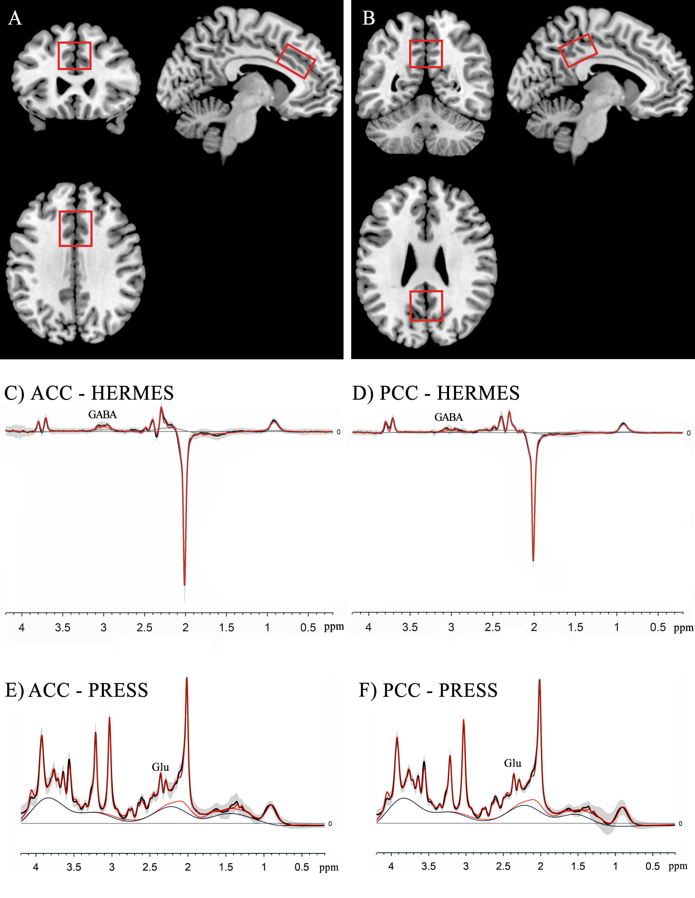
Participants were scanned using a 3.0 Tesla MR device (Ingenia CX, Philips), which had a 32-channel head coil. For obtaining structural information, we applied a 3D T1-weighted method. The levels of Glu and GABA+ in the anterior cingulate cortex (ACC) and the posterior cingulate cortex (PCC) were evaluated using two distinct techniques: the PRESS sequence and the HERMES sequence. Using Gannet 3.119, we processed the HERMES data that suppressed macromolecules by implementing a 3 Hz line expansion and using a Gaussian curve to adjust GABA peaks situated at 3 ppm. For determining Glu concentrations, the PRESS data were assessed using LCModel (version 6.3-1 M) 20.
Pearson partial correlations analyses were used to analyze the correlation between GABA+ and Glu, controlling for age and gender, and analyze the correlation between Glu/GABA+ and cognitive functions, controlling for age, gender, and educational attainment. Paired t-tests were used to compare Glu/GABA ratios in the ACC and the PCC.
Result
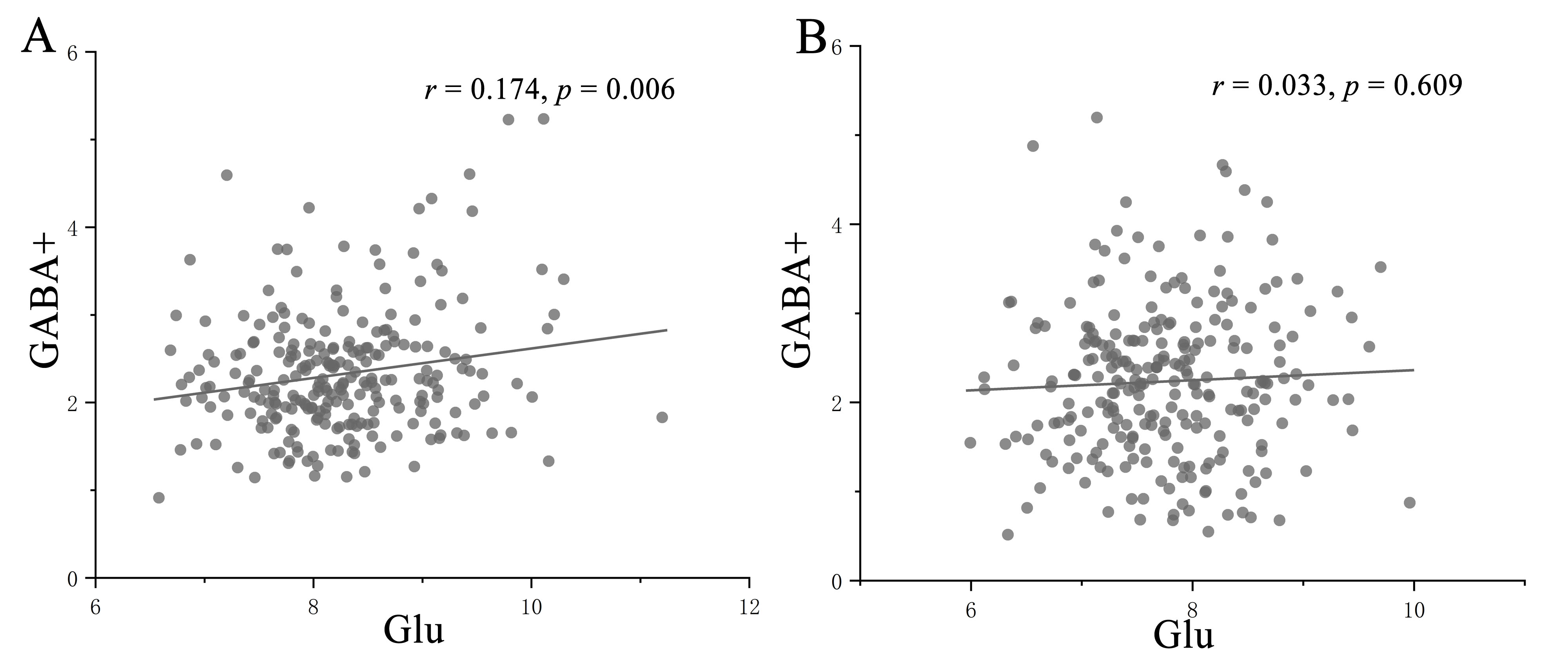
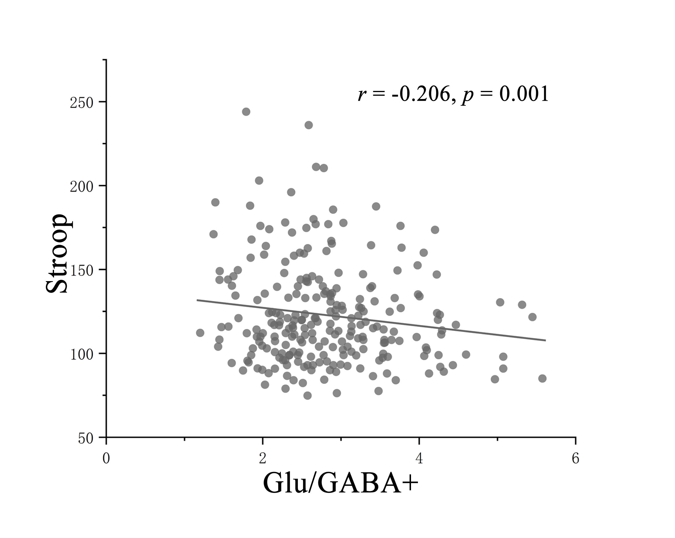
A positive correlation between Glu and GABA+ was found in the ACC (r = 0.174, p = 0.006), but not in the PCC (r = 0.033, p = 0.609). The Glu/GABA ratio was observed to be higher in the PCC compared to the ACC (p = 0.004). Besides, positive correlation between the ACC Glu/GABA+ ratio and Stroop was found (r = -0.206, p = 0.001). No correlations between Glu/GABA+ ratio and other cognitive functions have been identified in the ACC.Discussion
We observed a strong correlation between the MRS measures of GABA+ and Glu in the ACC with a sizable sample of healthy adults, suggesting a balance between inhibitory and excitatory levels in the human brain under normal conditions. This correlation was independent of variations in data acquisition methods across different sites, scanner vendors, subject demographics, or spectral quality. However, we did not observe a significant correlation between GABA and Glu in the PCC. This lack of correlation could be attributed to the elevated metabolism of the posterior cingulate gyrus, rendering it the most active region in the cerebral cortex during resting conditions21. This heightened activity leads to an increase in Glu levels22, consequently disturbing the balance between excitatory and inhibitory neurotransmission in this particular brain region. Interestingly, our discovery of higher Glu/GABA ratios in the PCC in comparison to the ACC supports our hypothesis.Interestingly, we observed a remarkable correlation between the Glu/GABA ratio in the anterior cingulate gyrus and the Stroop scores, suggesting that individuals with higher excitatory-inhibitory (E/I) ratios demonstrate superior working memory capacity. This finding is particularly noteworthy as the anterior cingulate gyrus is known to be involved in memory-related processes. Additionally, previous studies have shown that individuals with higher prefrontal E/I ratios exhibit improved volitional control 23, implying that heightened prefrontal excitation enhances cognitive performance.
Conclusion
This study enhances our understanding of the excitatory-inhibitory balance at the neurotransmitter level and identifies a correlation between the level of excitatory inhibition and cognitive function. These findings provide valuable insights into the impact of excitatory inhibition on cognitive function.Acknowledgements
This work was supported by the National Natural Science Foundation of China (Nos. 81601479), Taishan Scholars Project (No. tsqn201812147), Shandong Provincial Natural Science Foundation of China (Nos. ZR2021MH030).References
1 Palop, J. J. et al. Aberrant excitatory neuronal activity and compensatory remodeling of inhibitory hippocampal circuits in mouse models of Alzheimer's disease. 55, 697-711 (2007).
2 Sun, B. et al. Imbalance between GABAergic and glutamatergic transmission impairs adult neurogenesis in an animal model of Alzheimer's disease. 5, 624-633 (2009).
3 Bi, D., Wen, L., Wu, Z., Shen, Y. J. A. s. & Dementia. GABAergic dysfunction in excitatory and inhibitory (E/I) imbalance drives the pathogenesis of Alzheimer's disease. 16, 1312-1329 (2020).
4 Lauterborn, J. C. et al. Increased excitatory to inhibitory synaptic ratio in parietal cortex samples from individuals with Alzheimer’s disease. 12, 2603 (2021).
5 Bradford, H. J. P. i. n. Glutamate, GABA and epilepsy. 47, 477-511 (1995).
6 Olsen, R. W. & Avoli, M. J. E. GABA and epileptogenesis. 38, 399-407 (1997).
7 Biria, M. et al. Cortical glutamate and GABA are related to compulsive behaviour in individuals with obsessive compulsive disorder and healthy controls. 14, 3324 (2023).
8 Steel, A., Mikkelsen, M., Edden, R. A. & Robertson, C. E. J. N. Regional balance between glutamate+ glutamine and GABA+ in the resting human brain. 220, 117112 (2020).
9 Rideaux. No balance between glutamate+ glutamine and GABA+ in visual or motor cortices of the human brain: A magnetic resonance spectroscopy study. 237, 118191 (2021).
10 Rideaux, R. et al. On the relationship between GABA+ and glutamate across the brain. 257, 119273 (2022).
11 Shibata, K. et al. Overlearning hyperstabilizes a skill by rapidly making neurochemical processing inhibitory-dominant. 20, 470-475 (2017).
12 Takei, Y. et al. The inhibition/excitation ratio related to task-induced oscillatory modulations during a working memory task: a multtimodal-imaging study using MEG and MRS. 128, 302-315 (2016).
13 Nasreddine, Z. S. et al. The Montreal Cognitive Assessment, MoCA: a brief screening tool for mild cognitive impairment. J Am Geriatr Soc 53, 695-699, doi:10.1111/j.1532-5415.2005.53221.x (2005).
14 Zhao, Q., Lv, Y., Zhou, Y., Hong, Z. & Guo, Q. Short-term delayed recall of auditory verbal learning test is equivalent to long-term delayed recall for identifying amnestic mild cognitive impairment. PLoS One 7, e51157, doi:10.1371/journal.pone.0051157 (2012).
15 Harand, C., Mondou, A., Chevanne, D., Bocca, M. L. & Defer, G. Evidence of attentional impairments using virtual driving simulation in multiple sclerosis. Mult Scler Relat Disord 25, 251-257, doi:10.1016/j.msard.2018.08.005 (2018).
16 Perianez, J. A., Lubrini, G., Garcia-Gutierrez, A. & Rios-Lago, M. Construct Validity of the Stroop Color-Word Test: Influence of Speed of Visual Search, Verbal Fluency, Working Memory, Cognitive Flexibility, and Conflict Monitoring. Arch Clin Neuropsychol 36, 99-111, doi:10.1093/arclin/acaa034 (2021). 17 Wang, K. et al. Genome-wide association study identified INSC gene associated with Trail Making Test Part A and Alzheimer's disease related cognitive phenotypes. Prog Neuropsychopharmacol Biol Psychiatry 111, 110393, doi:10.1016/j.pnpbp.2021.110393 (2021).
18 Shin, M. S., Park, S. Y., Park, S. R., Seol, S. H. & Kwon, J. S. J. N. P. Clinical and empirical applications of the Rey-Osterrieth Complex Figure Test. 1, 892 (2006).
19 Edden, R. A., Puts, N. A., Harris, A. D., Barker, P. B. & Evans, C. J. Gannet: A batch-processing tool for the quantitative analysis of gamma-aminobutyric acid-edited MR spectroscopy spectra. J Magn Reson Imaging 40, 1445-1452, doi:10.1002/jmri.24478 (2014).
20 Provencher, S. W. Estimation of metabolite concentrations from localized in vivo proton NMR spectra. Magnetic resonance in medicine 30, 672-679 (1993).
21 Gusnard, D. A., Raichle, M. E. & Raichle, M. E. J. N. r. n. Searching for a baseline: functional imaging and the resting human brain. 2 (2001).
22 Rothman, D. L., Behar, K. L., Hyder, F. & Shulman, R. G. J. A. R. P. In vivo NMR studies of the glutamate neurotransmitter flux and neuroenergetics: implications for brain function. 65, 401-427 (2003).
23 Koizumi, A., Lau, H., Shimada, Y., Kondo, H. M. J. C. & Cognition. The effects of neurochemical balance in the anterior cingulate cortex and dorsolateral prefrontal cortex on volitional control under irrelevant distraction. 104 (2018).
Figures